
Main menu

  • Home
  • Content
    • Current
    • Ahead of print
    • Past Issues
    • JNM Supplement
    • SNMMI Annual Meeting Abstracts
    • Continuing Education
    • JNM Podcasts
  • Subscriptions
    • Subscribers
    • Institutional and Non-member
    • Rates
    • Journal Claims
    • Corporate & Special Sales
  • Authors
    • Submit to JNM
    • Information for Authors
    • Assignment of Copyright
    • AQARA requirements
  • Info
    • Reviewers
    • Permissions
    • Advertisers
  • About
    • About Us
    • Editorial Board
    • Contact Information
  • More
    • Alerts
    • Feedback
    • Help
    • SNMMI Journals
  • SNMMI
    • JNM
    • JNMT
    • SNMMI Journals
    • SNMMI

User menu

  • Subscribe
  • My alerts
  • Log in
  • Log out
  • My Cart

Search

  • Advanced search
Journal of Nuclear Medicine
  • SNMMI
    • JNM
    • JNMT
    • SNMMI Journals
    • SNMMI
  • Subscribe
  • My alerts
  • Log in
  • Log out
  • My Cart
Journal of Nuclear Medicine

Advanced Search

  • Home
  • Content
    • Current
    • Ahead of print
    • Past Issues
    • JNM Supplement
    • SNMMI Annual Meeting Abstracts
    • Continuing Education
    • JNM Podcasts
  • Subscriptions
    • Subscribers
    • Institutional and Non-member
    • Rates
    • Journal Claims
    • Corporate & Special Sales
  • Authors
    • Submit to JNM
    • Information for Authors
    • Assignment of Copyright
    • AQARA requirements
  • Info
    • Reviewers
    • Permissions
    • Advertisers
  • About
    • About Us
    • Editorial Board
    • Contact Information
  • More
    • Alerts
    • Feedback
    • Help
    • SNMMI Journals
  • View or Listen to JNM Podcast
  • Visit JNM on Facebook
  • Join JNM on LinkedIn
  • Follow JNM on Twitter
  • Subscribe to our RSS feeds
Research ArticleClinical Investigations

Prognostic Value of Myocardial Perfusion SPECT After Intravenous Bolus Administration of Nicorandil in Patients with Acute Ischemic Heart Failure

Yoshimitsu Fukushima, Shin-ichiro Kumita, Yukichi Tokita and Naoki Sato
Journal of Nuclear Medicine March 2016, 57 (3) 385-391; DOI: https://doi.org/10.2967/jnumed.115.162420
Yoshimitsu Fukushima
1Department of Radiology, Nippon Medical School Hospital, Tokyo, Japan; and
  • Find this author on Google Scholar
  • Find this author on PubMed
  • Search for this author on this site
Shin-ichiro Kumita
1Department of Radiology, Nippon Medical School Hospital, Tokyo, Japan; and
  • Find this author on Google Scholar
  • Find this author on PubMed
  • Search for this author on this site
Yukichi Tokita
2Department of Cardiovascular Medicine, Nippon Medical School Hospital, Tokyo, Japan
  • Find this author on Google Scholar
  • Find this author on PubMed
  • Search for this author on this site
Naoki Sato
2Department of Cardiovascular Medicine, Nippon Medical School Hospital, Tokyo, Japan
  • Find this author on Google Scholar
  • Find this author on PubMed
  • Search for this author on this site
  • Article
  • Figures & Data
  • Info & Metrics
  • PDF
Loading

Article Figures & Data

Figures

  • Tables
  • FIGURE 1.
    • Download figure
    • Open in new tab
    • Download powerpoint
    FIGURE 1.

    Kaplan–Meier curve in reference to MACE stratified by MBR-IR value y-axis represents cumulative event-free rate (log-rank test, P < 0.001).
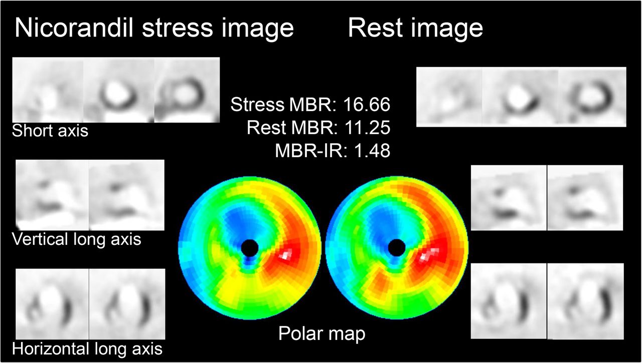
  • FIGURE 2.
    • Download figure
    • Open in new tab
    • Download powerpoint
    FIGURE 2.

    A 77-y-old man with acute ischemic heart failure in high–MBR-IR group. Rest myocardial perfusion SPECT images show anteroseptal–apical and distal inferior perfusion defects, and perfusion improvement is observed for anteroseptal wall in nicorandil-stress images, excluding central area of previous myocardial infarction site. MBR-IR is relatively high for an AIHF patient, with a value of 1.48. This patient remained MACE-free for entire 5-y follow-up period.

  • FIGURE 3.
    • Download figure
    • Open in new tab
    • Download powerpoint
    FIGURE 3.

    A 69-y-old woman with acute ischemic heart failure in low–MBR-IR group. Nicorandil stress and rest myocardial perfusion SPECT images show anteroseptal–apical perfusion defects, and perfusion improvement is observed only in marginal zone of previous myocardial infarction site in nicorandil-stress images. MBR-IR is low, with a value of 1.09. This patient was hospitalized due to deterioration of heart failure 189 d after initial onset.

Tables

  • Figures
    • View popup
    TABLE 1

    Patient Characteristics

    Characteristicn or mean ± SD
    No. of patients22
    Age (y)70 ± 12
    Male16 (73%)
    NYHA class (I/II/III/IV)0/2/2/18
    BNP (pg/mL)524 (258–744)
    Coronary risk factors
     Hypertension21 (95%)
     Dyslipidemia15 (68%)
     Diabetes mellitus13 (59%)
    Interval (d)
     Onset to first MPI8 (6–12)
     Nicorandil-stress and rest3 (3–5)
     Onset to CAG7 (4–15)
    Previous MI20 (91%)
    1VD/2VD/3VD2/7/13
    • NYHA = New York Heart Association; BNP = brain natriuretic peptide; MPI = myocardial perfusion imaging; MI = myocardial infarction; VD = vessel disease.

    • Categorical data are expressed as numbers, followed by percentages in parentheses, unless otherwise indicated; continuous data are expressed as mean ± SD or median (25th–75th).

    • View popup
    TABLE 2

    Cardiovascular Parameter Variations Between Nicorandil-Stress and Rest Conditions

    Cardiovascular parameterNicorandil-stressRestP
    Systolic BP (mm Hg)98 ± 16123 ± 15<0.001
    Diastolic BP (mm Hg)60 ± 670 ± 150.004
    Heart rate (bpm)78 ± 1567 ± 11<0.001
    LVEF (%)40 (31–44)36 (30–40)0.106
    LVEDV (mL)154 (118–191)161 (105–196)0.516
    LVESV (mL)96 (71–128)108 (63–131)0.276
    • BP = blood pressure.

    • Data are expressed as mean ± SD or median (25th–75th).

    • View popup
    TABLE 3

    Comparison of Clinical Profiles Between High– and Low–MBR-IR Groups

    CharacteristicHigh MBR-IR (n = 11 [50%])Low MBR-IR (n = 11 [50%])P
    Age (y)75 ± 965 ± 130.046
    Male7 (64%)9 (82%)0.635
    NYHA class0/0/2/90/2/0/90.883
    BNP (pg/mL)534 (189–965)513 (412–704)0.844
    Hypertension10 (91%)11 (100%)1.000
    Dyslipidemia6 (55%)9 (82%)0.361
    Diabetes mellitus5 (45%)8 (73%)0.387
    Previous myocardial infarction10 (91%)10 (91%)1.000
    SSS17 (15–24)25 (23–28)0.139
    SRS19 (16–28)27 (24–29)0.144
    Rest LVEF (%)38 (32–53)31 (30–38)0.216
    Rest LVEDV (mL)136 (90–165)186 (156–201)0.111
    Rest LVESV (mL)83 (43–116)118 (103–140)0.102
    Nicorandil-stress MBR29.49 (26.57–37.44)26.70 (16.96–31.93)0.151
    Rest MBR18.62 (16.62–26.80)25.39 (16.17–30.71)0.507
    MBR-IR1.55 (1.34–1.61)1.08 (1.02–1.10)<0.001
    MBR-IR in infarcted region1.06 (1.00–1.06)0.98 (0.97–1.00)0.149
    MBR-IR in ischemic region1.57 (1.41–1.74)1.18 (1.16–1.22)0.002
    • NYHA = New York Heart Association; BNP = brain natriuretic peptide; SSS = summed stress score; SRS = summed rest score.

    • Categorical data are expressed as counts (%), and continuous data are expressed as mean ± SD or median (25th–75th).

    • View popup
    TABLE 4

    Comparison of Clinical Profiles of All Patients Who Did and Did Not Experience MACE

    CharacteristicMACE (n = 12 [55%])No MACE (n = 10 [45%])P
    Age (y)70 ± 1071 ± 140.950
    Male9 (75%)7 (70%)1.000
    NYHA class0/0/1/110/2/1/70.185
    BNP (pg/mL)664 (420–794)408 (184–687)0.277
    Hypertension12 (100%)9 (90%)0.455
    Dyslipidemia9 (75%)6 (60%)0.652
    Diabetes mellitus7 (58%)6 (60%)1.000
    Previous myocardial infarction12 (100%)8 (80%)0.195
    SSS25 (21–28)20 (15–25)0.209
    SRS27 (23–29)22 (15–29)0.427
    Low MBR-IR10 (83%)1 (10%)0.002
    Rest LVEF (%)31(30–39)38 (31–53)0.477
    Treatments after myocardial perfusion imaging
     PCI3 (25%)1 (10%)0.594
     CABG1 (8%)1 (10%)1.000
     Nicorandil7 (58%)5 (50%)1.000
     ARB11 (92%)6 (60%)0.135
     β-blocker10 (83%)9 (90%)1.000
     Statin10 (83%)6 (60%)0.348
     Pacemaker implantation2 (17%)0 (0%)0.481
     ICD implantation1 (8%)0 (0%)1.000
    • NYHA = New York Heart Association; BNP = brain natriuretic peptide; SSS = summed stress score; SRS = summed rest score; PCI = percutaneous coronary intervention; CABG = coronary artery bypass grafting; ARB = angiotensin receptor blocker; ICD = implantable cardioverter-defibrillator.

    • Categorical data are expressed as counts (%), and continuous data are expressed as mean ± SD or median (25th–75th).

    • View popup
    TABLE 5

    Univariate and Multivariate Cox Regression Analysis for Occurrence of MACE

    UnivariateMultivariate
    CharacteristicHazard ratio95% confidence intervalPHazard ratio95% confidence intervalP
    Age0.9990.967–1.0320.945
    NYHA class IV1.5890.536–4.3120.404
    BNP1.0000.999–1.0010.1601.0010.999–1.0020.086
    Hypertension1.8330.246–13.6770.554
    Dyslipidemia1.2590.512–3.0970.615
    Diabetes mellitus0.9470.404–2.2200.901
    Previous myocardial infarction1.9200.446–8.2610.381
    SSS1.0090.966–1.0550.683
    SRS1.0030.964–1.0440.866
    Low MBR-IR3.6401.456–9.1030.0063.9131.552–9.8680.004
    Rest LVEF0.9920.964–1.0200.576
    Treatment after MPI
     PCI1.6010.538–4.7610.398
     CABG0.7710.179–3.3090.726
     Nicorandil1.0910.471–2.5290.839
     ARB1.6390.598–4.4950.337
     β-blocker1.0130.298–3.4510.983
     Statin1.3590.528–3.5010.525
    • NYHA = New York Heart Association; BNP = brain natriuretic peptide; SSS = summed stress score; SRS = summed rest score; MPI = myocardial perfusion imaging; PCI = percutaneous coronary intervention; CABG = coronary artery bypass grafting; ARB = angiotensin receptor blocker; ICD = implantable cardioverter-defibrillator.

PreviousNext
Back to top

In this issue

Journal of Nuclear Medicine: 57 (3)
Journal of Nuclear Medicine
Vol. 57, Issue 3
March 1, 2016
  • Table of Contents
  • Table of Contents (PDF)
  • About the Cover
  • Index by author
Print
Download PDF
Article Alerts
Sign In to Email Alerts with your Email Address
Email Article

Thank you for your interest in spreading the word on Journal of Nuclear Medicine.

NOTE: We only request your email address so that the person you are recommending the page to knows that you wanted them to see it, and that it is not junk mail. We do not capture any email address.

Enter multiple addresses on separate lines or separate them with commas.
Prognostic Value of Myocardial Perfusion SPECT After Intravenous Bolus Administration of Nicorandil in Patients with Acute Ischemic Heart Failure
(Your Name) has sent you a message from Journal of Nuclear Medicine
(Your Name) thought you would like to see the Journal of Nuclear Medicine web site.
Citation Tools
Prognostic Value of Myocardial Perfusion SPECT After Intravenous Bolus Administration of Nicorandil in Patients with Acute Ischemic Heart Failure
Yoshimitsu Fukushima, Shin-ichiro Kumita, Yukichi Tokita, Naoki Sato
Journal of Nuclear Medicine Mar 2016, 57 (3) 385-391; DOI: 10.2967/jnumed.115.162420

Citation Manager Formats

  • BibTeX
  • Bookends
  • EasyBib
  • EndNote (tagged)
  • EndNote 8 (xml)
  • Medlars
  • Mendeley
  • Papers
  • RefWorks Tagged
  • Ref Manager
  • RIS
  • Zotero
Share
Prognostic Value of Myocardial Perfusion SPECT After Intravenous Bolus Administration of Nicorandil in Patients with Acute Ischemic Heart Failure
Yoshimitsu Fukushima, Shin-ichiro Kumita, Yukichi Tokita, Naoki Sato
Journal of Nuclear Medicine Mar 2016, 57 (3) 385-391; DOI: 10.2967/jnumed.115.162420
Twitter logo Facebook logo LinkedIn logo Mendeley logo
  • Tweet Widget
  • Facebook Like
  • Google Plus One
Bookmark this article

Jump to section

  • Article
    • Abstract
    • MATERIALS AND METHODS
    • RESULTS
    • DISCUSSION
    • CONCLUSION
    • DISCLOSURE
    • Acknowledgments
    • Footnotes
    • REFERENCES
  • Figures & Data
  • Info & Metrics
  • PDF

Related Articles

  • This Month in JNM
  • PubMed
  • Google Scholar

Cited By...

  • No citing articles found.
  • Google Scholar

More in this TOC Section

  • Feasibility of Ultra-Low-Activity 18F-FDG PET/CT Imaging Using a Long–Axial-Field-of-View PET/CT System
  • Cardiac Presynaptic Sympathetic Nervous Function Evaluated by Cardiac PET in Patients with Chronotropic Incompetence Without Heart Failure
  • Validation and Evaluation of a Vendor-Provided Head Motion Correction Algorithm on the uMI Panorama PET/CT System
Show more Clinical Investigations

Similar Articles

Keywords

  • acute ischemic heart failure
  • nicorandil-stress myocardial perfusion imaging
  • single photon emission computed tomography
  • myocardium-to-background ratio increasing rate
  • major adverse cardiac event
SNMMI

© 2025 SNMMI

Powered by HighWire