
Main menu

  • Home
  • Content
    • Current
    • Ahead of print
    • Past Issues
    • JNM Supplement
    • SNMMI Annual Meeting Abstracts
    • Continuing Education
    • JNM Podcasts
  • Subscriptions
    • Subscribers
    • Institutional and Non-member
    • Rates
    • Journal Claims
    • Corporate & Special Sales
  • Authors
    • Submit to JNM
    • Information for Authors
    • Assignment of Copyright
    • AQARA requirements
  • Info
    • Reviewers
    • Permissions
    • Advertisers
  • About
    • About Us
    • Editorial Board
    • Contact Information
  • More
    • Alerts
    • Feedback
    • Help
    • SNMMI Journals
  • SNMMI
    • JNM
    • JNMT
    • SNMMI Journals
    • SNMMI

User menu

  • Subscribe
  • My alerts
  • Log in
  • My Cart

Search

  • Advanced search
Journal of Nuclear Medicine
  • SNMMI
    • JNM
    • JNMT
    • SNMMI Journals
    • SNMMI
  • Subscribe
  • My alerts
  • Log in
  • My Cart
Journal of Nuclear Medicine

Advanced Search

  • Home
  • Content
    • Current
    • Ahead of print
    • Past Issues
    • JNM Supplement
    • SNMMI Annual Meeting Abstracts
    • Continuing Education
    • JNM Podcasts
  • Subscriptions
    • Subscribers
    • Institutional and Non-member
    • Rates
    • Journal Claims
    • Corporate & Special Sales
  • Authors
    • Submit to JNM
    • Information for Authors
    • Assignment of Copyright
    • AQARA requirements
  • Info
    • Reviewers
    • Permissions
    • Advertisers
  • About
    • About Us
    • Editorial Board
    • Contact Information
  • More
    • Alerts
    • Feedback
    • Help
    • SNMMI Journals
  • View or Listen to JNM Podcast
  • Visit JNM on Facebook
  • Join JNM on LinkedIn
  • Follow JNM on Twitter
  • Subscribe to our RSS feeds
Research ArticleBasic Science Investigations

Tumor Uptake of Anti-CD20 Fabs Depends on Tumor Perfusion

Claudia Theresa Mendler, Annette Feuchtinger, Irina Heid, Michaela Aichler, Calogero D’Alessandria, Sabine Pirsig, Birgit Blechert, Hans-Jürgen Wester, Rickmer Braren, Axel Walch, Arne Skerra and Markus Schwaiger
Journal of Nuclear Medicine December 2016, 57 (12) 1971-1977; DOI: https://doi.org/10.2967/jnumed.116.176784
Claudia Theresa Mendler
1Nuklearmedizinische Klinik und Poliklinik, Klinikum rechts der Isar, Technische Universität München, München, Germany
2Munich Center for Integrated Protein Science (CIPS-M) and Lehrstuhl für Biologische Chemie, Technische Universität München, Freising (Weihenstephan), Germany
  • Find this author on Google Scholar
  • Find this author on PubMed
  • Search for this author on this site
Annette Feuchtinger
3Research Unit Analytical Pathology, Institute of Pathology, Helmholtz Zentrum München, Neuherberg, Germany
  • Find this author on Google Scholar
  • Find this author on PubMed
  • Search for this author on this site
Irina Heid
4Institute of Radiology, Klinikum rechts der Isar, Technische Universität München, München, Germany; and
  • Find this author on Google Scholar
  • Find this author on PubMed
  • Search for this author on this site
Michaela Aichler
3Research Unit Analytical Pathology, Institute of Pathology, Helmholtz Zentrum München, Neuherberg, Germany
  • Find this author on Google Scholar
  • Find this author on PubMed
  • Search for this author on this site
Calogero D’Alessandria
1Nuklearmedizinische Klinik und Poliklinik, Klinikum rechts der Isar, Technische Universität München, München, Germany
  • Find this author on Google Scholar
  • Find this author on PubMed
  • Search for this author on this site
Sabine Pirsig
1Nuklearmedizinische Klinik und Poliklinik, Klinikum rechts der Isar, Technische Universität München, München, Germany
  • Find this author on Google Scholar
  • Find this author on PubMed
  • Search for this author on this site
Birgit Blechert
1Nuklearmedizinische Klinik und Poliklinik, Klinikum rechts der Isar, Technische Universität München, München, Germany
  • Find this author on Google Scholar
  • Find this author on PubMed
  • Search for this author on this site
Hans-Jürgen Wester
5Pharmaceutical Radiochemistry, Technische Universität München, Garching, Germany
  • Find this author on Google Scholar
  • Find this author on PubMed
  • Search for this author on this site
Rickmer Braren
4Institute of Radiology, Klinikum rechts der Isar, Technische Universität München, München, Germany; and
  • Find this author on Google Scholar
  • Find this author on PubMed
  • Search for this author on this site
Axel Walch
3Research Unit Analytical Pathology, Institute of Pathology, Helmholtz Zentrum München, Neuherberg, Germany
  • Find this author on Google Scholar
  • Find this author on PubMed
  • Search for this author on this site
Arne Skerra
2Munich Center for Integrated Protein Science (CIPS-M) and Lehrstuhl für Biologische Chemie, Technische Universität München, Freising (Weihenstephan), Germany
  • Find this author on Google Scholar
  • Find this author on PubMed
  • Search for this author on this site
Markus Schwaiger
1Nuklearmedizinische Klinik und Poliklinik, Klinikum rechts der Isar, Technische Universität München, München, Germany
  • Find this author on Google Scholar
  • Find this author on PubMed
  • Search for this author on this site
  • Article
  • Figures & Data
  • Supplemental
  • Info & Metrics
  • PDF
Loading

Article Figures & Data

Figures

  • Additional Files
  • FIGURE 1.
    • Download figure
    • Open in new tab
    • Download powerpoint
    FIGURE 1.

    Autoradiography and biodistribution studies on Granta-519 and SUDHL-4 tumors. (A) Autoradiography of tumor sections from immunocompromised mice was performed 24 h after injection of 125I-Fab-ABD or 125I-Fab-PAS200 or 1 h after injection of 18F-FDG. (B and C) Biodistribution studies of Fab-ABD and Fab-PAS200 labeled with 125I were conducted 24 h after injection. Tumor-to-organ ratios are plotted for both αCD20 Fabs (mean ± SD; n = 5). (D) No strong correlation between tumor uptake and tumor weight is found for SUDHL-4 tumors 24 h after injection, irrespective of the Fab radiotracer used (Pearson coefficient of −0.32 for Fab-PAS200 and −0.36 for Fab-ABD).

  • FIGURE 2.
    • Download figure
    • Open in new tab
    • Download powerpoint
    FIGURE 2.

    Histologic and immunohistochemical characterization of Granta-519 and SUDHL-4 tumors. (A) Explanted tumors were stained with hematoxylin and eosin (H&E) or immunostained for the target antigen CD20, apoptosis marker caspase 3 (Casp3), proliferation marker Mib1, or endothelial vessel marker CD31 (scale bar, 50 μm). (B) Example is shown of H&E-stained SUDHL-4 tumor comprising an area of 6 × 10 mm. (C) Quantification of Mib1 staining reveals significantly stronger proliferation for Granta-519 tumors than for SUDHL-4 tumors (P = 0.01). (D) Quantification of vessel area (n = 5) reveals significantly higher values for Granta-519 tumors than for SUDHL-4 tumors (P < 0.01).

  • FIGURE 3.
    • Download figure
    • Open in new tab
    • Download powerpoint
    FIGURE 3.

    Visualization of vessels and αCD20 Cy5-Fab-PAS200 distribution in Granta-519 and SUDHL-4 tumors. Both xenograft models were stained in vivo with fluorescence-labeled αCD20 Fab-PAS200 (24 h before sacrifice) and fluorescence-labeled B. (G.) simplicifolia-isolectin 1 for vessels (5 min before sacrifice) and imaged by light-sheet fluorescence microscopy. (A) Both xenograft models show extensive vascularization (red) throughout. Heterogeneous penetration and distribution of labeled Fab-PAS200 (green) is observed across entire Granta-519 tumor, whereas for the SUDHL-4 tumor nearly no Fab-PAS200 outside vessels is detected. Heat map visualization confirms this different distribution pattern of Granta-519 and SUDHL-4 tumors (scale bar, 1,500 μm). (B) Different localization of labeled Fab-PAS200 (green) in relation to lectin-labeled vessels (red) is clearly visible in magnified images of the maximum-intensity projection of 25 virtual single optical slices. Granta-519 tumor shows perivascular distribution of Fab-PAS200, whereas SUDHL-4 tumor shows confinement of Fab-PAS200 to intravascular space (arrows).
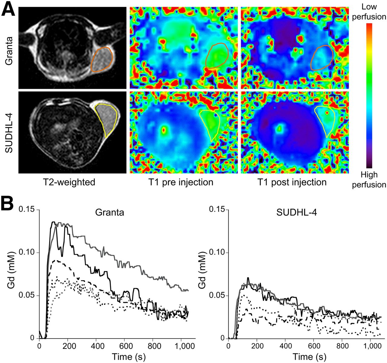
  • FIGURE 4.
    • Download figure
    • Open in new tab
    • Download powerpoint
    FIGURE 4.

    DCE MRI perfusion study of NHL xenograft models. (A) T2-weighted anatomic images are shown of an example Granta-519 tumor and an example SUDHL-4 tumor and their corresponding gadolinium-DTPA T1-weighted maps with color scale. (B) Gadolinium-concentration curves show higher wash-in pattern in individual Granta-519 tumors than in SUDHL-4 tumors despite considerable interindividual variation.
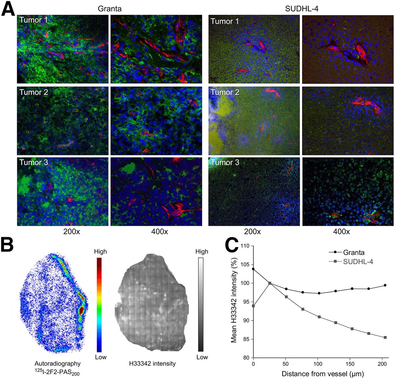
  • FIGURE 5.
    • Download figure
    • Open in new tab
    • Download powerpoint
    FIGURE 5.

    H33342 perfusion and penetration in Granta-519 and SUDHL-4 tumors. (A) Perfusion dye H33342 was injected 24 h after radioiodinated Fab-ABD or Fab-PAS200 and 1 min before mice were sacrificed. Afterward, CD20 (green) and CD31 (red) were detected in tumor cryosections using immunofluorescence. H33342 perfusion of 3 different SUDHL-4 and Granta-519 tumors is represented in blue. (B) Autoradiography and corresponding H33342 intensity is shown for an example SUDHL-4 tumor. (C) Mean H33342 penetration into tissue was quantified using distance maps based on CD31-positive vessels, with distance of zero equivalent to vessel lumen. H33342 intensity was normalized with regard to first distance value (25 μm) after vessel. 2F2 = CD20-specific Fab, derived from ofatumumab.

Additional Files

  • Figures
  • Supplemental Data

    Files in this Data Supplement:

    • Supplemental Data
    • Supplemental Video 1
    • Supplemental Video 2
    • Supplemental Video 3
    • Supplemental Video 4
    • Supplemental Video 5
PreviousNext
Back to top

In this issue

Journal of Nuclear Medicine: 57 (12)
Journal of Nuclear Medicine
Vol. 57, Issue 12
December 1, 2016
  • Table of Contents
  • Table of Contents (PDF)
  • About the Cover
  • Index by author
Print
Download PDF
Article Alerts
Sign In to Email Alerts with your Email Address
Email Article

Thank you for your interest in spreading the word on Journal of Nuclear Medicine.

NOTE: We only request your email address so that the person you are recommending the page to knows that you wanted them to see it, and that it is not junk mail. We do not capture any email address.

Enter multiple addresses on separate lines or separate them with commas.
Tumor Uptake of Anti-CD20 Fabs Depends on Tumor Perfusion
(Your Name) has sent you a message from Journal of Nuclear Medicine
(Your Name) thought you would like to see the Journal of Nuclear Medicine web site.
Citation Tools
Tumor Uptake of Anti-CD20 Fabs Depends on Tumor Perfusion
Claudia Theresa Mendler, Annette Feuchtinger, Irina Heid, Michaela Aichler, Calogero D’Alessandria, Sabine Pirsig, Birgit Blechert, Hans-Jürgen Wester, Rickmer Braren, Axel Walch, Arne Skerra, Markus Schwaiger
Journal of Nuclear Medicine Dec 2016, 57 (12) 1971-1977; DOI: 10.2967/jnumed.116.176784

Citation Manager Formats

  • BibTeX
  • Bookends
  • EasyBib
  • EndNote (tagged)
  • EndNote 8 (xml)
  • Medlars
  • Mendeley
  • Papers
  • RefWorks Tagged
  • Ref Manager
  • RIS
  • Zotero
Share
Tumor Uptake of Anti-CD20 Fabs Depends on Tumor Perfusion
Claudia Theresa Mendler, Annette Feuchtinger, Irina Heid, Michaela Aichler, Calogero D’Alessandria, Sabine Pirsig, Birgit Blechert, Hans-Jürgen Wester, Rickmer Braren, Axel Walch, Arne Skerra, Markus Schwaiger
Journal of Nuclear Medicine Dec 2016, 57 (12) 1971-1977; DOI: 10.2967/jnumed.116.176784
Twitter logo Facebook logo LinkedIn logo Mendeley logo
  • Tweet Widget
  • Facebook Like
  • Google Plus One
Bookmark this article

Jump to section

  • Article
    • Abstract
    • MATERIALS AND METHODS
    • RESULTS
    • DISCUSSION
    • CONCLUSION
    • DISCLOSURE
    • Acknowledgments
    • Footnotes
    • REFERENCES
  • Figures & Data
  • Supplemental
  • Info & Metrics
  • PDF

Related Articles

  • This Month in JNM
  • PubMed
  • Google Scholar

Cited By...

  • New Proline, Alanine, Serine Repeat Sequence for Pharmacokinetic Enhancement of Anti-VEGF Single-Domain Antibody
  • Google Scholar

More in this TOC Section

  • PET/MRI of Hypoxic Atherosclerosis Using 64Cu-ATSM in a Rabbit Model
  • How Sensitive Is the Upper Gastrointestinal Tract to 90Y Radioembolization? A Histologic and Dosimetric Analysis in a Porcine Model
Show more Basic Science Investigations

Similar Articles

Keywords

  • 3D light-sheet fluorescence microscopy
  • DCE MRI
  • Fab fragment
  • lymphoma
  • tumor perfusion
SNMMI

© 2025 SNMMI

Powered by HighWire