
Main menu

  • Home
  • Content
    • Current
    • Ahead of print
    • Past Issues
    • JNM Supplement
    • SNMMI Annual Meeting Abstracts
    • Continuing Education
    • JNM Podcasts
  • Subscriptions
    • Subscribers
    • Institutional and Non-member
    • Rates
    • Journal Claims
    • Corporate & Special Sales
  • Authors
    • Submit to JNM
    • Information for Authors
    • Assignment of Copyright
    • AQARA requirements
  • Info
    • Reviewers
    • Permissions
    • Advertisers
  • About
    • About Us
    • Editorial Board
    • Contact Information
  • More
    • Alerts
    • Feedback
    • Help
    • SNMMI Journals
  • SNMMI
    • JNM
    • JNMT
    • SNMMI Journals
    • SNMMI

User menu

  • Subscribe
  • My alerts
  • Log in
  • Log out
  • My Cart

Search

  • Advanced search
Journal of Nuclear Medicine
  • SNMMI
    • JNM
    • JNMT
    • SNMMI Journals
    • SNMMI
  • Subscribe
  • My alerts
  • Log in
  • Log out
  • My Cart
Journal of Nuclear Medicine

Advanced Search

  • Home
  • Content
    • Current
    • Ahead of print
    • Past Issues
    • JNM Supplement
    • SNMMI Annual Meeting Abstracts
    • Continuing Education
    • JNM Podcasts
  • Subscriptions
    • Subscribers
    • Institutional and Non-member
    • Rates
    • Journal Claims
    • Corporate & Special Sales
  • Authors
    • Submit to JNM
    • Information for Authors
    • Assignment of Copyright
    • AQARA requirements
  • Info
    • Reviewers
    • Permissions
    • Advertisers
  • About
    • About Us
    • Editorial Board
    • Contact Information
  • More
    • Alerts
    • Feedback
    • Help
    • SNMMI Journals
  • View or Listen to JNM Podcast
  • Visit JNM on Facebook
  • Join JNM on LinkedIn
  • Follow JNM on Twitter
  • Subscribe to our RSS feeds
Research ArticleBasic Science Investigations

Counting Rate Characteristics and Image Distortion in Preclinical PET Imaging During Radiopharmaceutical Therapy

Emma Mellhammar, Magnus Dahlbom, Johan Axelsson and Sven-Erik Strand
Journal of Nuclear Medicine December 2016, 57 (12) 1964-1970; DOI: https://doi.org/10.2967/jnumed.116.175539
Emma Mellhammar
1Division of Oncology and Pathology, Department of Clinical Sciences–Lund, Lund University, Lund, Sweden
  • Find this author on Google Scholar
  • Find this author on PubMed
  • Search for this author on this site
Magnus Dahlbom
2Department of Molecular and Medical Pharmacology, David Geffen School of Medicine, UCLA, Los Angeles, California
  • Find this author on Google Scholar
  • Find this author on PubMed
  • Search for this author on this site
Johan Axelsson
3Department of Physics, LTH, Lund, Sweden; and
  • Find this author on Google Scholar
  • Find this author on PubMed
  • Search for this author on this site
Sven-Erik Strand
1Division of Oncology and Pathology, Department of Clinical Sciences–Lund, Lund University, Lund, Sweden
4Division of Oncology and Pathology, Department of Clinical Sciences–Lund, Lund University, Lund, Sweden
  • Find this author on Google Scholar
  • Find this author on PubMed
  • Search for this author on this site
  • Article
  • Figures & Data
  • Info & Metrics
  • PDF
Loading

Article Figures & Data

Figures

  • Tables
  • FIGURE 1.
    • Download figure
    • Open in new tab
    • Download powerpoint
    FIGURE 1.

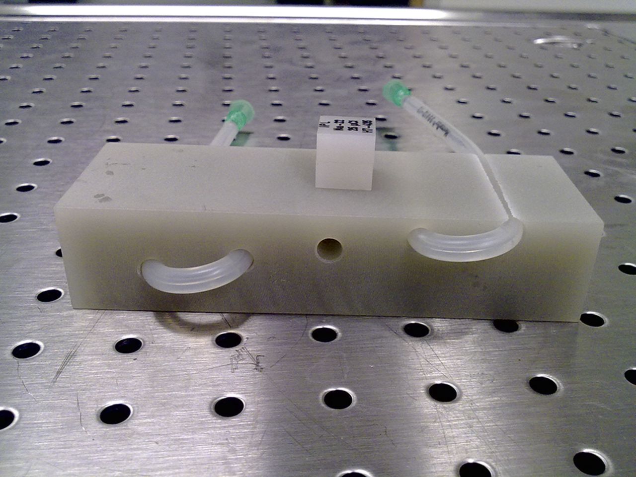
    In-house–made mouse phantom with thin silicone tube drawn through the channels and point source placed on top.

  • FIGURE 2.
    • Download figure
    • Open in new tab
    • Download powerpoint
    FIGURE 2.

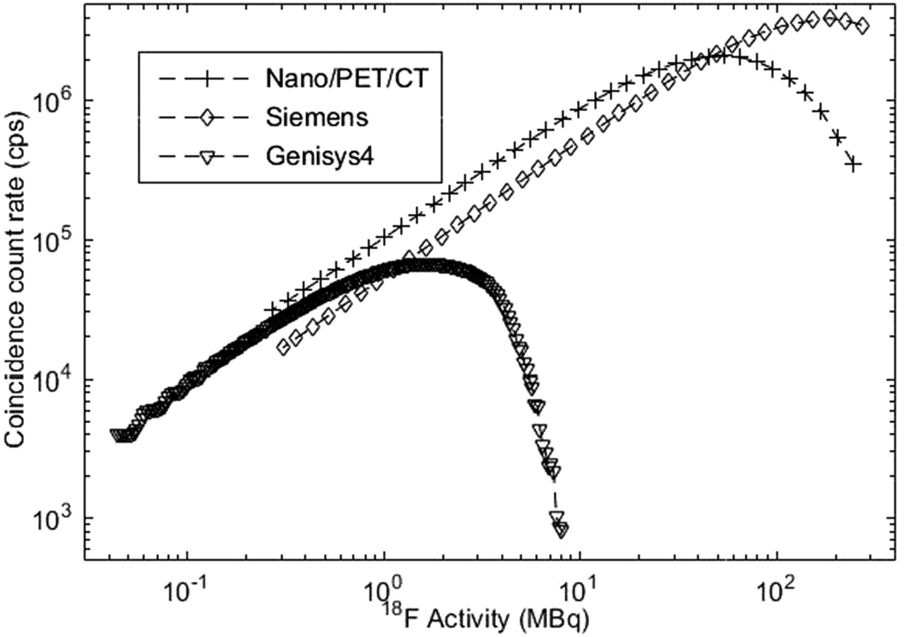
    True coincidence counting rates detected on Genisys4, Inveon, and NanoPET/CT when high activities of 18F were allowed to decay in PET system in Study 1. (On NanoPET/CT, available counting rates were plotted.)

  • FIGURE 3.
    • Download figure
    • Open in new tab
    • Download powerpoint
    FIGURE 3.

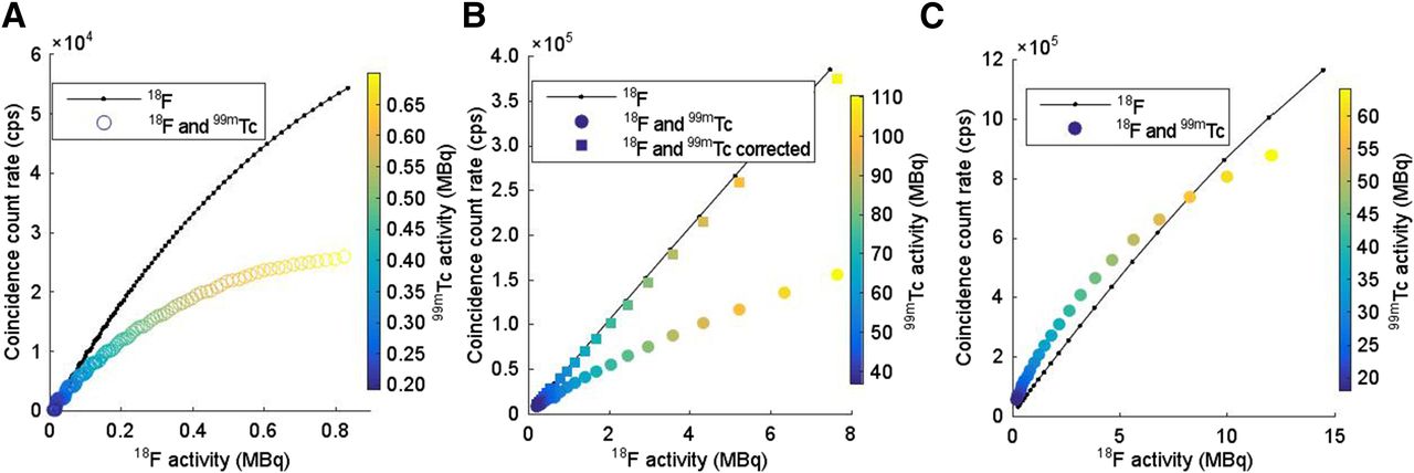
    Coincidence counting rates when 22Na point source was imaged together with decaying 99mTc (Study 2A) for Genisys4 (A), Inveon (B), and NanoPET/CT (C). B also presents corrected counting rate for Study 2A for Inveon. C also presents coincidence counting rates for decaying 99mTc (Study 2B) and 22Na point source imaged together with decaying 177Lu (Study 2C) on NanoPET/CT.

  • FIGURE 4.
    • Download figure
    • Open in new tab
    • Download powerpoint
    FIGURE 4.

    Volume resolution for Genisys4 (A), Inveon (B), and NanoPET/CT (C) measured over 22Na point source imaged together with decaying 99mTc (Study 2A). C also presents volume resolution measured over 22Na point source together with decaying 177Lu for NanoPET/CT (Study 2C). FWHM = full width at half maximum; FWTM = full width at tenth maximum.

  • FIGURE 5.
    • Download figure
    • Open in new tab
    • Download powerpoint
    FIGURE 5.

    Comparison of true coincidence counting rates detected for 18F decaying alone (Study 1) or together with 99mTc (Study 3) on Genisys4 (A), Inveon (B), and NanoPET/CT (C). (On NanoPET/CT, available counting rates were plotted.) B also presents corrected counting rate for Study 3 for Inveon.

Tables

  • Figures
    • View popup
    TABLE 1

    PET System Characteristics

    CharacteristicInveonNanoPET/CTGenisys4
    Detector materialLSOLYSO:CeBGO
    Crystal dimensions (mm3)1.5 × 1.5 × 101.12 × 1.12 × 131.8 × 1.8 × 7
    Axial field of view (mm)1279594
    Transaxial field of view (mm)10012345
    Energy window (keV)350–650400–600150–650
    Coincidence window (ns)3.4520
    Sensitivity (%)6.77.714
    Image spatial resolution (mm)1.811.4
    Energy resolution (511 keV) (%)14.61918.0
    Crystal decay time (ns)4741300
    Number of detectors64124
    Number of crystals per detector4001,2003,159
    • BGO = bismuth germanate; LSO = lutetium oxyorthosilicate; LYSO = lutetium yttrium orthosilicate.

    • View popup
    TABLE 2

    Specific Activities and Phantoms Used

    Activity (MBq)
    StudyPhantomInveonGenisys4NanoPET/CT
    1Sphere*/NEMA270 (18F)11 (18F)250 (18F)
    2APoint source and block†500 (99mTc), 0.074 (22Na)170 (99mTc), 0.074 (22Na)550 (99mTc), 0.43 (22Na)
    2BSphere——480 (99mTc)
    2CPoint source and block†——82 (177Lu), 0.43 (22Na)
    3Sphere‡/NEMA7.7 (18F), 110 (99mTc)0.85 (18F), 0.71 (99mTc)12 (18F), 64 (99mTc)
    • ↵* 0.5 mL.

    • ↵† 22Na point source placed in center channel; silicon tube pulled through surrounding 4 channels.

    • ↵‡ 1.0 mL for 99mTc and 0.5 mL for 18F.

    • View popup
    TABLE 3

    Radionuclides Used in This Report

    IsotopeHalf-lifeDecay modeQ-value (keV)Branching (%)γ-emission (keV)γ-yield (%)
    177Lu6.734 dβ−498.31001136.4
    20811.0
    18F109.77 me, β+1,655.53.27——
    633.596.73
    99mTc6.01 hIT—100140.51189
    142.6280.0187
    22Na2.6019 ye, β+2,842.21001,274.5399.944
    • e = electron capture; IT = isomeric transition.

PreviousNext
Back to top

In this issue

Journal of Nuclear Medicine: 57 (12)
Journal of Nuclear Medicine
Vol. 57, Issue 12
December 1, 2016
  • Table of Contents
  • Table of Contents (PDF)
  • About the Cover
  • Index by author
Print
Download PDF
Article Alerts
Sign In to Email Alerts with your Email Address
Email Article

Thank you for your interest in spreading the word on Journal of Nuclear Medicine.

NOTE: We only request your email address so that the person you are recommending the page to knows that you wanted them to see it, and that it is not junk mail. We do not capture any email address.

Enter multiple addresses on separate lines or separate them with commas.
Counting Rate Characteristics and Image Distortion in Preclinical PET Imaging During Radiopharmaceutical Therapy
(Your Name) has sent you a message from Journal of Nuclear Medicine
(Your Name) thought you would like to see the Journal of Nuclear Medicine web site.
Citation Tools
Counting Rate Characteristics and Image Distortion in Preclinical PET Imaging During Radiopharmaceutical Therapy
Emma Mellhammar, Magnus Dahlbom, Johan Axelsson, Sven-Erik Strand
Journal of Nuclear Medicine Dec 2016, 57 (12) 1964-1970; DOI: 10.2967/jnumed.116.175539

Citation Manager Formats

  • BibTeX
  • Bookends
  • EasyBib
  • EndNote (tagged)
  • EndNote 8 (xml)
  • Medlars
  • Mendeley
  • Papers
  • RefWorks Tagged
  • Ref Manager
  • RIS
  • Zotero
Share
Counting Rate Characteristics and Image Distortion in Preclinical PET Imaging During Radiopharmaceutical Therapy
Emma Mellhammar, Magnus Dahlbom, Johan Axelsson, Sven-Erik Strand
Journal of Nuclear Medicine Dec 2016, 57 (12) 1964-1970; DOI: 10.2967/jnumed.116.175539
Twitter logo Facebook logo LinkedIn logo Mendeley logo
  • Tweet Widget
  • Facebook Like
  • Google Plus One
Bookmark this article

Jump to section

  • Article
    • Abstract
    • MATERIALS AND METHODS
    • RESULTS
    • DISCUSSION
    • CONCLUSION
    • DISCLOSURE
    • Footnotes
    • REFERENCES
  • Figures & Data
  • Info & Metrics
  • PDF

Related Articles

  • This Month in JNM
  • PubMed
  • Google Scholar

Cited By...

  • Preserving Preclinical PET Quality During Intratherapeutic Imaging in Radionuclide Therapy with Rose Metal Shielding Reducing Photon Flux
  • Google Scholar

More in this TOC Section

  • Tumor Uptake of Anti-CD20 Fabs Depends on Tumor Perfusion
  • How Sensitive Is the Upper Gastrointestinal Tract to 90Y Radioembolization? A Histologic and Dosimetric Analysis in a Porcine Model
  • 11C-Methionine PET of Myocardial Inflammation in a Rat Model of Experimental Autoimmune Myocarditis
Show more Basic Science Investigations

Similar Articles

Keywords

  • PET
  • radionuclide therapy
  • Dead time
  • Pile up
  • Image distortion
SNMMI

© 2025 SNMMI

Powered by HighWire