


Article Figures & Data
Figures
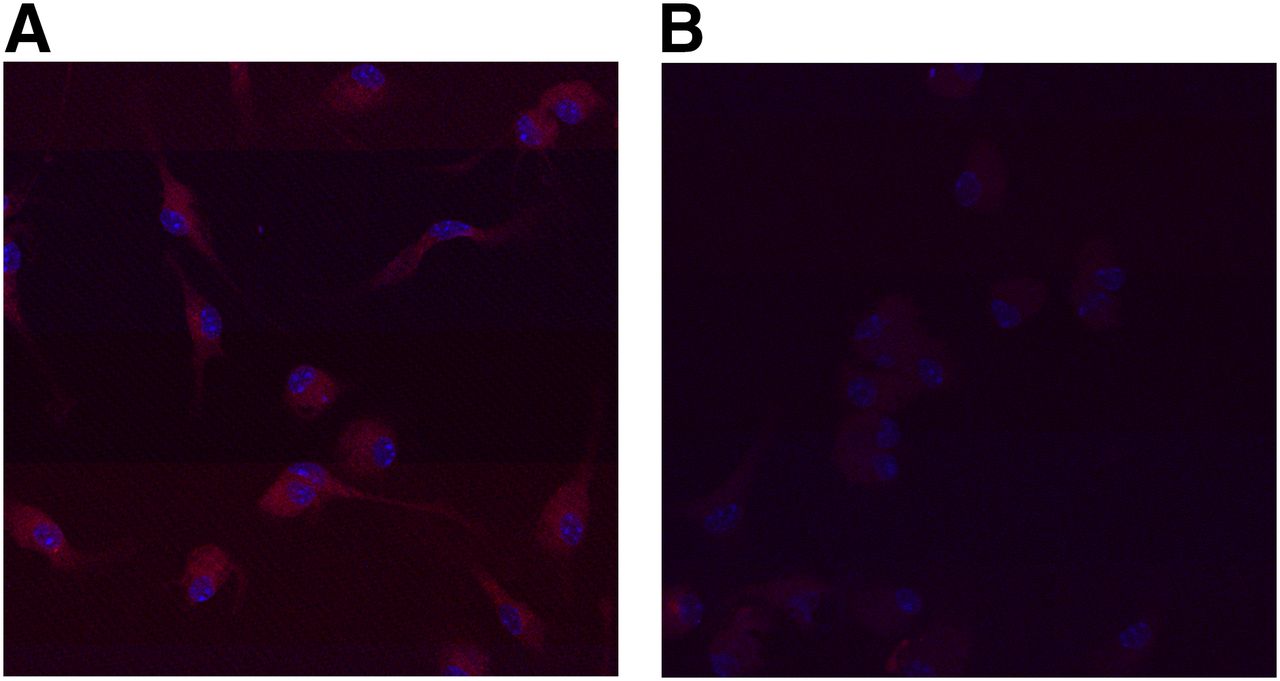
- FIGURE 1.
Confocal microscopic image showing MR-specific binding of MSA to peritoneal macrophages derived from a C57/BL6 mouse. (A) Macrophages incubated with RITC-MSA and control immunoglobulin, both at concentrations of 10 μg/mL, show red fluorescence. (B) Macrophages preincubated with anti-CD206 MR (10 μg/mL) blocking antibody show markedly diminished fluorescence on incubation with 10 μg of RITC-MSA per mL.
- FIGURE 2.
Representative images illustrating RITC-MSA binding to MRs in aortic plaque of atherosclerotic rabbit. (A) Differential interference contrast microscopy image of an atherosclerotic plaque in the rabbit aorta. (B) DAPI-stained nuclei (blue) visualized in the same fields by switching to fluorescence confocal microscopy. (C) Red fluorescent image of same atherosclerotic plaque and medial layers of aorta after incubation with RITC-MSA. (D) IX81-ZDC focus drift-compensating microscope showing faint green fluorescence of FITC over atherosclerotic lesion, with definite fluorescence over medial smooth muscle layers (excitation and emission wavelengths of 480 and 520 nm for FITC and 545 and 595 nm for RITC, respectively). (E) Overlay of fluorescent images of atherosclerotic aorta, with yellow arrows indicating presence of M2 macrophages. (F) Immunohistochemistry photograph (magnification, 100×) of aortic atherosclerotic lesion labeled with antirabbit macrophage monoclonal antibody RAM11 at adjacent portion of aorta shown in representative fluorescence images. Arrows indicate presence of macrophages. Presence of lipid-laden macrophages in atherosclerotic rabbit is demonstrated in Supplemental Figure 2.
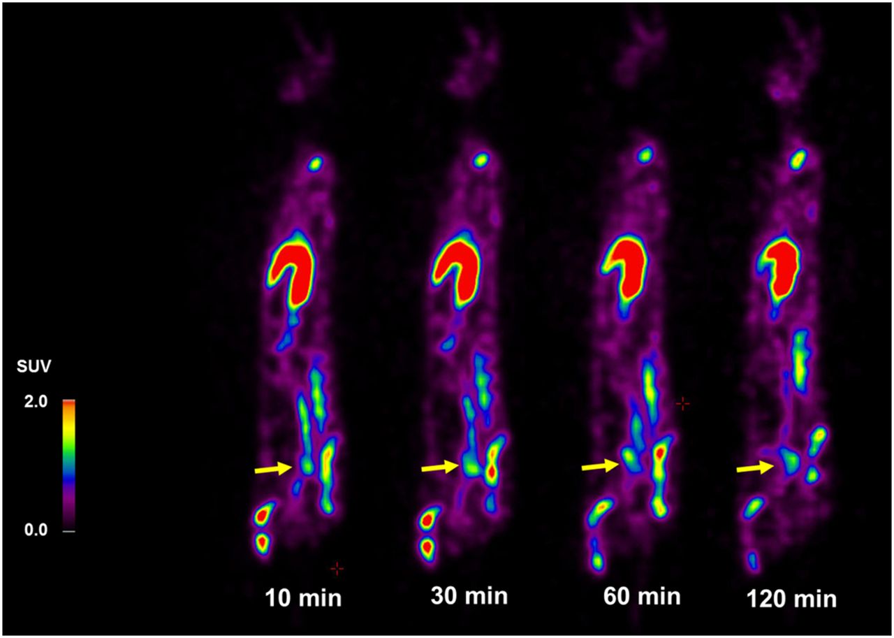
- FIGURE 3.
Sequential 68Ga-NOTA-MSA PET images of atherosclerosis at 10, 30, 60, and 120 min after tracer injection confirm persistence of radiopharmaceutical by visualization of atherosclerotic aorta (yellow arrow) until 120 min in cholesterol-fed atherosclerotic rabbit. Maximal SUVs of atherosclerotic aorta at 10, 30, 60, and 120 min were 1.7, 1.8, 1.5, and 1.2, respectively. Liver and bone marrows of spine, pelvis, and femur also showed high uptake of 68Ga-NOTA-MSA, indicating intensity of cells presenting MRs in reticuloendothelial system (17).
- FIGURE 4.
Representative examples of 18F-FDG and 68Ga-NOTA-MSA PET images in atherosclerotic rabbit fed cholesterol diet. Comparison of 18F-FDG and 68Ga-NOTA-MSA PET images of atherosclerotic aorta reveals visualization of aortic lesions with both tracers. Magnified uptake of 18F-FDG at 60 min after injection (A) and 68Ga-NOTA-MSA at 30 min after injection at abdominal aorta (red asterisks) (B). Vertebral column is visible in enlarged PET/CT images. PET images were acquired 60 min after intravenous injection of 18F-FDG and 30 min after injection of 68Ga-NOTA-MSA.
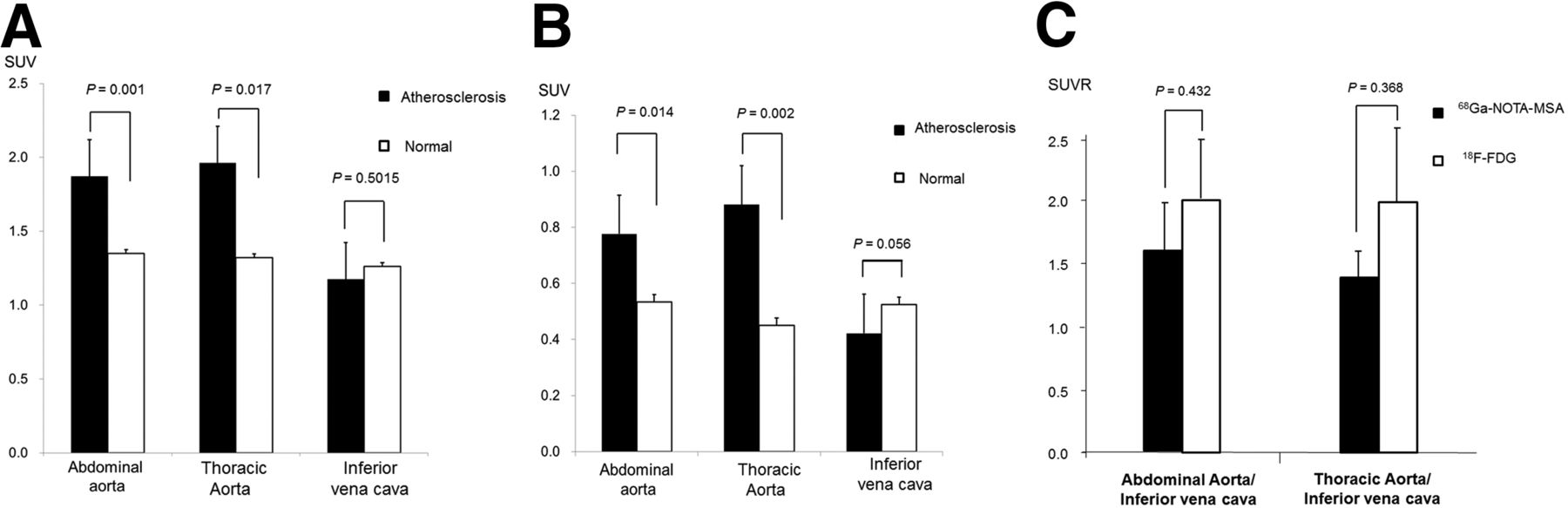
- FIGURE 5.
SUVs of atherosclerotic lesions and normal aorta with 68Ga-MSA (A) and 18F-FDG (B) and SUV ratio (SUVR) of atherosclerotic aorta to inferior vena cava (C) in PET images. Maximal SUVs were measured at aorta and mean SUVs at inferior vena cava. Maximal SUVs of 68Ga-NOTA-MSA were higher than those of 18F-FDG (A and B) in atherosclerotic aorta, but SUVR of aorta to inferior vena cava was not significantly different between 18F-FDG and 68Ga-NOTA-MSA images (C).


Additional Files
Supplemental Data
Files in this Data Supplement:


Jump to section
Related Articles
Cited By...
- Development of a CD163-Targeted PET Radiotracer That Images Resident Macrophages in Atherosclerosis
- Total-Body Multiparametric PET Quantification of 18F-FDG Delivery and Metabolism in the Study of Coronavirus Disease 2019 Recovery
- Phase I Study of [68Ga]Ga-Anti-CD206-sdAb for PET/CT Assessment of Protumorigenic Macrophage Presence in Solid Tumors (MMR Phase I)
- Total-Body Multiparametric PET Quantification of 18F-FDG Delivery and Metabolism in the Study of COVID-19 Recovery
- Biodistribution and Radiation Dosimetry of 124I-DPA-713, a PET Radiotracer for Macrophage-Associated Inflammation