


Article Figures & Data
Figures
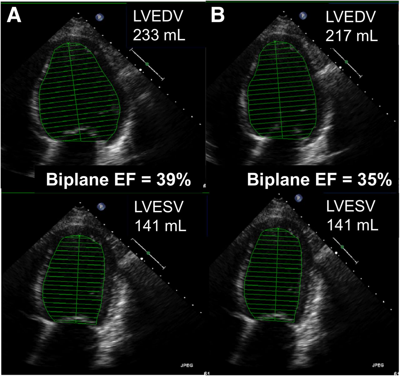
- FIGURE 1.
Calculation of LV volumes and EF from biplane Simpson’s formula using 2D echocardiography. Repeated measurement at same examination (A vs. B) shows that relatively minor (7%) difference in volume calculation translates to EF change sufficient to change a decision. LVEDV = LV end-diastolic volume; LVESV = LV end-systolic volume.
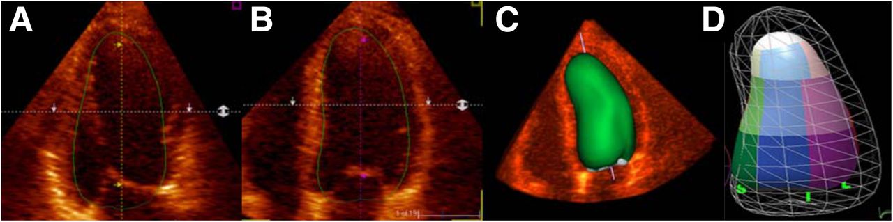
- FIGURE 2.
Use of 3D echocardiography allows LV assumptions to be discarded. Positions of LV contours can be confirmed from 2D images (A and B) to produce 3D volume (C), and segmental evaluation can be performed (D).
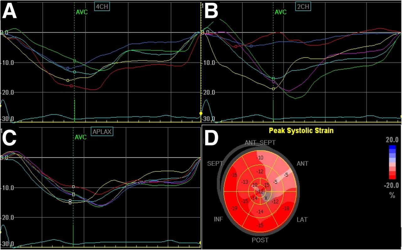
- FIGURE 3.
LV regional and global strain can be calculated from speckle-tracking of standard 2D images. Each LV segment is tracked throughout cycle to provide regional strain curve, and these are displayed in each apical view (4-chamber [A], 2-chamber [B], and long-axis [C]). Global strain is shown in polar map display (D). In this example of patient with anterior myocardial infarction, strain curves are abnormal in apical 2-chamber view and extent of infarction is illustrated in parametric display.
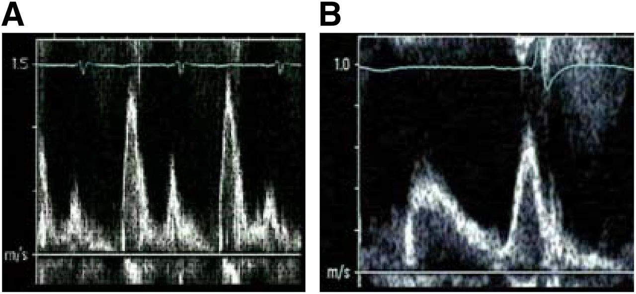
- FIGURE 4.
Load dependence of diastolic evaluation from transmitral flow. In this dialysis patient, imaging during fluid overload (A) showed restrictive filling pattern, reverting to delayed relaxation (B) after development of euvolemia.

Tables
- TABLE 1
Measurements, Indications, and Modalities Used in Echocardiographic Evaluation of HF
Measurement Indication Modality EF Classification, risk assessment; selection for implantable cardioverter defibrillator and cardiac resynchronization therapy 2D or 3D echocardiography, contrast Non-EF indices of function Risk assessment Doppler dP/dt Mitral regurgitant orifice area Quantification of mitral regurgitation Color and CW Doppler Transmitral and annular velocities Assessment of diastolic function and filling pressure Pulsed-wave tissue and blood-flow Doppler Left atrial volume Prognosis, diastolic evaluation 2D or 3D echocardiography RV assessment Prognosis, evaluation for assist device RV strain, tissue Doppler, tricuspid annular plane systolic excursion, fractional area change, etc. Regional function Assessment of CAD Wall motion analysis, strain Regional timing Site of greatest maximal delay Cardiac resynchronization therapy selection Situation Cause Fusion of E and A waves Tachycardia Unreliable measurement of E velocity Significant mitral regurgitation (>2+) Significant mitral stenosis Significant aortic regurgitation (>2+) Unreliable measurement of e′ velocity Severe mitral annular calcification Mitral valve repair or replacement Localized wall motion abnormalities Left bundle branch block Biventricular pacing Application Technical requirement Technique LV hypertrophy, infiltration High spatial resolution Cardiac MR LV synchrony High temporal resolution Tissue Doppler, strain LV volumes High contrast resolution Cardiac MR, contrast echocardiography Sequential follow-up High repeatability Cardiac MR, 3D echocardiography Subclinical cardiomyopathy Sensitivity to minor change Strain Parameter Measure Response at 6 mo Clinical Composite “Improved” New York Heart Association class Decrease by at least one class Minnesota Living with Heart Failure questionnaire ≥9-point decrease Exercise capacity 6-min walk ≥10% improvement (or any improvement if walked zero at baseline) LV features LV volume ≥15% decrease LV EF ≥5% increase LV mass Any decrease Tei index Any decrease Mitral valve Mitral regurgitation Decrease in mitral regurgitation severity Parameter Absolute difference Relative difference ∆ Left ventricular EF 8.1% ± 11.5% 17% ± 30% ∆ Left atrial area 4.0 ± 5.2 cm2 17% ± 23% ∆ Tissue Em 2.1 ± 2.7 cm/s 27% ± 36% ∆ E/e′ 5.0 ± 7.0 46% ± 64% Em = mitral annular tissue diastolic velocity.
- TABLE 6
Heart Failure Stages as Defined by Classification System of American College of Cardiology and American Heart Association
Stage Risk factors for HF Structural/functional heart disease History of HF Clinical congestive HF Normal No No No — Stage A Yes No No — Stage B — Yes No No Stage C1 — Yes No Yes Stage C2 — Yes Yes — Stage D — Yes Yes Yes Modality Accessibility Patient-friendliness Preclinical HF Accuracy Hemodynamics Regional function Reproducibility Echocardiography ++ ++ ++ + ++ + + Nuclear + + − + +/− +/− + CT + + − + − + + Cardiac MR +/− + ++ ++ + ++ ++ ++ = excellent; + = good; +/− = fair; − = poor.


