
Main menu

  • Home
  • Content
    • Current
    • Ahead of print
    • Past Issues
    • JNM Supplement
    • SNMMI Annual Meeting Abstracts
    • Continuing Education
    • JNM Podcasts
  • Subscriptions
    • Subscribers
    • Institutional and Non-member
    • Rates
    • Journal Claims
    • Corporate & Special Sales
  • Authors
    • Submit to JNM
    • Information for Authors
    • Assignment of Copyright
    • AQARA requirements
  • Info
    • Reviewers
    • Permissions
    • Advertisers
  • About
    • About Us
    • Editorial Board
    • Contact Information
  • More
    • Alerts
    • Feedback
    • Help
    • SNMMI Journals
  • SNMMI
    • JNM
    • JNMT
    • SNMMI Journals
    • SNMMI

User menu

  • Subscribe
  • My alerts
  • Log in
  • My Cart

Search

  • Advanced search
Journal of Nuclear Medicine
  • SNMMI
    • JNM
    • JNMT
    • SNMMI Journals
    • SNMMI
  • Subscribe
  • My alerts
  • Log in
  • My Cart
Journal of Nuclear Medicine

Advanced Search

  • Home
  • Content
    • Current
    • Ahead of print
    • Past Issues
    • JNM Supplement
    • SNMMI Annual Meeting Abstracts
    • Continuing Education
    • JNM Podcasts
  • Subscriptions
    • Subscribers
    • Institutional and Non-member
    • Rates
    • Journal Claims
    • Corporate & Special Sales
  • Authors
    • Submit to JNM
    • Information for Authors
    • Assignment of Copyright
    • AQARA requirements
  • Info
    • Reviewers
    • Permissions
    • Advertisers
  • About
    • About Us
    • Editorial Board
    • Contact Information
  • More
    • Alerts
    • Feedback
    • Help
    • SNMMI Journals
  • View or Listen to JNM Podcast
  • Visit JNM on Facebook
  • Join JNM on LinkedIn
  • Follow JNM on Twitter
  • Subscribe to our RSS feeds
Research ArticleBasic Science Investigations

Trimeric Radiofluorinated Sulfonamide Derivatives to Achieve In Vivo Selectivity for Carbonic Anhydrase IX–Targeted PET Imaging

Joseph Lau, Zhibo Liu, Kuo-Shyan Lin, Jinhe Pan, Zhengxing Zhang, Daniela Vullo, Claudiu T. Supuran, David M. Perrin and François Bénard
Journal of Nuclear Medicine September 2015, 56 (9) 1434-1440; DOI: https://doi.org/10.2967/jnumed.114.153288
Joseph Lau
1Department of Molecular Oncology, BC Cancer Agency, Vancouver, British Columbia, Canada
  • Find this author on Google Scholar
  • Find this author on PubMed
  • Search for this author on this site
Zhibo Liu
2Chemistry Department, University of British Columbia, Vancouver, British Columbia, Canada; and
  • Find this author on Google Scholar
  • Find this author on PubMed
  • Search for this author on this site
Kuo-Shyan Lin
1Department of Molecular Oncology, BC Cancer Agency, Vancouver, British Columbia, Canada
  • Find this author on Google Scholar
  • Find this author on PubMed
  • Search for this author on this site
Jinhe Pan
1Department of Molecular Oncology, BC Cancer Agency, Vancouver, British Columbia, Canada
  • Find this author on Google Scholar
  • Find this author on PubMed
  • Search for this author on this site
Zhengxing Zhang
1Department of Molecular Oncology, BC Cancer Agency, Vancouver, British Columbia, Canada
  • Find this author on Google Scholar
  • Find this author on PubMed
  • Search for this author on this site
Daniela Vullo
3Università degli Studi di Firenze, Dipartimento Neurofarba and Laboratorio di Chimica Bioinorganica, Florence, Italy
  • Find this author on Google Scholar
  • Find this author on PubMed
  • Search for this author on this site
Claudiu T. Supuran
3Università degli Studi di Firenze, Dipartimento Neurofarba and Laboratorio di Chimica Bioinorganica, Florence, Italy
  • Find this author on Google Scholar
  • Find this author on PubMed
  • Search for this author on this site
David M. Perrin
2Chemistry Department, University of British Columbia, Vancouver, British Columbia, Canada; and
  • Find this author on Google Scholar
  • Find this author on PubMed
  • Search for this author on this site
François Bénard
1Department of Molecular Oncology, BC Cancer Agency, Vancouver, British Columbia, Canada
  • Find this author on Google Scholar
  • Find this author on PubMed
  • Search for this author on this site
  • Article
  • Figures & Data
  • Supplemental
  • Info & Metrics
  • PDF
Loading

Article Figures & Data

Figures

  • Tables
  • Additional Files
  • FIGURE 1.
    • Download figure
    • Open in new tab
    • Download powerpoint
    FIGURE 1.

    Reported radiolabeled small-molecule inhibitors for CA-IX imaging. Compounds A–M are sulfonamide-based derivatives, whereas compound N is a coumarin-based inhibitor.
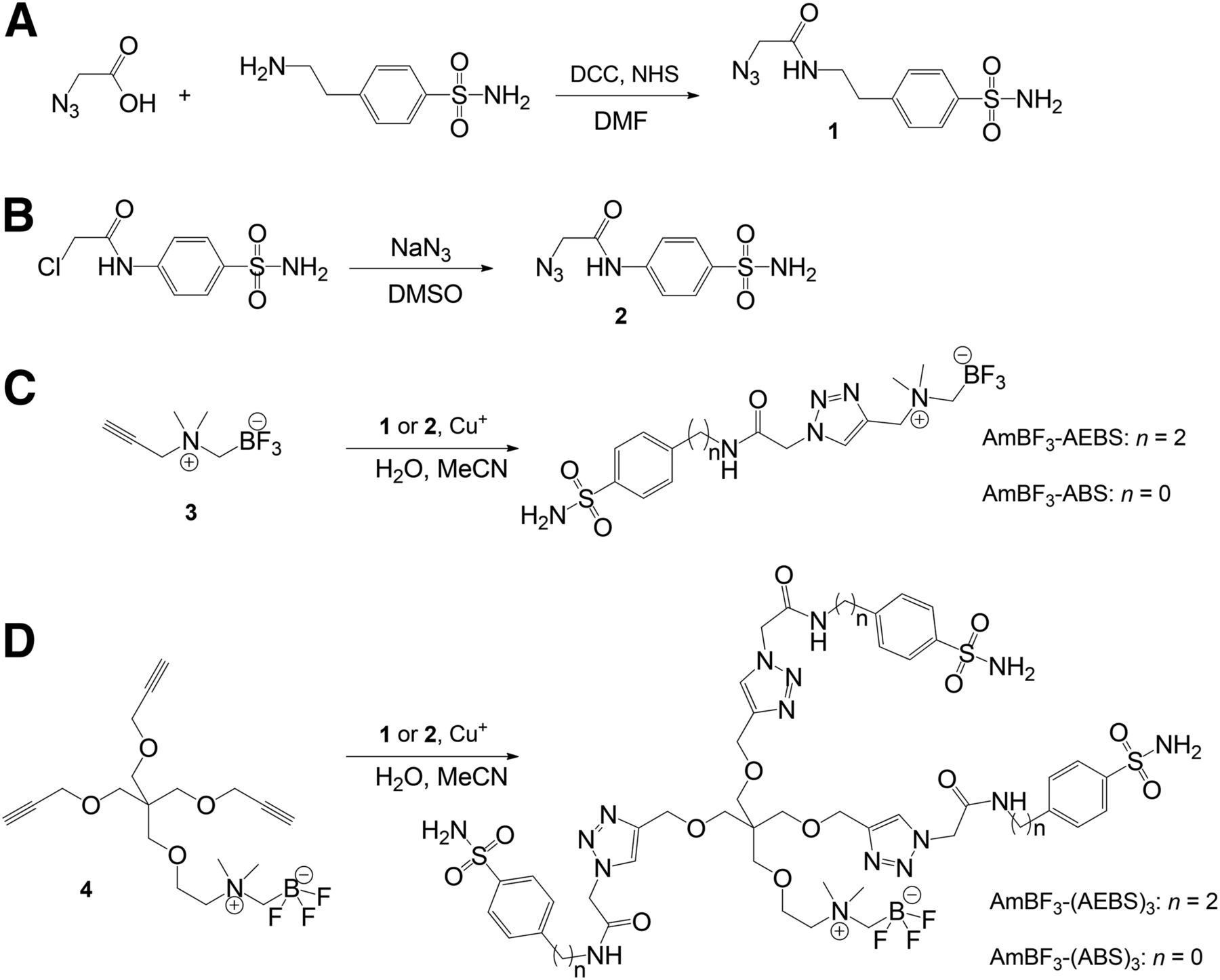
  • FIGURE 2.
    • Download figure
    • Open in new tab
    • Download powerpoint
    FIGURE 2.

    Synthesis of azidoacetyl-AEBS 1 (A), azidoacetyl-ABS 2 (B), AmBF3-AEBS and AmBF3-ABS (C), and AmBF3-(AEBS)3 and AmBF3-(ABS)3 (D). DCC = N,N′-dicyclohexylcarbodiimine; NHS = N-hydroxysuccinimde.
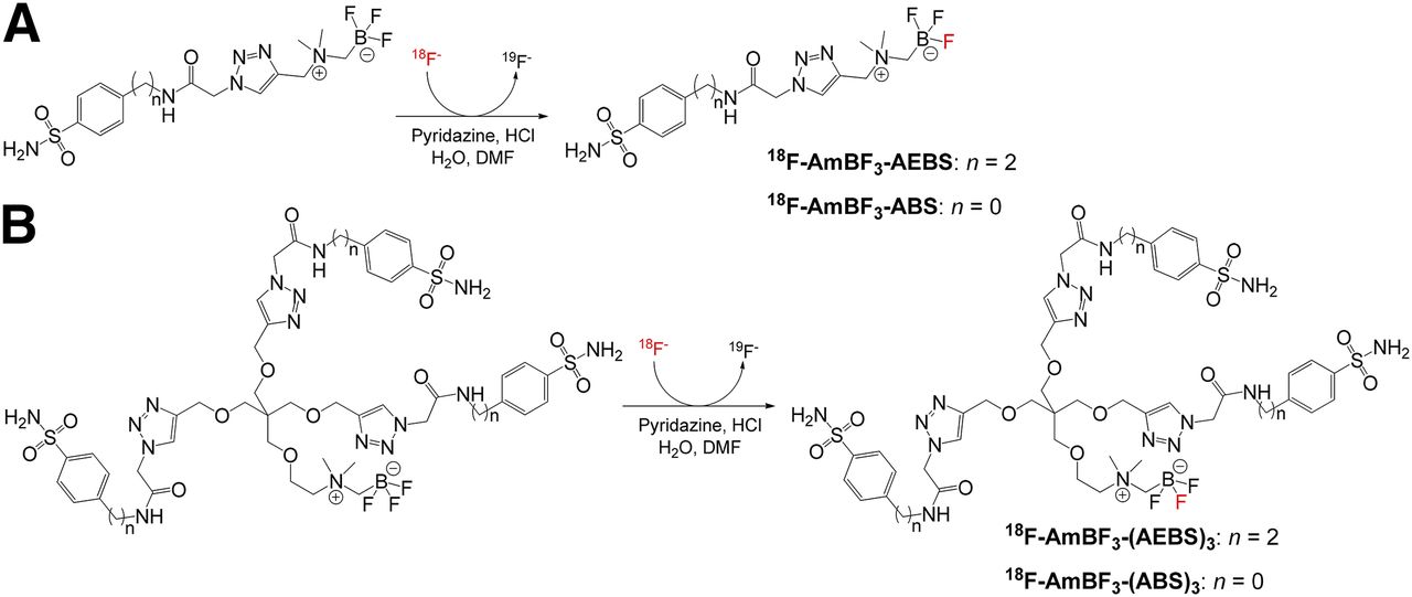
  • FIGURE 3.
    • Download figure
    • Open in new tab
    • Download powerpoint
    FIGURE 3.

    Radiosynthesis of 18F-AmBF3-AEBS and 18F-AmBF3-ABS (A) and 18F-AmBF3-(AEBS)3 and 18F-AmBF3-(ABS)3 (B) via 18F-19F isotope exchange reaction.
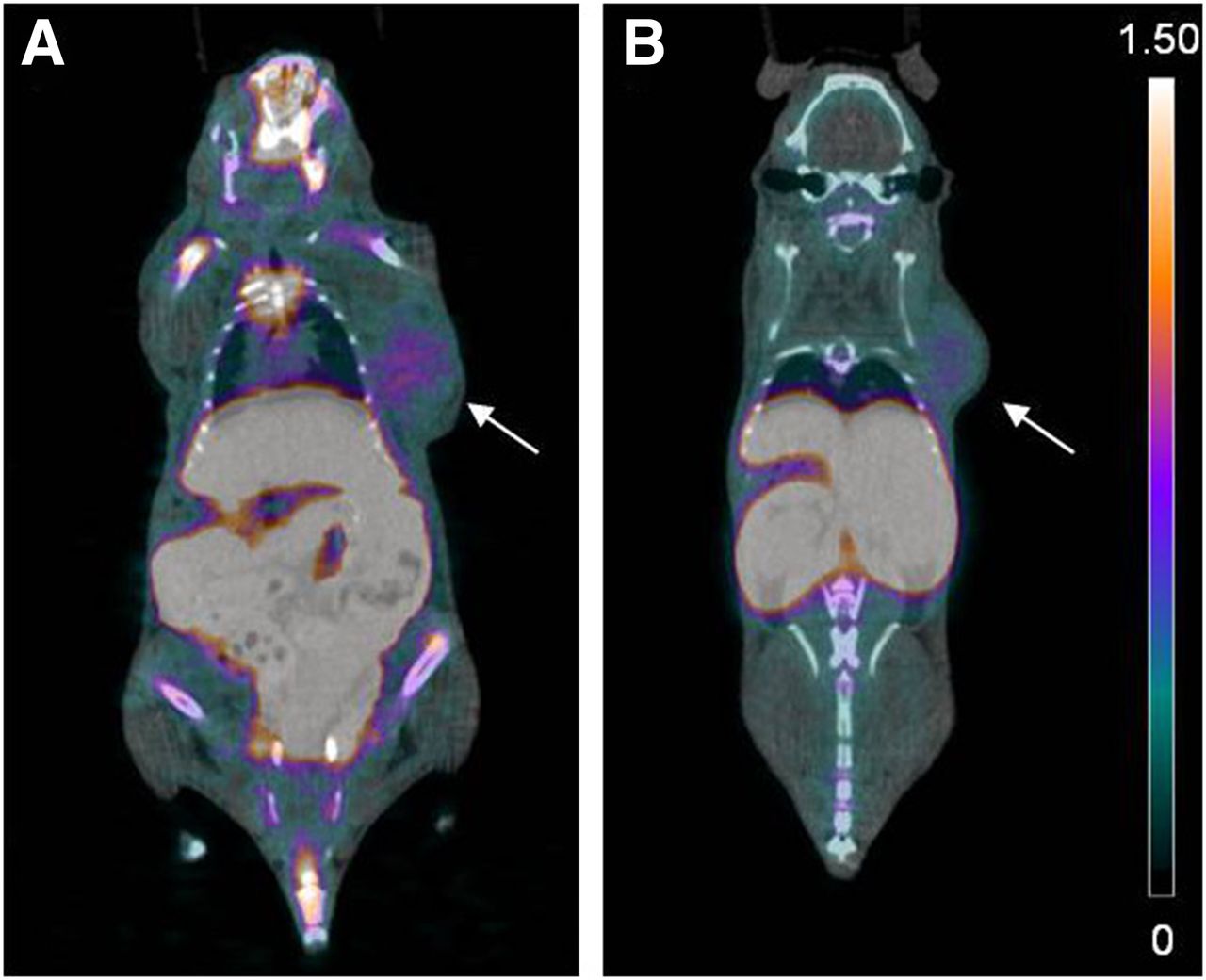
  • FIGURE 4.
    • Download figure
    • Open in new tab
    • Download powerpoint
    FIGURE 4.

    PET/CT images of monomeric 18F-sulfonamides at 1 h after injection of 18F-AmBF3-AEBS (A) and 18F-AmBF3-ABS (B). Tumors are indicated by arrows. Scale bar unit is %ID/g.
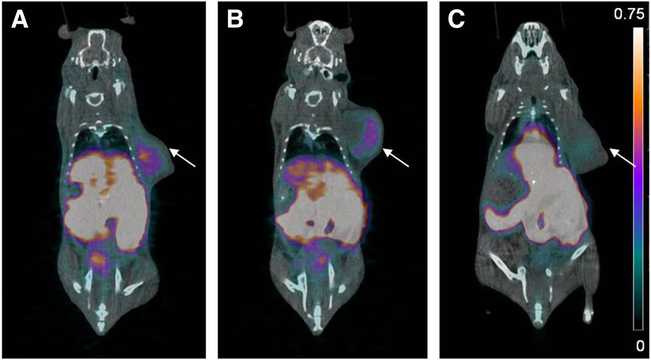
  • FIGURE 5.
    • Download figure
    • Open in new tab
    • Download powerpoint
    FIGURE 5.

    PET/CT images of trimeric 18F-sulfonamides at 1 h after injection of 18F-AmBF3-(AEBS)3 (A), 18F-AmBF3-(ABS)3 (B), and 18F-AmBF3-(ABS)3 (C) preblocking with acetazolamide. Tumors are indicated by arrows. Scale bar unit is %ID/g.

Tables

  • Figures
  • Additional Files
    • View popup
    TABLE 1

    Summary of Biologic Evaluation Data of Radiolabeled CA-IX Imaging Probes Shown in Figure 1

    Biodistribution
    CompoundBinding affinity Ki (nM)Tumor modelTumor uptake (%ID/g)Tumor-to-blood ratioTumor-to-muscle ratioImaging dataReference
    ANANANANANANA16
    B58HT-290.13 at 0.5 h0.75 at 0.5 hNANA19
    C59HT-290.2–0.1 at 0.5–4 h0.0 at 0.5–4 hNANA18
    D66HT-290.5–0.1 at 0.5–4 h0.4–0.5 at 0.5–4 hNANA18
    ENAHT-29≤0.1 at 0.5–4 h≤1.0 at 0.5–4 hNANA18
    FNAHT-29≤0.2 at 0.5–4 h0.2–1.0 at 0.5–4 hNANA18
    G0.9 (R = 3-NO2)NANANANANA22
    5.4 (R = 4-Ac)NANANANANA22
    0.3 (R = 2-CN)NANANANANA22
    H45HT-290.83 at 1 h<1.0 at 1 h<1.0 at 1 hNA21
    I124HT-29, U373<0.25 at 2–4 hNANANo tumor visualization39
    J9.0NANANANANA23
    K9.3 (R = Me)HT-290.51 at 1 h∼1.0 at 1 h∼1.0 at 1 hNo tumor visualization20
    9.6 (R = Ac)HT-290.59 at 1 h∼1.0 at 1 h∼1.0 at 1 hNo tumor visualization20
    9.1 (R = Cl)HT-290.98 at 1 h∼1.0 at 1 h∼1.0 at 1 hNo tumor visualization20
    L5.2NANANANANA24
    M7.0NANANANANA24
    N70HT-291.16 at 1 h<1.0 at 1 h∼1.0 at 1 hNo tumor visualization21
    • NA = not available.

    • View popup
    TABLE 2

    CA-I, -II, -IX, and -XII Inhibition Data

    Ki (nM)*
    CompoundCA-ICA-IICA-IXCA-XII
    AmBF3-AEBS13727.58.00.76
    AmBF3-ABS65.659.86.60.49
    AmBF3-(AEBS)334.826.535.78.6
    AmBF3-(ABS)3100.38.68.58.6
    Acetazolamide25012256.0
    • ↵* Errors in range of 5%–10% of reported values from 3 different assays.

    • View popup
    TABLE 3

    Biodistribution and Tumor-to-Nontarget Ratios for 18F-AmBF3 Sulfonamides

    18F-AmBF3-(ABS)3
    Organ18F-AmBF3-AEBS, 1 h after injection18F-AmBF3-ABS, 1 h after injection18F-AmBF3-(AEBS)3, 1 h after injection0.5 h after injection1 h after injection, unblocked1 h after injection, blocked*2 h after injection
    Blood0.56 ± 0.050.51 ± 0.050.19 ± 0.200.26 ± 0.020.09 ± 0.050.17 ± 0.170.07 ± 0.01
    Fat0.08 ± 0.030.08 ± 0.030.04 ± 0.050.16 ± 0.080.02 ± 0.010.03 ± 0.040.02 ± 0.00
    Testes0.14 ± 0.050.23 ± 0.150.04 ± 0.050.20 ± 0.110.04 ± 0.010.03 ± 0.020.03 ± 0.00
    Stomach0.54 ± 0.392.32 ± 2.141.03 ± 0.274.66 ± 4.061.90 ± 1.620.26 ± 0.360.44 ± 0.26
    Spleen0.38 ± 0.030.54 ± 0.310.55 ± 0.730.68 ± 0.240.37 ± 0.340.19 ± 0.240.19 ± 0.05
    Liver10.87 ± 0.5313.64 ± 2.490.98 ± 0.679.64 ± 3.660.97 ± 0.270.34 ± 0.18†0.58 ± 0.19
    Pancreas0.59 ± 0.070.57 ± 0.180.07 ± 0.050.22 ± 0.100.07 ± 0.050.08 ± 0.110.05 ± 0.03
    Adrenals0.32 ± 0.100.54 ± 0.270.34 ± 0.500.97 ± 1.020.21 ± 0.150.08 ± 0.030.26 ± 0.14
    Kidney74.33 ± 19.6452.70 ± 14.090.94 ± 0.3218.63 ± 3.411.78 ± 0.490.14 ± 0.04†5.86 ± 0.86
    Lungs0.90 ± 0.181.97 ± 0.110.48 ± 0.542.75 ± 0.520.41 ± 0.290.23 ± 0.140.24 ± 0.04
    Heart0.29 ± 0.040.27 ± 0.020.10 ± 0.070.32 ± 0.050.08 ± 0.040.04 ± 0.030.08 ± 0.04
    Muscle0.18 ± 0.050.32 ± 0.110.07 ± 0.030.26 ± 0.080.04 ± 0.020.03 ± 0.010.10 ± 0.05
    Bone2.05 ± 0.360.85 ± 0.110.18 ± 0.120.52 ± 0.090.21 ± 0.090.10 ± 0.040.34 ± 0.02
    Brain0.05 ± 0.020.04 ± 0.000.02 ± 0.020.09 ± 0.020.02 ± 0.010.01 ± 0.010.02 ± 0.01
    Tumor0.56 ± 0.110.64 ± 0.080.30 ± 0.100.70 ± 0.130.33 ± 0.070.06 ± 0.01†0.24 ± 0.05
    Tumor-to-liver0.05 ± 0.010.05 ± 0.010.37 ± 0.140.07 ± 0.030.35 ± 0.070.19 ± 0.04†0.42 ± 0.07
    Tumor-to-blood1.01 ± 0.251.24 ± 0.122.88 ± 1.812.74 ± 0.683.93 ± 1.261.08 ± 1.03†3.53 ± 0.55
    Tumor-to-muscle3.18 ± 0.632.15 ± 0.664.94 ± 2.762.87 ± 1.439.55 ± 2.961.95 ± 0.52†2.78 ± 1.44
    • ↵* Blocked by preinjection of 10 mg of acetazolamide per kilogram 1 h before radiotracer administration.

    • ↵† Preinjection significantly reduced uptake of same organ/ratio (P < 0.05).

    • Values are mean ± SD (n ≥ 4).

Additional Files

  • Figures
  • Tables
  • Supplemental Data

    Files in this Data Supplement:

    • Supplemental Data
PreviousNext
Back to top

In this issue

Journal of Nuclear Medicine: 56 (9)
Journal of Nuclear Medicine
Vol. 56, Issue 9
September 1, 2015
  • Table of Contents
  • Table of Contents (PDF)
  • About the Cover
  • Index by author
Print
Download PDF
Article Alerts
Sign In to Email Alerts with your Email Address
Email Article

Thank you for your interest in spreading the word on Journal of Nuclear Medicine.

NOTE: We only request your email address so that the person you are recommending the page to knows that you wanted them to see it, and that it is not junk mail. We do not capture any email address.

Enter multiple addresses on separate lines or separate them with commas.
Trimeric Radiofluorinated Sulfonamide Derivatives to Achieve In Vivo Selectivity for Carbonic Anhydrase IX–Targeted PET Imaging
(Your Name) has sent you a message from Journal of Nuclear Medicine
(Your Name) thought you would like to see the Journal of Nuclear Medicine web site.
Citation Tools
Trimeric Radiofluorinated Sulfonamide Derivatives to Achieve In Vivo Selectivity for Carbonic Anhydrase IX–Targeted PET Imaging
Joseph Lau, Zhibo Liu, Kuo-Shyan Lin, Jinhe Pan, Zhengxing Zhang, Daniela Vullo, Claudiu T. Supuran, David M. Perrin, François Bénard
Journal of Nuclear Medicine Sep 2015, 56 (9) 1434-1440; DOI: 10.2967/jnumed.114.153288

Citation Manager Formats

  • BibTeX
  • Bookends
  • EasyBib
  • EndNote (tagged)
  • EndNote 8 (xml)
  • Medlars
  • Mendeley
  • Papers
  • RefWorks Tagged
  • Ref Manager
  • RIS
  • Zotero
Share
Trimeric Radiofluorinated Sulfonamide Derivatives to Achieve In Vivo Selectivity for Carbonic Anhydrase IX–Targeted PET Imaging
Joseph Lau, Zhibo Liu, Kuo-Shyan Lin, Jinhe Pan, Zhengxing Zhang, Daniela Vullo, Claudiu T. Supuran, David M. Perrin, François Bénard
Journal of Nuclear Medicine Sep 2015, 56 (9) 1434-1440; DOI: 10.2967/jnumed.114.153288
Twitter logo Facebook logo LinkedIn logo Mendeley logo
  • Tweet Widget
  • Facebook Like
  • Google Plus One
Bookmark this article

Jump to section

  • Article
    • Abstract
    • MATERIALS AND METHODS
    • RESULTS
    • DISCUSSION
    • CONCLUSION
    • DISCLOSURE
    • Acknowledgments
    • Footnotes
    • REFERENCES
  • Figures & Data
  • Supplemental
  • Info & Metrics
  • PDF

Related Articles

  • This Month in JNM
  • PubMed
  • Google Scholar

Cited By...

  • One-Step 18F-Labeling and Preclinical Evaluation of Prostate-Specific Membrane Antigen Trifluoroborate Probes for Cancer Imaging
  • Evaluation of Nonpeptidic Ligand Conjugates for the Treatment of Hypoxic and Carbonic Anhydrase IX-Expressing Cancers
  • Google Scholar

More in this TOC Section

  • Counting Rate Characteristics and Image Distortion in Preclinical PET Imaging During Radiopharmaceutical Therapy
  • Design and Fabrication of Kidney Phantoms for Internal Radiation Dosimetry Using 3D Printing Technology
  • Synthesis and Biologic Evaluation of Novel 18F-Labeled Probes Targeting Prostate-Specific Membrane Antigen for PET of Prostate Cancer
Show more Basic Science Investigations

Similar Articles

Keywords

  • Positron Emission Tomography
  • hypoxia
  • carbonic anhydrase IX
  • one-step 18F-labeling
  • 18F-AmBF3
SNMMI

© 2025 SNMMI

Powered by HighWire