


Article Figures & Data
Figures
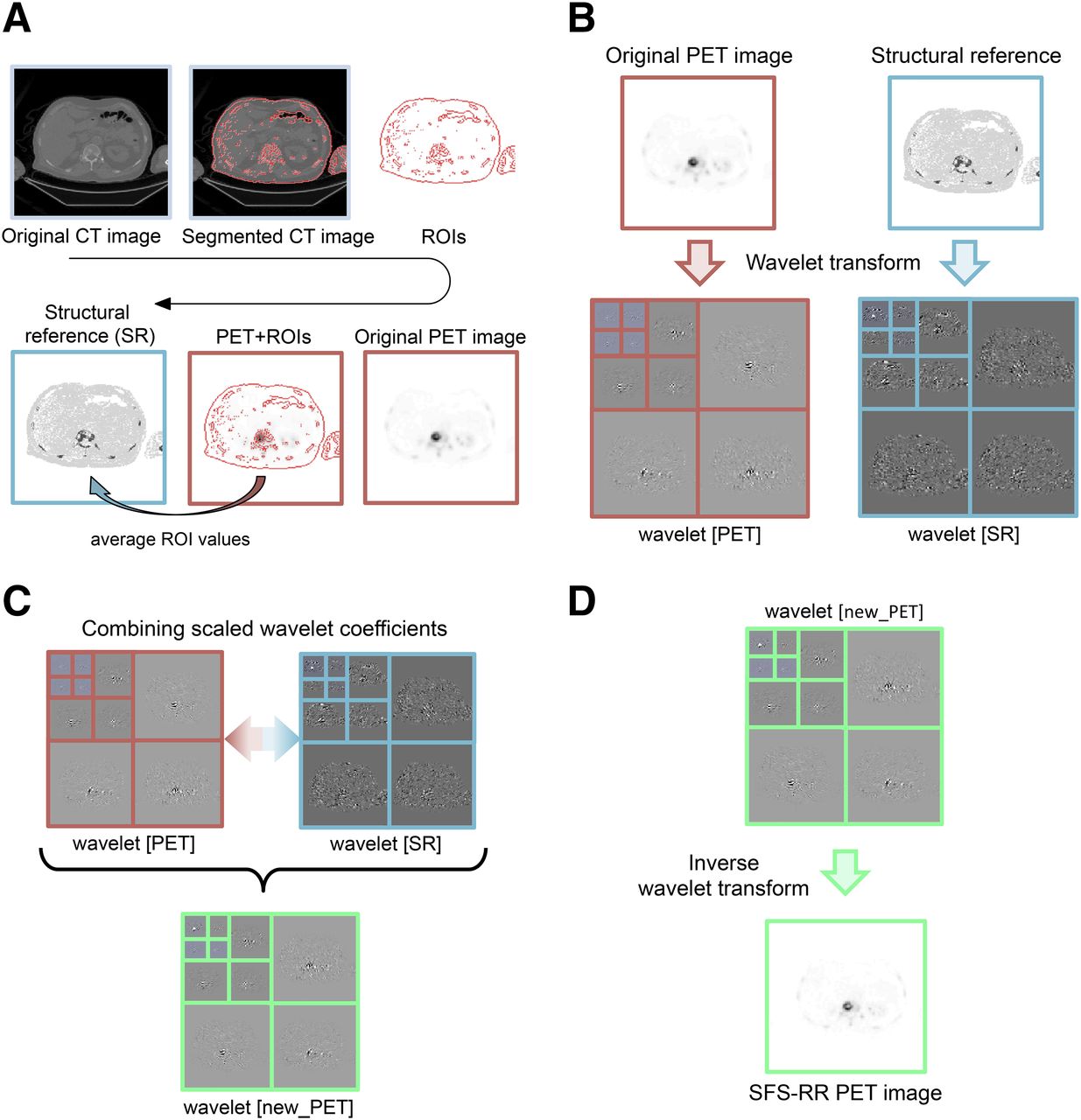
- FIGURE 1.
Graphical representation of SFS-RR algorithm. (A) Structural reference image required by SFS-RR algorithm is computed from CT and PET images. (B) Wavelet decomposition of functional and structural images. (C) Functional and structural wavelet coefficients are combined to get new high-resolution PET coefficients. (D) Inverse wavelet transform of coefficients obtained from step C resulting in the new high-resolution SFS-RR PET image. For detailed mathematic formulation, refer to supplemental materials.
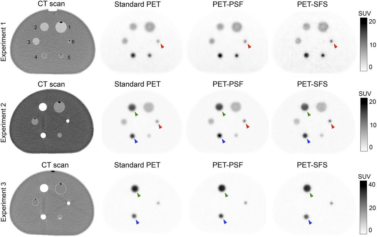
- FIGURE 2.
18F-fluoride PET/CT transaxial images of 3 different phantom experiment acquisitions (1 for each line). Alongside CT image (first column) are 3 different types of functional images: standard PET images (second column), images resulting from inclusion of PSF model into reconstruction (third column), and images resulting after application of SFS resolution recovery algorithm (fourth column). For detailed information on lesion volume, CM, and 18F-FDG concentrations, refer to Table 1. Green markers highlight sphere 2, blue markers highlight sphere 4, and red markers highlight sphere 6.
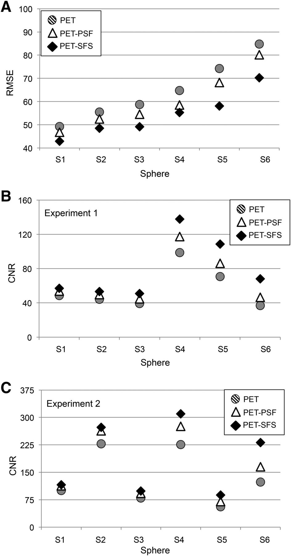
- FIGURE 3.
RMSE and noise analysis. For each sphere (S1–S6), 3 values corresponding to images obtained with different modalities are reported: standard PET (dashed circle), PET with PSF reconstruction (white triangle), and PET corrected with SFS-RR algorithm (black diamond). (A) RMSE for 6 spheres obtained as average among 3 phantom experiments. (B and C) CNR computed for each sphere against uniform region in phantom background. Only experiments 1 and 2 are reported for consistency reasons (in experiment 3, three spheres have zero activity).
- FIGURE 4.
Maximum-intensity projection and transaxial views of representative subject (patient 01). (Left) Standard PET. (Right) PET corrected with SFS-RR algorithm. Red and blue markers highlight 2 representative lesions (spine and rib, respectively) that appear sharper in PET-SFS image than in standard PET 1. Dashed lines indicate slice position of transaxial views reported below maximum-intensity projection.
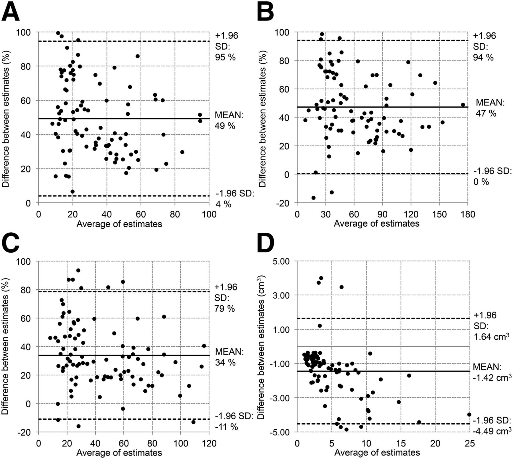
- FIGURE 5.
Bland–Altman plots showing differences in MATV and activity quantification when estimates are computed in images obtained with standard PET and PET corrected with SFS-RR algorithm. Each gray circle represents specific lesion; all lesions of all patients are reported. Differences between estimates for SUVmean (A), SUVmax (B), and SUVpeak (C) are reported as relative percentage difference. MATV (D) is reported as absolute difference in cm3.


Tables
Experiment 1 Experiment 2 Experiment 3 Phantom compartments Iodine contrast medium (mg/mL) 18F-FDG (kBq/mL) Iodine contrast medium (mg/mL) 18F-FDG (kBq/mL) Iodine contrast medium (mg/mL) 18F-FDG (kBq/mL) Sphere volume (mL) Background 1.08* 4.56* 1.20* 5.29* 1.20* 5.70* 9,700 S1 6.00† 53.20† 6.00† 66.50† 1.20* 5.70* 26.52 S2 6.00† 53.20† 42.00‡ 187.00‡ 41.20‡ 227.00‡ 11.49 S3 6.00† 53.20† 6.00† 66.50† 1.20* 5.70* 5.57 S4 1.00* 148.50‡ 42.00‡ 187.00‡ 41.20‡ 227.00‡ 2.57 S5 1.00* 148.50‡ 6.00† 66.50† 1.20* 5.70* 1.15 S6 1.00* 148.50‡ 42.00‡ 187.00‡ 41.20‡ 227.00‡ 0.52 ↵* Concentration resulting in image contrast comparable to normal soft tissue.
↵† Concentration resulting in image contrast comparable to normal bone.
↵‡ Concentration resulting in image contrast comparable to metastatic bone.
Concentrations of iodine (from Omnipaque300) and 18F-FDG injected in all phantom compartments for each experiment. Compartment volumes are also reported. Spheres 4–6, experiment 1, and spheres 1, 3, and 5, experiment 3, are filled with same radioactivity concentration as background; as a result they are indiscernible in PET image.
SUVmean (g/mL) SUVmax (g/mL) SUVpeak (g/mL) MATV (cm3) Experiment PET PETPSF PETSFS PET PETPSF PETSFS PET PETPSF PETSFS Ground truth (g/mL) PET PETPSF PETSFS Ground truth 1 S1 5.66 5.95 6.44 8.45 8.84 10.03 8.03 8.17 8.19 9.60 32.22 30.81 30.44 26.52 S2 5.29 5.64 6.28 8.50 9.00 10.69 8.50 8.8 7.88 9.60 13.84 13.20 12.47 11.49 S3 4.54 5.01 5.73 7.73 8.58 9.37 7.73 7.87 7.97 9.60 7.46 6.72 6.75 5.57 S4 10.22 12.25 13.17 18.64 22.12 22.11 18.64 16.25 18.24 24.87 3.52 2.86 3.33 2.57 S5 7.72 9.51 12.60 14.82 18.55 23.56 14.81 10.91 13.75 24.87 1.98 1.56 1.37 1.15 S6 4.21 5.27 8.50 8.19 10.52 16.92 4.77 10.51 11.96 24.87 1.34 1.10 0.83 0.52 2 S1 4.65 4.87 5.16 6.85 7.08 7.78 6.62 6.69 6.54 8.67 34.28 32.96 31.03 26.52 S2 10.40 11.11 11.55 16.28 16.83 17.16 15.63 15.87 14.92 24.38 14.06 13.54 13.52 11.49 S3 3.87 4.25 4.60 6.74 7.25 7.57 6.09 6.61 6.39 8.67 7.53 6.89 6.53 5.57 S4 9.93 11.42 12.40 17.57 20.21 19.76 13.95 15.90 17.20 24.38 3.35 3.03 3.20 2.57 S5 2.49 3.08 4.02 4.97 6.05 7.71 3.14 3.57 4.76 8.67 2.32 1.71 1.37 1.15 S6 4.56 6.16 8.57 8.79 11.76 16.14 5.02 7.60 10.03 24.38 1.39 0.98 0.86 0.52 3 S2 24.59 25.95 26.63 38.11 38.01 37.90 37.20 36.82 34.87 40.61 13.52 13.42 13.59 11.49 S4 16.68 19.08 20.74 29.92 34.12 33.97 23.78 26.89 29.19 40.61 3.35 3.08 3.25 2.57 S6 7.36 9.78 13.61 13.77 18.33 25.52 8.02 14.08 19.15 40.61 1.25 0.90 0.79 0.52 SUVmean, SUVmax, SUVpeak, and MATV estimates computed for phantom spheres after automated segmentation for all experiments 1–3. Values are reported for estimates obtained with 3 different modalities (standard PET, PET reconstructed with a PSF model, and PET corrected with SFS-RR algorithm) alongside corresponding ground truth values.
Supplemental Data
Files in this Data Supplement:


Jump to section
Related Articles
Cited By...
- No citing articles found.