


Article Figures & Data
Figures
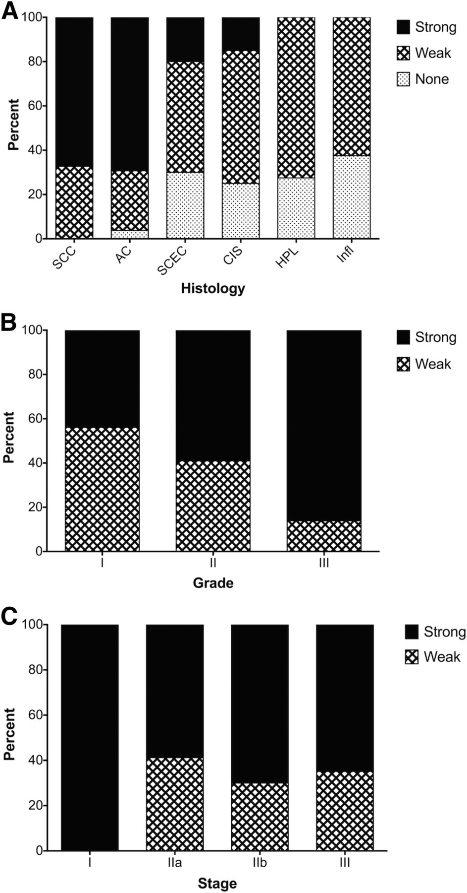
- FIGURE 1.
Immunohistochemical staining of esophageal tissue arrays for periostin. (A) Almost all of the tissue samples of SSC and adenocarcinoma stained positive for periostin, with most stained strongly for periostin. Only 15% of carcinoma in situ stained strongly for periostin, and none of the hyperplasia or inflammation samples stained strongly for periostin. (B) Among primary SCC tissue samples, periostin staining significantly changed with tumor grade, with higher-grade tumors exhibiting significantly stronger periostin staining. (C) There was no significant change of periostin staining with changes in tumor stage. AC = adenocarcinoma; CIS = carcinoma in situ; HPL = hyperplasia; Infl = inflammation; SCEC = small cell esophageal cancer.
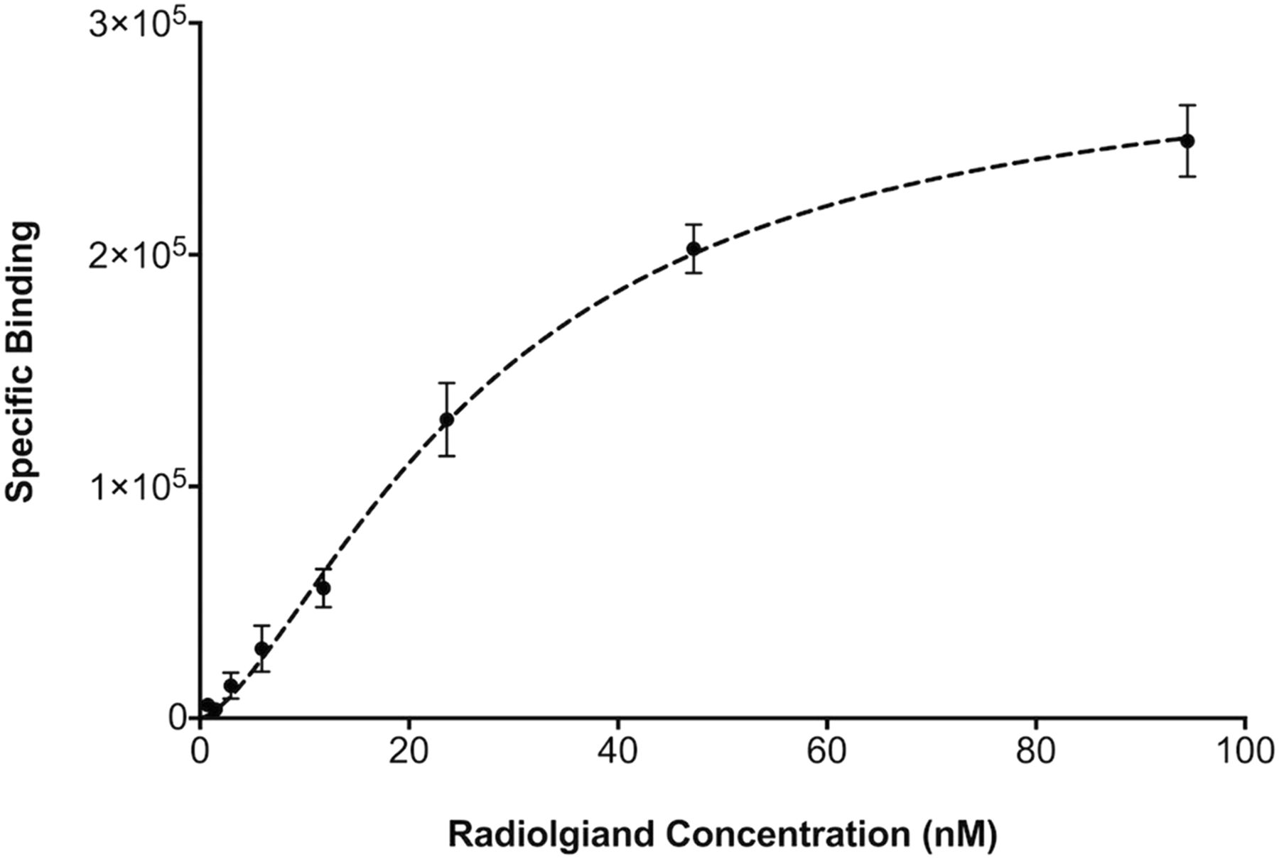
- FIGURE 2.
Direct (saturation) radioligand binding assays showed that 64Cu-DOTA-antiperiostin-F(ab′)2 bound specifically to human recombinant periostin fixed to HisLink Protein Purification Resin. The Kd value for 64Cu-DOTA-antiperiostin- F(ab′)2 was 29.2 ± 3.0 nM.
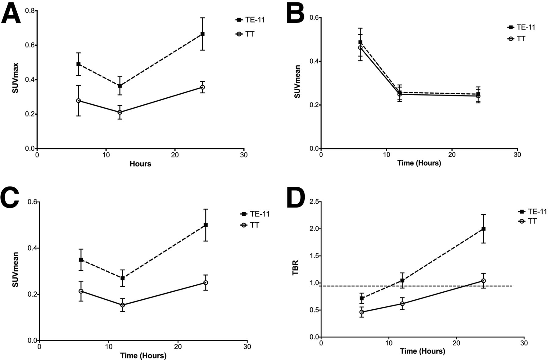
- FIGURE 3.
Quantitative PET imaging of xenografts TE-11 and TT at different time points after intravenous injection of probe. (A–C) Blood-pool activity decreases over time, and uptake in tumors increases over 24 h after injection of 64Cu-DOTA-antiperiostin-F(ab′)2 probe. (D) TBR linearly increases in both xenografts but at greater slope in TE-11 xenografts, compared with TT tumors. TE-11 TBR reached approximately 2 at 24 h after injection. SUVmax = maximum standardized uptake value.
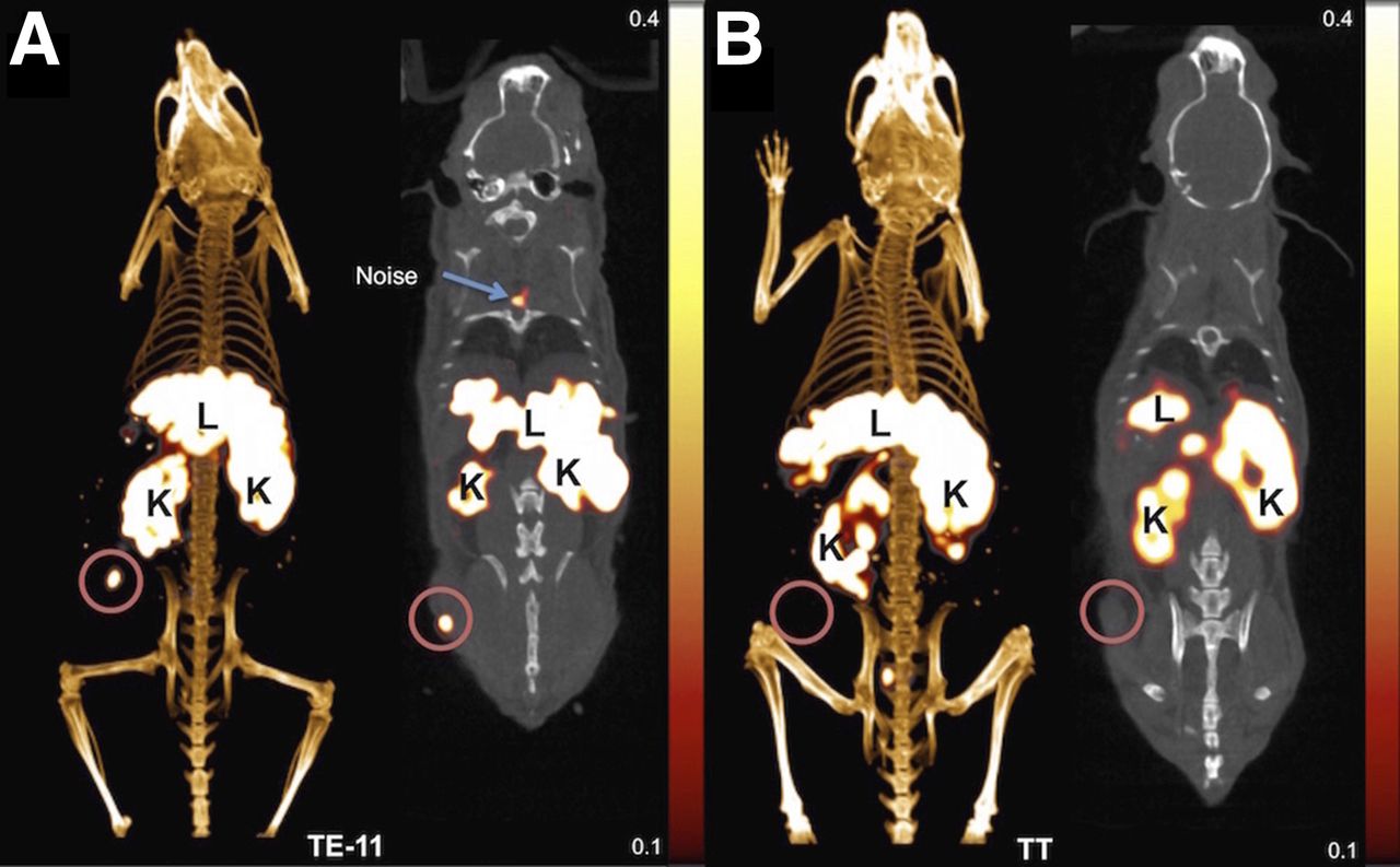
- FIGURE 4.
Representative PET/CT images using 64Cu-DOTA-antiperiostin-F(ab′)2 show high uptake of probe in subcutaneously implanted human esophageal squamous cell TE-11 tumors (A) and minimal uptake in TT tumors (B). K = kidney; L = liver.
- FIGURE 5.
Representative18F-FDG PET/CT scan of L2Cre-p120loxp/loxp mouse shows tumor in gastroesophageal junction (A and B), whereas PET/CT imaging with 64Cu-DOTA-antiperiostin-F(ab′)2 shows specific uptake in same location of high uptake with no increased uptake in adjacent organs such as aorta (C and D). B = bladder; H = heart; K = kidney.
- FIGURE 6.
Biodistribution studies 24 h after injection confirmed our results, with PET/CT imaging demonstrating significantly higher accumulation of 64Cu-DOTA-antiperiostin-F(ab′)2 in TE-11 tumors and compared with negative control, TT tumors. PET probe is cleared from other organs and excreted through kidney and liver.


Tables
Region of interest Time point TE-11 TT P Tumor 6 0.490 ± 0.063 0.278 ± 0.088 0.0082* Maximum SUV 12 0.364 ± 0.052 0.210 ± 0.039 0.0031* 24 0.665 ± 0.093 0.356 ± 0.033 0.0008* Tumor (SUVmean) 6 0.350 ± 0.046 0.214 ± 0.043 0.005* 12 0.271 ± 0.036 0.154 ± 0.028 0.002* 24 0.501 ± 0.069 0.251 ± 0.033 0.0006* Blood (SUVmean) 6 0.488 ± 0.064 0.463 ± 0.060 0.589 12 0.258 ± 0.034 0.249 ± 0.033 0.717 24 0.250 ± 0.033 0.241 ± 0.031 0.704 Tumor to background 6 0.717 ± 0.095 0.463 ± 0.093 0.0087* 12 1.047 ± 0.140 0.617 ± 0.111 0.0009* 24 2.000 ± 0.263 1.041 ± 0.136 0.0006* ↵* Statistically significant; provided values represent mean ± SD.
Supplemental Data
Files in this Data Supplement:


