


Article Figures & Data
Figures
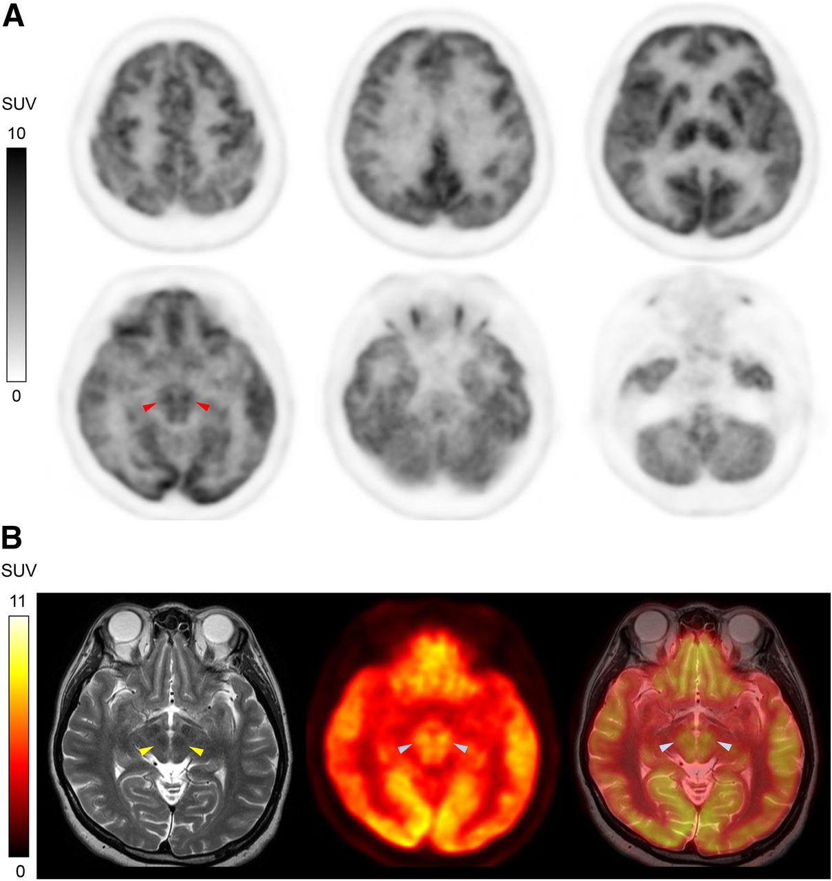
- FIGURE 1.
A representative case. (A) Bilateral RN indicates areas of higher metabolism (arrowheads). (B) Coregistration with T2-weighted MR images confirmed that high 18F-FDG uptake corresponded to low-signal-intensity areas in midbrain, indicating RN (arrowheads).
- FIGURE 2.
Metabolic correlations between right RN and surface brain areas (A) and left RN and surface brain areas (B). Degree of correlation is represented by z score.
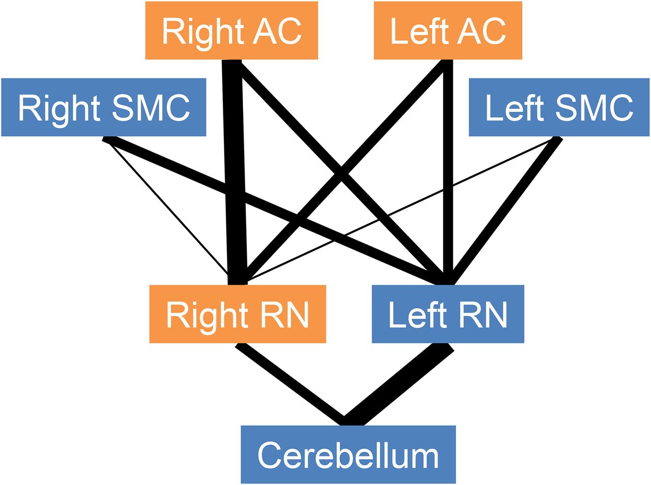
- FIGURE 3.
Scheme of possible neural networks. Width of lines represents degree of association between 2 regions. Right RN is more involved in social–emotional networks (orange regions), whereas left RN is more involved in motor function networks (blue regions). Right AC is known to be more responsible for social–emotional and alerting/arousal vigilance functions than left AC. AC = association cortex; SMC = sensorimotor cortex.


Tables
Right hemisphere Left hemisphere Talairach coordinate (mm) Talairach coordinate (mm) Region BA Maximum z score (Pearson R) x y z BA Maximum z score (Pearson R) x y z Frontal association cortex 6,9,10*,11*,25*, 44*,45*,46,47* 3.34 (0.69) 39.3 55.1 15.8 10† 3.60 (0.72) −41.6 52.9 −4.5 Parietal cortex 40† 3.78 (0.75) 61.9 −37.1 31.5 N/A N/A N/A N/A N/A Temporal cortex 21†,22*,38 3.52 (0.71) 59.6 −7.9 −15.8 20 2.58 (0.56) −57.3 −16.9 −22.5 Occipital cortex N/A N/A N/A N/A N/A N/A N/A N/A N/A N/A Sensorimotor cortex N/A N/A N/A N/A N/A N/A N/A N/A N/A N/A Limbic lobe 23*,25,29‡, 30*,34†,35 4.24 (0.80) 1.1 −43.9 13.5 23,24,25†,30, 31,34*,35* 3.57 (0.72) −1.1 −1.1 −2.2 Cerebellum N/A 4.01 (0.77)‡ 5.6 −59.6 −42.8 N/A 3.43 (0.70)† −1.1 −37.1 −20.3 Right hemisphere Left hemisphere Talairach coordinate (mm) Talairach coordinate (mm) Region BA Maximum z score (Pearson R) x y z BA Maximum z score (Pearson R) x y z Frontal association cortex 6‡,45‡,46† 3.91 (0.76) 57.4 25.9 11.3 6*,10*,46,47 3.35 (0.69) −34.9 59.6 11.3 Parietal cortex N/A N/A N/A N/A N/A 43 2.65 (0.58) −61.9 −5.6 15.8 Temporal cortex 22 2.77 (0.60) 57.4 1.1 −2.3 N/A N/A N/A N/A N/A Occipital cortex N/A N/A N/A N/A N/A N/A N/A N/A N/A N/A Sensorimotor cortex 4† 3.43 (0.70) 59.6 −1.1 15.8 4* 3.02 (0.64) −61.9 −5.6 24.8 Limbic lobe 25*,34*,35*,36 3.28 (0.68) 25.9 −23.6 −22.5 25†,31*,34* 3.44 (0.70) −1.1 −3.4 −2.3 Cerebellum N/A 3.82 (0.75)‡ 1.1 −41.6 −22.5 N/A 4.48 (0.83)‡ −1.1 −41.6 −20.3


