


Article Figures & Data
Figures
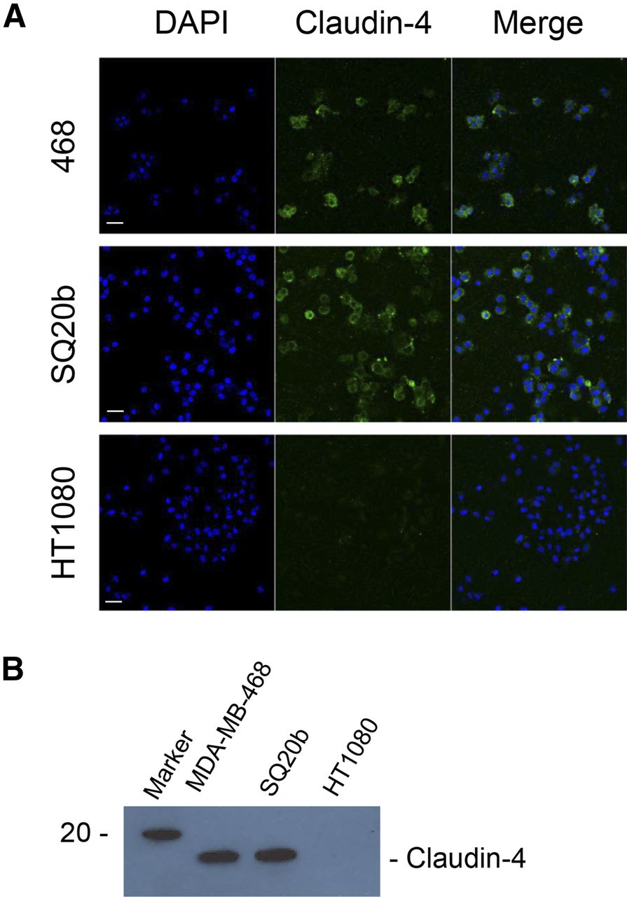
- FIGURE 1.
(A) MDA-MB-468, SQ20b, and HT1080 cells were stained for claudin-4. Scale bar, 20 μm. (B) Western blot demonstrating presence or absence of claudin-4 in whole cell lysates obtained from MDA-MB-468, SQ20b, and HT1080 cells.
- FIGURE 2.
(A) MDA-MB-468 cells were exposed for 1 h at 4°C to increasing concentrations of 111In-labeled cCPE.GST or GST, and extent of cell-binding was determined. (B) Increasing amounts of unlabeled cCPE.GST were used to block binding of 111In-cCPE.GST to MDA-MB-468 cells.
- FIGURE 3.
(A) Representative SPECT/CT images of mice carrying tumor xenografts (white circles) of MDA-MB-468 (claudin-4–positive) or HT1080 (claudin-4–negative) cells, 24 h after intravenous administration of 111In-cCPE.GST or 111In-GST. 111In uptake was also observed in shoulder joints (S), liver (L), intestines (I), and knee joints (K). Coronal sections through tumor are shown. (B) Sections obtained from MDA-MB-468 or HT1080 xenograft tumors were stained using anticlaudin-4 antibodies. Scale bar, 20 μm. (C) Biodistribution results, 24 h after intravenous administration of 111In-cCPE.GST or 111In-GST. Tumor-to-blood and tumor-to-muscle ratios were calculated. Each group contained at least 3 animals. *P < 0.05. **P < 0.01. ***P < 0.001. ****P < 0.0001.
- FIGURE 4.
(A) Sections obtained from tumors harvested from 139-d-old BALB/neuT mouse were stained using anti–claudin-4 antibodies. Scale bar, 20 μm. (B) Representative SPECT/CT images of BALB/neuT mice, aged 139 d, bearing mammary tumors (white circles), 24 h after intravenous administration of 111In-cCPE.GST or 111In-GST. Overt tumors were clearly visible on CT images. Coronal sections through tumor are shown. (C) Volume-of-interest analysis of BALB/neuT mice bearing overt tumors 3 or 24 h after injection of 111In-cCPE.GST or 111In-GST. Each group contained at least 3 animals. *P < 0.05.
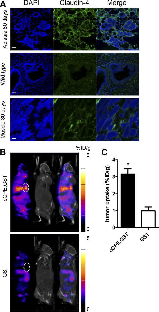
- FIGURE 5.
(A) Sections obtained from aplastic lesion or muscle harvested from 80-d-old BALB/neuT mouse or ductal tissue obtained from a wild-type BALB/c mouse were stained using anti–claudin-4 antibodies. Scale bar, 20 μm. (B) Representative SPECT/CT images of BALB/neuT mice, aged 80 d, bearing aplastic lesions (white circles), 24 h after intravenous administration of 111In-cCPE.GST or 111In-GST. Functional SPECT imaging was compared with anatomic CT imaging. Aplastic lesion could not be detected on CT images. Coronal sections through mammary fat pads are shown. (C) Volume-of-interest analysis of 80-d-old BALB/neuT mice bearing aplastic 24 h after injection of 111In-cCPE.GST or 111In-GST. Each group contained at least 3 animals. *P < 0.05.


Additional Files
Supplemental Data
Files in this Data Supplement:


Jump to section
Related Articles
Cited By...
- Cryo-EM Structures of Clostridium perfringens Enterotoxin Bound to its Human Receptor, Claudin-4
- Radiolabeled cCPE Peptides for SPECT Imaging of Claudin-4 Overexpression in Pancreatic Cancer
- Creation of a Claudin-2 Binder and Its Tight Junction-Modulating Activity in a Human Intestinal Model
- Claudin-5-Binders Enhance Permeation of Solutes across the Blood-Brain Barrier in a Mammalian Model
- Database-augmented Mass Spectrometry Analysis of Exosomes Identifies Claudin 3 as a Putative Prostate Cancer Biomarker
- Dual-Targeting Nanoparticles for In Vivo Delivery of Suicide Genes to Chemotherapy-Resistant Ovarian Cancer Cells