
Main menu

  • Home
  • Content
    • Current
    • Ahead of print
    • Past Issues
    • JNM Supplement
    • SNMMI Annual Meeting Abstracts
    • Continuing Education
    • JNM Podcasts
  • Subscriptions
    • Subscribers
    • Institutional and Non-member
    • Rates
    • Journal Claims
    • Corporate & Special Sales
  • Authors
    • Submit to JNM
    • Information for Authors
    • Assignment of Copyright
    • AQARA requirements
  • Info
    • Reviewers
    • Permissions
    • Advertisers
  • About
    • About Us
    • Editorial Board
    • Contact Information
  • More
    • Alerts
    • Feedback
    • Help
    • SNMMI Journals
  • SNMMI
    • JNM
    • JNMT
    • SNMMI Journals
    • SNMMI

User menu

  • Subscribe
  • My alerts
  • Log in
  • My Cart

Search

  • Advanced search
Journal of Nuclear Medicine
  • SNMMI
    • JNM
    • JNMT
    • SNMMI Journals
    • SNMMI
  • Subscribe
  • My alerts
  • Log in
  • My Cart
Journal of Nuclear Medicine

Advanced Search

  • Home
  • Content
    • Current
    • Ahead of print
    • Past Issues
    • JNM Supplement
    • SNMMI Annual Meeting Abstracts
    • Continuing Education
    • JNM Podcasts
  • Subscriptions
    • Subscribers
    • Institutional and Non-member
    • Rates
    • Journal Claims
    • Corporate & Special Sales
  • Authors
    • Submit to JNM
    • Information for Authors
    • Assignment of Copyright
    • AQARA requirements
  • Info
    • Reviewers
    • Permissions
    • Advertisers
  • About
    • About Us
    • Editorial Board
    • Contact Information
  • More
    • Alerts
    • Feedback
    • Help
    • SNMMI Journals
  • View or Listen to JNM Podcast
  • Visit JNM on Facebook
  • Join JNM on LinkedIn
  • Follow JNM on Twitter
  • Subscribe to our RSS feeds
Research ArticleClinical Investigations

Methods of CT Dose Estimation in Whole-Body 18F-FDG PET/CT

Yusuke Inoue, Kazunori Nagahara, Yoshihito Tanaka, Hiroki Miyatake, Hirofumi Hata and Toshimasa Hara
Journal of Nuclear Medicine May 2015, 56 (5) 695-700; DOI: https://doi.org/10.2967/jnumed.114.153718
Yusuke Inoue
1Department of Diagnostic Radiology, Kitasato University School of Medicine, Sagamihara, Kanagawa, Japan; and
  • Find this author on Google Scholar
  • Find this author on PubMed
  • Search for this author on this site
Kazunori Nagahara
2Department of Radiology, Kitasato University Hospital, Sagamihara, Japan
  • Find this author on Google Scholar
  • Find this author on PubMed
  • Search for this author on this site
Yoshihito Tanaka
2Department of Radiology, Kitasato University Hospital, Sagamihara, Japan
  • Find this author on Google Scholar
  • Find this author on PubMed
  • Search for this author on this site
Hiroki Miyatake
2Department of Radiology, Kitasato University Hospital, Sagamihara, Japan
  • Find this author on Google Scholar
  • Find this author on PubMed
  • Search for this author on this site
Hirofumi Hata
2Department of Radiology, Kitasato University Hospital, Sagamihara, Japan
  • Find this author on Google Scholar
  • Find this author on PubMed
  • Search for this author on this site
Toshimasa Hara
1Department of Diagnostic Radiology, Kitasato University School of Medicine, Sagamihara, Kanagawa, Japan; and
  • Find this author on Google Scholar
  • Find this author on PubMed
  • Search for this author on this site
  • Article
  • Figures & Data
  • Info & Metrics
  • PDF
Loading

Article Figures & Data

Figures

  • Tables
  • FIGURE 1.
    • Download figure
    • Open in new tab
    • Download powerpoint
    FIGURE 1.

    Division of scan range into 6 anatomic regions (head, neck, chest, abdomen, pelvis, and proximal thigh) is shown on display of CT-Expo software for male subject.
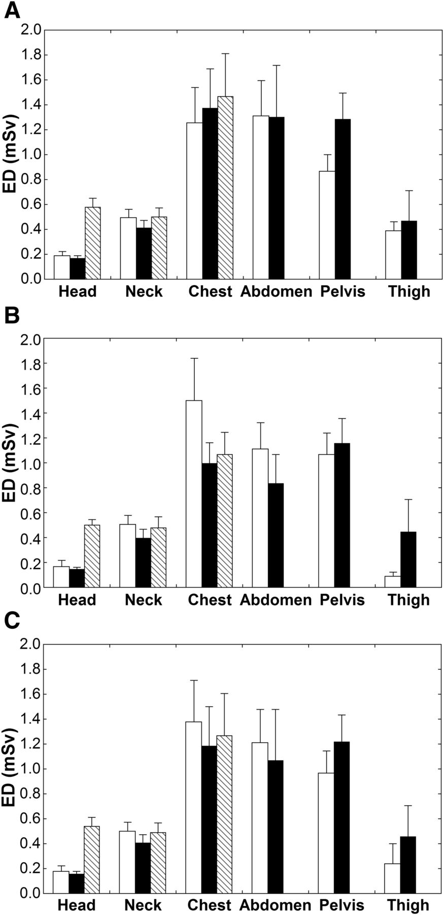
  • FIGURE 2.
    • Download figure
    • Open in new tab
    • Download powerpoint
    FIGURE 2.

    Regional ED values in group A estimated by CT-Expo method (white bars), regional DLP method (black bars), and simple DLP method (shaded bars). Means and SDs in men (A), women (B), and all patients (C) are presented.

  • FIGURE 3.
    • Download figure
    • Open in new tab
    • Download powerpoint
    FIGURE 3.

    Relationships of EDs estimated by different methods in group A. Estimates of regional DLP method (A) and simple DLP method (B) are plotted against those of CT-Expo method. Line of identity is also shown. ○ = data for men; △ = data for women.
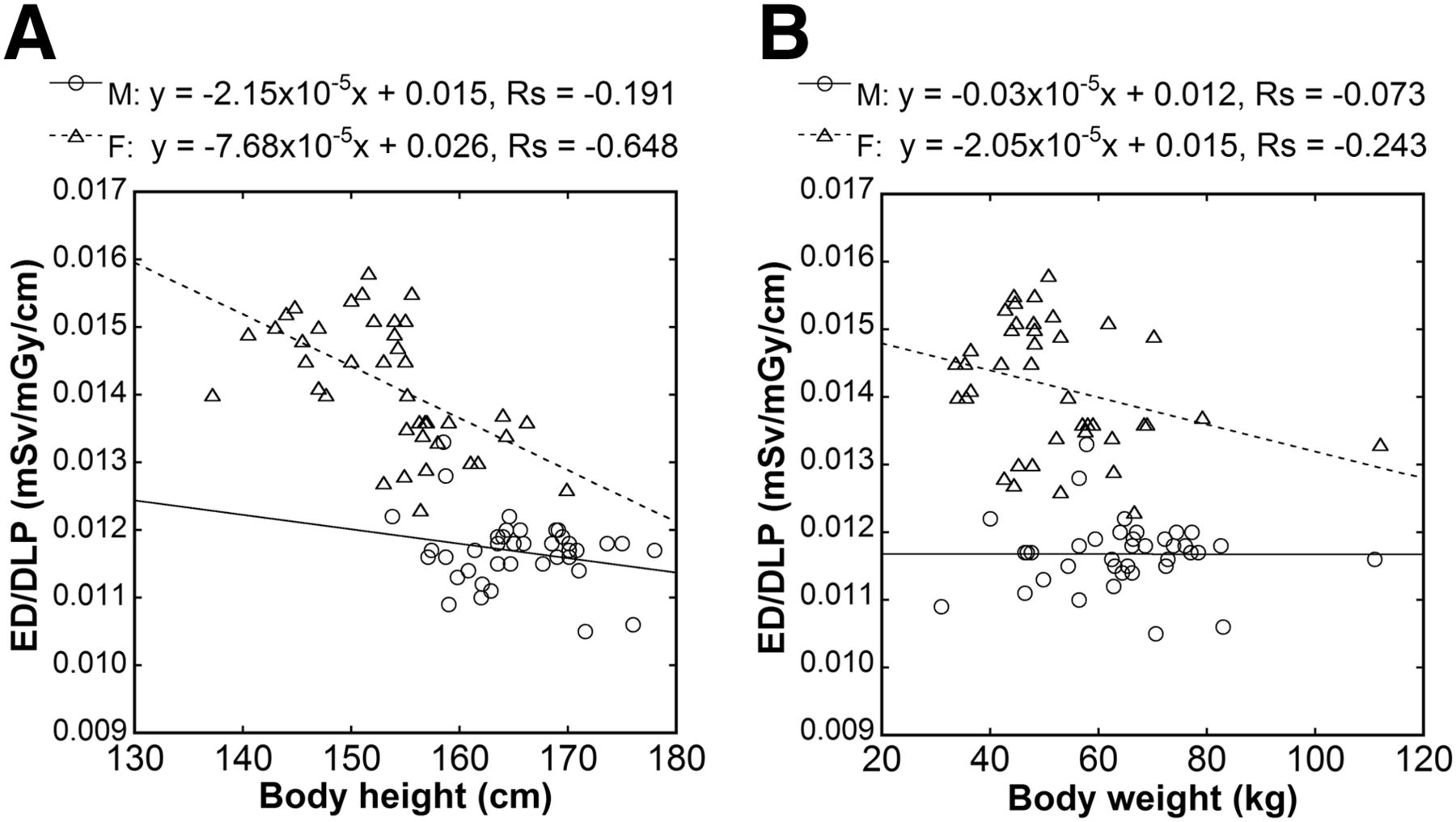
  • FIGURE 4.
    • Download figure
    • Open in new tab
    • Download powerpoint
    FIGURE 4.

    Relationships of ED/DLP with body height (A) and body weight (B). Linear regression lines, regression equations, and Spearman correlation coefficients are shown. ○ = data for men; △ = data for women.

Tables

  • Figures
    • View popup
    TABLE 1

    Patient Characteristics

    Group AGroup B
    CharacteristicMaleFemaleAllMaleFemaleAll
    Age (y)65.2 ± 13.161.9 ± 15.963.6 ± 14.568.5 ± 9.064.5 ± 11.966.5 ± 10.6
    Body height (cm)166.8 ± 6.1154.1 ± 6.8160.4 ± 9.1164.4 ± 4.9152.9 ± 7.3158.7 ± 8.4
    Body weight (kg)64.6 ± 12.849.1 ± 8.856.8 ± 13.465.8 ± 15.855.6 ± 18.360.7 ± 17.7
    • Values are mean ± SD.

    • View popup
    TABLE 2

    Whole-Body ED Values in Group A

    ED (mSv)
    MethodMaleFemaleAll
    CT-Expo4.49 ± 0.814.40 ± 0.724.44 ± 0.76
    Regional DLP5.01 ± 1.043.98 ± 0.814.49 ± 1.06
    Simple DLP5.78 ± 1.124.69 ± 0.875.23 ± 1.14
    • Data are mean ± SD.

    • View popup
    TABLE 3

    ED/DLP (mSv/mGy/cm)

    SubjectMaleFemaleAll
    Group A0.0117 ± 0.00050.0142 ± 0.00100.0129 ± 0.0015
    Group A + B0.0117 ± 0.00050.0141 ± 0.00090.0129 ± 0.0015
    • Data are mean ± SD. Group A + B represents subjects pooled from groups A and B.

    • View popup
    TABLE 4

    Whole-Body ED Values in Group B

    ED (mSv)
    MethodConversion factorMaleFemaleAll
    CT-Expo4.69 ± 1.105.01 ± 1.474.85 ± 1.29
    Simple DLPOriginal5.93 ± 1.395.26 ± 1.685.60 ± 1.56
    Simple DLPModified5.14 ± 1.214.56 ± 1.464.85 ± 1.35
    Simple DLPSex-specific4.74 ± 1.114.91 ± 1.574.83 ± 1.35
    • Data are mean ± SD.

PreviousNext
Back to top

In this issue

Journal of Nuclear Medicine: 56 (5)
Journal of Nuclear Medicine
Vol. 56, Issue 5
May 1, 2015
  • Table of Contents
  • Table of Contents (PDF)
  • About the Cover
  • Index by author
Print
Download PDF
Article Alerts
Sign In to Email Alerts with your Email Address
Email Article

Thank you for your interest in spreading the word on Journal of Nuclear Medicine.

NOTE: We only request your email address so that the person you are recommending the page to knows that you wanted them to see it, and that it is not junk mail. We do not capture any email address.

Enter multiple addresses on separate lines or separate them with commas.
Methods of CT Dose Estimation in Whole-Body 18F-FDG PET/CT
(Your Name) has sent you a message from Journal of Nuclear Medicine
(Your Name) thought you would like to see the Journal of Nuclear Medicine web site.
Citation Tools
Methods of CT Dose Estimation in Whole-Body 18F-FDG PET/CT
Yusuke Inoue, Kazunori Nagahara, Yoshihito Tanaka, Hiroki Miyatake, Hirofumi Hata, Toshimasa Hara
Journal of Nuclear Medicine May 2015, 56 (5) 695-700; DOI: 10.2967/jnumed.114.153718

Citation Manager Formats

  • BibTeX
  • Bookends
  • EasyBib
  • EndNote (tagged)
  • EndNote 8 (xml)
  • Medlars
  • Mendeley
  • Papers
  • RefWorks Tagged
  • Ref Manager
  • RIS
  • Zotero
Share
Methods of CT Dose Estimation in Whole-Body 18F-FDG PET/CT
Yusuke Inoue, Kazunori Nagahara, Yoshihito Tanaka, Hiroki Miyatake, Hirofumi Hata, Toshimasa Hara
Journal of Nuclear Medicine May 2015, 56 (5) 695-700; DOI: 10.2967/jnumed.114.153718
Twitter logo Facebook logo LinkedIn logo Mendeley logo
  • Tweet Widget
  • Facebook Like
  • Google Plus One
Bookmark this article

Jump to section

  • Article
    • Abstract
    • MATERIALS AND METHODS
    • RESULTS
    • DISCUSSION
    • CONCLUSION
    • DISCLOSURE
    • Acknowledgments
    • Footnotes
    • REFERENCES
  • Figures & Data
  • Info & Metrics
  • PDF

Related Articles

  • This Month in JNM
  • PubMed
  • Google Scholar

Cited By...

  • Feasibility of an Ultra-Low-Dose PET Scan Protocol with CT-Based and LSO-TX-Based Attenuation Correction Using a Long-Axial-Field-of-View PET/CT Scanner
  • Establishment of National DRL for CT in Hybrid Imaging Studies (The Second Phase of the National NM CT (PET) Dose Audit for Kuwait Population -2019)
  • Google Scholar

More in this TOC Section

  • Feasibility of Ultra-Low-Activity 18F-FDG PET/CT Imaging Using a Long–Axial-Field-of-View PET/CT System
  • Cardiac Presynaptic Sympathetic Nervous Function Evaluated by Cardiac PET in Patients with Chronotropic Incompetence Without Heart Failure
  • Validation and Evaluation of a Vendor-Provided Head Motion Correction Algorithm on the uMI Panorama PET/CT System
Show more Clinical Investigations

Similar Articles

Keywords

  • PET/CT
  • CT
  • radiation exposure
  • effective dose
  • dose-length product
SNMMI

© 2025 SNMMI

Powered by HighWire