


Abstract
The muscarinic M1 receptor (M1R) is highly involved in cognition, and selective M1 agonists have procognitive properties. Loss of M1R has been found in postmortem brain tissue for several neuropsychiatric disorders and may be related to symptoms of cognitive dysfunction. 123I-iododexetimide is used for imaging muscarinic acetylcholine receptors (mAchRs). Considering its high brain uptake and intense binding in M1R-rich brain areas, 123I-iododexetimide may be an attractive radiopharmaceutical to image M1R. To date, the binding affinity and selectivity of 123I-iododexetimide for the mAchR subtypes has not been characterized, nor has its brain distribution been studied intensively. Therefore, this study aimed to address these topics. Methods: The in vitro affinity and selectivity of 127I-iododexetimide (cold-labeled iododexetimide), as well as its functional antagonist properties (guanosine 5′-[γ-35S-thio]triphosphate [GTPγ35S] assay), were assessed on recombinant human M1R–M5R. Distributions of 127I-iododexetimide and 123I-iododexetimide in the brain were evaluated using liquid chromatography–mass spectrometry and storage phosphor imaging, respectively, ex vivo in rats, wild-type mice, and M1–M5 knock-out (KO) mice. Inhibition of 127I-iododexetimide and 123I-iododexetimide binding in M1R-rich brain areas by the M1R/M4R agonist xanomeline, or the antipsychotics olanzapine (M1R antagonist) and haloperidol (low M1R affinity), was assessed in rats ex vivo. Results: In vitro, 127I-iododexetimide displayed high affinity for M1R (pM range), with modest selectivity over other mAchRs. In biodistribution studies on rats, ex vivo 127I-iododexetimide binding was much higher in M1R-rich brain areas, such as the cortex and striatum, than in cerebellum (devoid of M1Rs). In M1 KO mice, but not M2–M5 KO mice, 127I-iododexetimide binding was strongly reduced in the frontal cortex compared with wild-type mice. Finally, acute administration of both an M1R/M4R agonist xanomeline and the M1R antagonist olanzapine was able to inhibit 123I-iododexetimide ex vivo, and 123I-iododexetimide binding in M1-rich brain areas in rats, whereas administration of haloperidol had no effect. Conclusion: The current results suggest that 123I-iododexetimide preferentially binds to M1R in vivo and can be displaced by M1R ligands. 123I-iododexetimide may therefore be a useful imaging tool as a way to further evaluate M1R changes in neuropsychiatric disorders, as a potential stratifying biomarker, or as a clinical target engagement biomarker to assess M1R.
Cholinergic neurotransmission plays an important role in maintaining cognitive function, as shown by the profound cognitive deficits caused by administration of muscarinic acetylcholine receptor (mAchR) antagonists (1) and the broad clinical use of cholinesterase inhibitors to maintain or improve cognitive function in mild to moderate Alzheimer disease, a disease characterized by loss of cholinergic neurons (2). Abnormalities in the cholinergic system have also been hypothesized to contribute to cognitive dysfunction in schizophrenia, as, for example, deficits in mAchR expression have been observed in postmortem tissue from a subset of schizophrenic patients (3), whereas xanomeline, a muscarinic agonist, was shown to improve cognitive function in a pilot study on patients with schizophrenia (4–6).
Of the 5 human mAchR subtypes (M1–M5), the M1 receptor (M1R) is most abundant in the brain (7–10), displaying the highest expression in regions relevant to cognition such as the hippocampus and cerebral cortex, as well as subcortical areas such as the striatum (11–13). Pharmacologic imaging studies, electrophysiologic studies, and behavioral animal studies further support a role for M1Rs in learning and memory (14–16). In this regard, recently developed drugs selectively targeting M1R, including M1 agonists and positive allosteric modulators, have helped to further define the role of this receptor subtype in cognition (5,8–10), providing further impetus for clinical development of therapies for diseases associated with cognitive deficits.
Thus, to probe changes in M1R expression in disease and potential patient stratification or target engagement biomarkers, imaging of M1R may be of considerable value. 123I-iododexetimide has been used to image mAchR in vivo with SPECT (17,18). However, this radiotracer has not been well characterized pharmacologically, and only a few studies have described its distribution in the rodent brain. Taking into account the high expression of M1R in the brain (7), the high binding of 123I-iododexetimide in brain areas expressing M1R (19,20), and its high brain uptake (7%–8%) (19), we were interested in establishing whether the in vivo binding of this tracer predominantly reflects binding to the M1R subtype. Therefore, we have characterized 123I-iododexetimide binding to mAchRs by studying, first, the in vitro binding selectivity across recombinant human M1R–M5R; second, ex vivo binding in rats, wild-type mice, and M1R–M5R knockout (KO) mice using liquid chromatography with mass spectroscopy (LC-MS/MS) and storage phosphor imaging; and last, the effects of an M1 agonist and antipsychotics with high and low affinity for M1R (olanzapine and haloperidol, respectively) on ex vivo 123I-iododexetimide binding in rats. We anticipate that our data will support the hypothesis that in vivo binding of 123/127I-iododexetimide predominantly reflects binding to M1R.
MATERIALS AND METHODS
127I-iododexetimide was prepared by Eli Lilly and Co. For the 127I-iododexetimide experiments, male Sprague–Dawley rats were purchased from Harlan (weight range, 200–300 g). Male C57Bl6J mice (15–34 g) and muscarinic KO animals for M1R (line 1784), M2R (line 1454), M3R (line 1455), M4R (line 1456), and M5R (line 1457) (15–47 g) were purchased from Taconic. For the 123I-iododexetimide studies, male Wistar rats (200–250 g) were obtained from Harlan. 3H-n-methylscopolamine (specific activity, 3.16 TBq/mmol) and Chinese hamster ovary cell membranes expressing human M1–M5 mAChR were obtained from Perkin Elmer.
All animals were group-housed and provided with food and water ad libitum. A normal 12 h/12 h light/dark cycle was applied with lights on at 7:00 am.
In Vitro Competitive Binding Experiments
To investigate binding selectivity, the affinity of 127I-iododexetimide was examined across mAchR subtypes M1–M5. Chinese hamster ovary cell membranes overexpressing human mAchR M1–M5 subtypes were incubated with 3H-n-methylscopolamine in the presence or absence of 11 different concentrations of 127I-iododexetimide. The concentration–effect data were curve-fit to derive the potency (inhibitory concentration of 50% [IC50]) of 127I-iododexetimide. The inhibition constant (Ki) of 127I-iododexetimide was then calculated by the Cheng–Prusoff equation: Ki = IC50/(1 + ([L]/Kd)), where L is concentration of free radioligand (21). Further details are provided in the supplemental materials (available at http://jnm.snmjournals.org).
To assess functional antagonism of 127I-iododexetimide, further in vitro profiling was performed using a GTPγ35S binding methodology. Membranes acquired from Chinese hamster ovary cells overexpressing human mAchR subtypes were used at 20 mg/well. An 80% effective acetylcholine concentration (20 μM) was used to assess antagonism. Further details are provided in the supplemental materials.
LC-MS/MS Biodistribution Studies
To examine the brain distribution of 127I-iododexetimide, a 10 μg/kg bolus of 127I-iododexetimide was injected into the lateral tail vein of awake rats (n = 4 per group), who were sacrificed by cervical dislocation after 5, 10, 20, and 60 min. The frontal cortex, striatum, cerebellum, and plasma were collected from each animal. To evaluate specific binding in vivo, groups of rats (n = 3–4) received a dose of the M1R/M4R agonist xanomeline (1–60 mg/kg subcutaneously) or vehicle. Thirty minutes later, the animals received a 10 μg/kg bolus of intravenous 127I-iododexetimide. Forty minutes afterward, the animals were sacrificed and the same tissues as for the brain distribution study were collected.
To further investigate the composition of specific binding to mAchR subtypes, we administered intravenous 127I-iododexetimide to control C57Bl6 (n = 5) and KO mice (n = 5 except for M3, where n = 3). The KO mice included M1, M2, M3, M4, and M5 animals. For this experiment, mice received a 30 μg/kg dose of 127I-iododexetimide (dose volume of 5 mL/kg of body weight) via the lateral tail vein and were sacrificed 40 min afterward.
Calculations, curve fitting, and graphics of the in vitro competitive binding experiments and ex vivo LC-MS/MS biodistribution studies were done using GraphPad software, version 4.0.
Storage Phosphor Imaging Experiments
To quantify the influence of acute and chronic release of acetylcholine and the effects of competitive binding to M1R on 123I-iododexetimide binding, phosphor storage imaging was conducted concurrently with acute and chronic administration of haloperidol and olanzapine. Haloperidol is a dopamine D2 receptor blocker that leads to increased acetylcholine release (22) but has a low affinity for the mAchR M1R (Ki = 1,475 nM) (23). Olanzapine blocks the D2 receptor but has a high affinity for M1Rs (Ki = 1.9 nM) (23). If 123I-iododexetimide preferentially binds to M1R, 123I-iododexetimide binding will be influenced significantly by olanzapine but only marginally by haloperidol (indirectly by acetylcholine release, which may compete for binding). In total, 54 male Wistar rats (n = 8–10 per group) participated.
Drugs and Treatment Protocol
Two experiments were conducted, one to measure acute effects and one to assess the effects of chronic or subchronic administration of haloperidol and olanzapine on 123I-iododexetimide binding. For the acute experiments, a single intraperitoneal injection of haloperidol (1 mg/kg of body weight), olanzapine (2.5 mg/kg of body weight), or placebo (saline) was administered. The chronic experiment lasted 2 wk, during which the animals were injected intraperitoneally twice a day with haloperidol (1 mg/kg), olanzapine (2.5 mg/kg), or placebo. Both haloperidol and olanzapine were dissolved in saline.
Storage Phosphor Imaging
One hour after drug injection in the acute group, and 24 h after the final injection in the chronic group, the animals were anesthetized via intraperitoneal administration of ketamine and xylazine (2:1) and injected with approximately 37 MBq of 123I-iododexetimide (specific activity > 185 MBq/nmol; radiochemical purity > 95%) in the tail vein. The radiosynthesis was described earlier (24). This dose corresponds to a tracer dose rather than a pharmacologic dose; details and discussion are provided in the supplemental materials. Two hours after injection of the radiotracer, the anesthetized animals were sacrificed by cardiac puncture followed by dislocation of the spine. The brains were quickly removed, frozen in nitrogen, and sectioned horizontally into 50-μm slices. Every fifth slice was placed on a phosphor imaging plate (Hypercassette; Amersham Biosciences) for 24 h (25). Subsequently, the plates were scanned at a 25-μm resolution with a 16-bit pixel depth using a Typhoon FLA 7000 phosphor imager (GE Healthcare) (26).
For analysis purposes, regions of interest (ROIs) were drawn for the M1R-rich prefrontal cortex, hippocampus, and striatum (11,14). The cerebellum was chosen to assess nonspecific binding and was analyzed using AIDA (Automatic Image Data Analysis) image analysis as described earlier (27). Outcome values were ratios of ROI binding to cerebellum binding minus 1 (binding potential).
All storage phosphor imaging experiments were approved by the local Animal Welfare Committee at the Academic Medical Center in Amsterdam and were conducted according to Dutch regulations governing animal welfare and protection. All animal research conducted by Eli Lilly was in accordance with the National Research Council Guide under protocols approved by the Animal Care and Use Committee of Eli Lilly and Co.
Statistical Analyses
One-way ANOVA was performed to compare differences in frontal cortex/cerebellum binding potential between the conditions in the liquid chromatography mass spectroscopy studies in KO mice (M1–M5 KO and control mice). When there was a significant main effect, a post hoc analysis was performed using the Tukey multiple-comparison test.
For the storage phosphor imaging experiments, differences in striatal, prefrontal, and hippocampal muscarinic receptor binding ratios among the 3 treatment conditions (olanzapine, haloperidol, and placebo treatment) were analyzed using 1-way multivariate ANOVA. Dependent variables were the separate ROI–cerebellum binding ratios. Treatment condition was the fixed factor. When there was a significant main effect, a post hoc analysis was performed. Statistical analyses for the storage phosphor imaging experiments were performed using the Statistical Package for the Social Sciences (SPSS, version 20.0; IBM).
RESULTS
In Vitro Competitive Binding Experiments
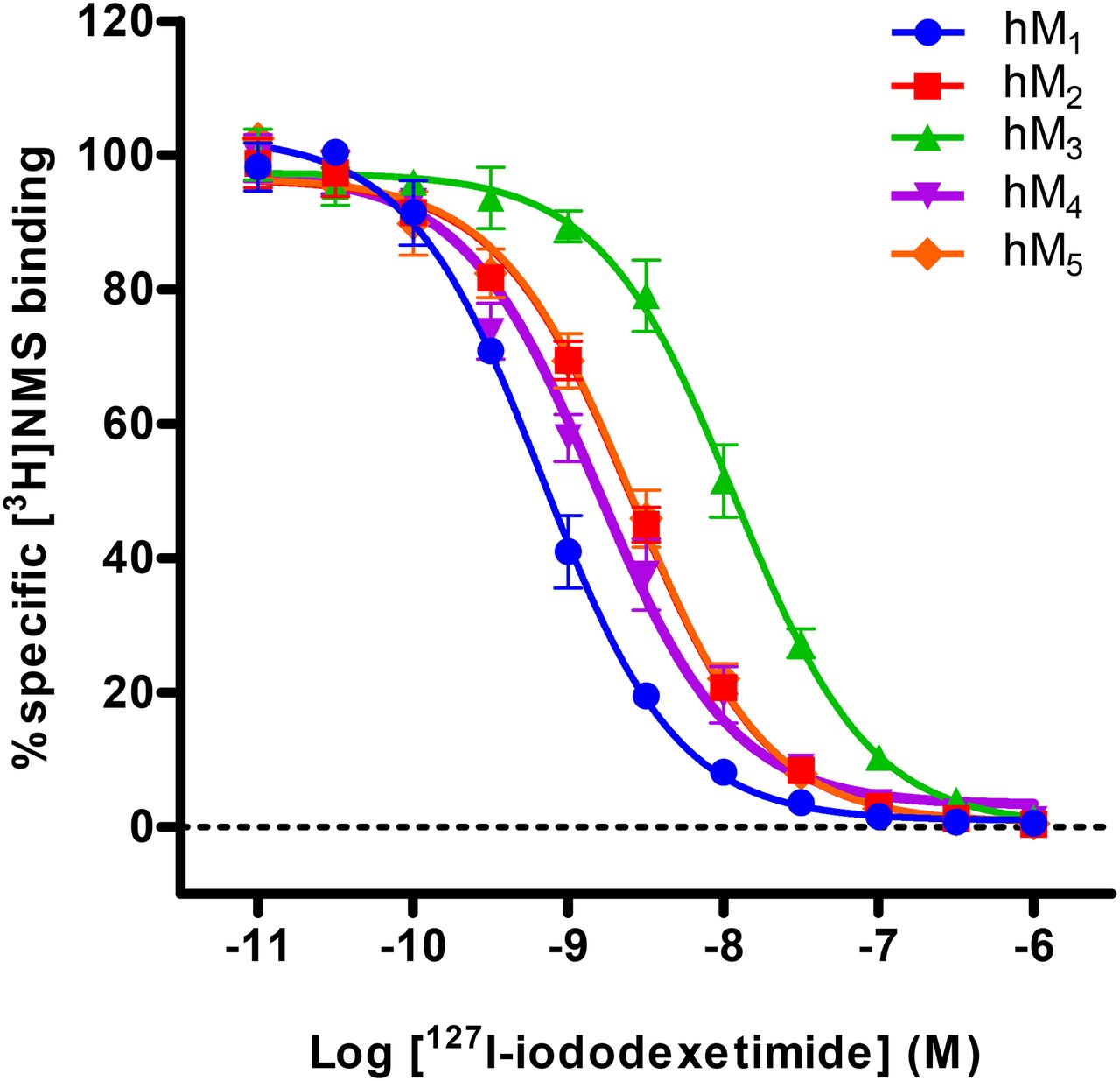
127I-iododexetimide displayed a high affinity for M1R (on average, 337 pM). The affinity for M1R was 1.9–16.9 times higher than for the other 4 mAchRs (Fig. 1; Table 1). Further, in vitro profiling was performed using a GTPγ35S binding assay to assess functional antagonism of 127I-iododexetimide on acetylcholine-activated M1R–M5R (Fig. 2). 127I-iododexetimide displayed an average IC50 of 31 nM for M1R. The IC50 was 2.6–20.7 times higher than for the other muscarinic receptor subtypes (Fig. 2; Table 2).

Displacement of 3H-n-methylscopolamine (3H-NMS) radioligand binding by 127I-iododexetimide across mAchR subtypes M1–M5. Data points represent mean specific binding ± SEM from 3 independent experiments each containing 3 replicates.
Ki Values for 127I-Iododexetimide
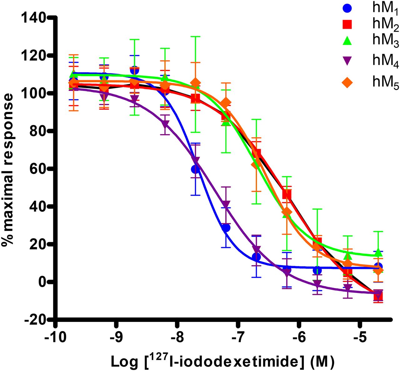
127I-iododexetimide in vitro muscarinic receptor selectivity profile by GTPγ35S. Data are expressed as mean ± SEM from 3 separate experiments.
IC50 Values for 127I-Iododexetimide
LC-MS/MS Ex Vivo Characterization Experiments
Ex vivo characterization of 127I-iododexetimide showed that binding was differentially distributed in wild-type rat brain, with a slow time course. 127I-iododexetimide levels continued to increase in the frontal cortex and striatum over the 1-h period. Cerebellar and plasma levels of 127I-iododexetimide began to wash out as early as 20 min after injection (Supplemental Fig. 1). 127I-iododexetimide had a specific binding signal at all time points measured in rats (Supplemental Fig. 1). There was a significant main effect in the M1 KO mice compared with controls; M3, M4, and M5 KO mice (P < 0.01); and M2 KO mice (P < 0.001) (Fig. 3.). Binding potential was significantly reduced in M1 KO mice.
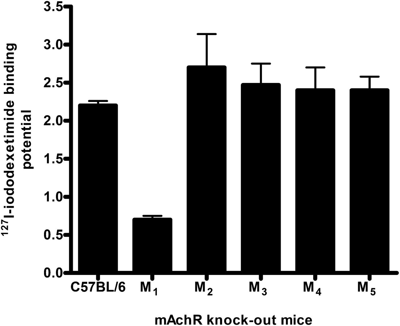
Validation of specific binding to M1Rs of 127I-iododexetimide in M1–M5 muscarinic receptor KO mice, 40 min after injection. Binding potential was calculated as specific binding in frontal cortex (total binding minus nonspecific binding) divided by nonspecific binding (measured in cerebellum).
To validate specific binding of 127I-iododexetimide to muscarinic receptors, groups of rats received doses of the M1/4R agonist xanomeline (Fig. 4). Xanomeline reduced the levels of 127I-iododexetimide in both rat frontal cortex and rat striatum in a dose-dependent manner; whereas no changes were observed in cerebellar and plasma 127I-iododexetimide levels.

Distribution of 127I-iododexetimide in cerebellum, frontal cortex, striatum, and plasma (n = 3–4) in response to dose increase of agonist xanomeline (1–60 mg/kg) 40 min after injection of 127I-iododexetimide. Mice were pretreated with vehicle (0) or xanomeline 30 min before injection of radiotracer. Data points represent mean specific binding ± SEM.
Storage Phosphor Imaging Experiments
One rat from the chronic experiment was excluded because of a binding ratio more than 2 SDs above the mean, resulting in a control group of 7 animals.
After a single administration of olanzapine, but not haloperidol, ex vivo 123I-iododexetimide binding ratios significantly decreased compared with placebo-treated rats (Figs. 5 and 6; Table 3). A significant main effect of the treatment condition in the acute experiment was found for all 3 ROIs (P < 0.001), and post hoc analyses showed that the effect was driven by differences between olanzapine and both haloperidol (P < 0.001) and placebo (P < 0.001). No differences were found between the haloperidol and placebo condition for any ROI (Table 3). After chronic administration of antipsychotics, no significant differences for treatment condition were found in any ROI (P = 0.425) (Supplemental Fig. 2).
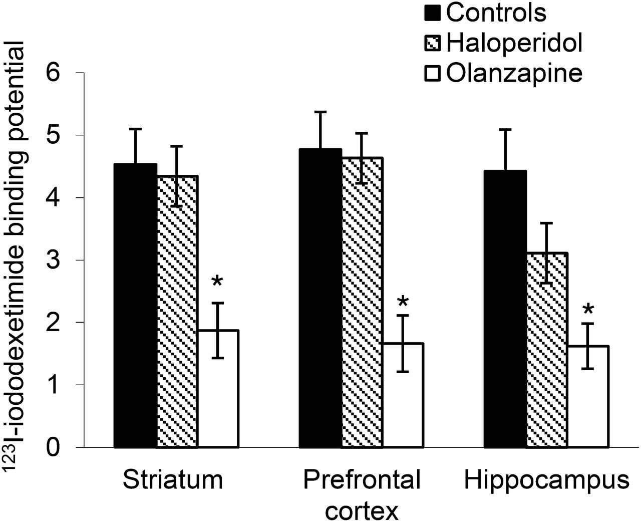
Binding potential of 123I-iododexetimide in ROIs of striatum, prefrontal cortex, and hippocampus. Binding potential was calculated as specific binding (total binding minus nonspecific binding) in ROI divided by nonspecific binding (measured in cerebellum). 123I-iododexetimide binding was measured 2 h after intravenous injection of 123I-iododexetimide. Rats (n = 10/group) were pretreated with 1 dose of saline, haloperidol (1 mg/kg), or olanzapine (2.5 mg/kg) 1 h before injection with radiotracer. *Statistically significantly lower as compared with control group.

Intense 123I-iododexetimide binding (2 h after injection of radiotracer) in cortical brain areas, striatum, and hippocampus in placebo-treated rat (A) and much lower binding in these brain areas (B) after acute olanzapine treatment. Rats (n = 10) were pretreated with 1 dose of saline or olanzapine (2.5 mg/kg) 1 h before injection with radiotracer.
Differences Between Acute and Chronic 123I-Iododexetimide Binding Potential for All ROIs
t tests were performed to check for possible differences in M1 binding between the acute and chronic treatment conditions for each ROI separately. After correction for multiple comparisons, the only significant differences were found in the olanzapine condition between acute and chronic administration in all 3 ROIs (Table 3).
DISCUSSION
The current study aimed to characterize 123I-iododexetimide binding to mAchRs by studying in vitro selectivity, ex vivo 127I-iododexetimide binding in rats and KO mice for the 5 different mAchR, the effects of an M1 agonist on ex vivo 127I-iododexetimide binding, and the effects of antipsychotics with high and low affinity for M1R on ex vivo 123I-iododexetimide binding in rats.
The in vitro experiments showed that 127I-iododexetimide has a high affinity for M1R and lower affinity for other mAchR subtypes (ranging from 1.9-fold to 16.9-fold). In the rat study, 127I-iododexetimide levels continued to increase in the frontal cortex and striatum over the period measured (1 h), and 127I-iododexetimide exhibited specific uptake in the frontal cortex and striatum.127I-iododexetimide binding was significantly reduced in the M1 KO mice, suggesting that most of the specific 127I-iododexetimide binding is attributed to M1R in vivo. Furthermore, the agonist xanomeline dose-dependently decreased 127I-iododexetimide binding in M1-rich brain areas. Finally, as opposed to haloperidol and placebo, 123I-iododexetimide binding was reduced significantly after a single administration of the antipsychotic drug olanzapine, which displays high affinity for M1R. No differences in 123I-iododexetimide binding were found among the 3 treatment conditions after chronic administration, from which one may infer no significant receptor up- or downregulation after chronic administration of olanzapine or haloperidol.
For two decades, 123I-iododexetimide has been used as a radiopharmaceutical to assess mAchR binding in neuropsychiatric disorders in humans (24,28,29). In the 1980s, 123I-iododexetimide was characterized by in vitro experiments, but the binding assays were performed on tissue homogenates that express all mAchRs (17). Later, techniques were developed to assess binding to subtypes of mAchR. To our knowledge, the current study is the first to characterize the binding selectivity and affinity of 123I-iododexetimide for each individual mAchR subtype. The radioligand binding experiments in the current study showed that 127I-iododexetimide displayed a very high affinity for the human M1R (mean Ki of 337 pM) but also high affinity for the other 4 muscarinic receptor subtypes. This was confirmed by the GTPγ35S assay, which showed that in human M1R membranes, 127I-iododexetimide had the greatest potency to reduce an agonist response evoked by acetylcholine (mean IC50 of 31 nM). This finding is also in line with earlier studies showing that 127I-iododexetimide is an antagonist (17). This information is crucial to the interpretation of results from 123I-iododexetimide SPECT imaging studies because agonists and antagonists may recognize different populations of the same receptor subtype (e.g., high- and low-affinity states (30)). Because antagonist radiotracers may bind to receptors in the high- as well as the low-affinity state, 123I-iododexetimide SPECT may be used to assess overall muscarinic M1R availability, whereas agonist tracers (which may bind predominantly to the receptor in its high-affinity state) may be more sensitive to detecting changes in acetylcholine concentrations.
Regarding selectivity, the radioligand binding experiments showed that 127I-iododexetimide has a 2-fold selectivity for human M1R over human M4R and a 4-fold selectivity over human M2R and human M5R. This preference for human M1R is also shown in the GTPγ35S assay, which shows a 2.6-fold selectivity over human M4R, a 5-fold selectivity over human M4R, and around a 20-fold selectivity over human M2R and human M5R. The biodistribution studies in both rats and wild-type mice showed that the highest binding of 127I-iododexetimide was in the frontal cortex and striatum, areas with high M1R densities. So, specific binding seems predominantly attributable to M1R, as is in line with our in vitro results. This selectivity for M1R is also confirmed by the experiments on KO mice in which only the KO of the M1 gene caused a significant decrease in 127I-iododexetimide binding in M1-rich brain areas, compared with the wild-type and other mAchR subtype KO mice.
In the rat, acute administration of the M1/M4 agonist xanomeline or the antipsychotic drug olanzapine, which is known to also block M1Rs, reduced 127I-iododexetimide and 123I-iododexetimide binding potential, respectively, whereas the relatively dopamine D2–selective antipsychotic haloperidol (31) did not affect 123I-iododexetimide binding significantly. This finding confirms that the in vivo binding of 123I-iododexetimide reflects binding to M1R; this finding also may indicate that caution is needed in applying 123I-iododexetimide in vivo to humans who are treated with olanzapine or other antipsychotics with a muscarinic or antimuscarinic profile, such as clozapine. Indeed, in a previous 123I-iododexetimide SPECT study, we showed that schizophrenic patients stabilized on olanzapine had significantly lower 123I-iododexetimide binding in the frontal cortex and striatum than patients stabilized on risperidone (24).
As expected, acute treatment with olanzapine did induce lower 123I-iododexetimide binding, representing occupancy of M1Rs by the drug. Interestingly, acute or chronic treatment with haloperidol did not affect 123I-iododexetimide binding significantly, possibly indicating that it is not necessary to discontinue use of haloperidol if one is interested in studying M1R expression. Although Damsma et al. (22) found increased acetylcholine release after administration of haloperidol in rats, these changes in acetylcholine release evoked by haloperidol may be too small to compete with 123I-iododexetimide binding in vivo. Also, no effects of chronic administration of olanzapine were found on 123I-iododexetimide binding potential after a washout period of 24 h, suggesting that only a relatively short washout period of this drug is necessary if one is interested in studying M1R expression.
The biodistribution study on rats using 127I-iododexetimide and liquid chromatography–mass spectroscopy analysis showed that uptake of 127I-iododexetimide was highest in the frontal and striatal tissues, with low specific binding in the cerebellum, a brain area with low levels of M1Rs, similar to the present findings with 123I-iododexetimide. An advantage of storage phosphor imaging is that the measurement is not influenced by metabolites of 123I-iododexetimide that are formed after injection. Both techniques showed high binding potential, suggesting that metabolites of 123I-iododexetimide formed in the brain are not a major issue in quantification of 123I-iododexetimide binding in clinical practice. As early as 1 h after injection of 123I-iododexetimide, the specific-to-nonspecific binding ratios were already 3. A study by Wilson et al. (17) equally showed a binding ratio of 3–4 at 1 h after injection in mice; our results are thus congruent with this ratio in the wild-type mice and the rat, advocating 123I-iododexetimide as an accurate tool for human applications.
Besides 123I-iododexetimide, other antagonist radiopharmaceuticals for muscarinic receptors (e.g., 18F-FP-TZTP [3-(4-(3-18F-fluoropropylthio)-1,2,5-thiadiazol-3-yl)-1-methyl-1,2,5,6-tetrahydropyridine] or 123I-QNB [(R,R)123I-iodoquinuclidinylbenzilate]) are available, but none of them is known to bind selectively to M1R. 123I-iododexetimide has some advantages over 123I-QNB; 123I-iododexetimide can be synthesized with high specific activity more easily and has a much higher synthetic yield, making it a preferable radioligand to assess M1R binding. In humans, 123I-iododexetimide showed a high nondisplaceable binding potential (>3 at 6 h after injection) (19). The current study provides the pharmacodynamic evidence that 123I-iododexetimide in humans most likely evaluates predominately the availability of M1R in the frontal cortex, striatum, and hippocampus (32). With respect to cognition, these regions are highly pertinent, and 123I-iododexetimide SPECT may be an important means to assess M1R expression in vivo.
Although 127I-iododexetimide had the highest affinity for M1R, the binding to the other 4 mAchRs was relatively high (Fig. 1). More specifically, the affinity of 127I-iododexetimide was in the picomolar range for both the M1 and M4R, although there was some selectivity (2 times higher binding to M1R). Interestingly, the studies on the KO mice showed that only KO of M1R reduced the 127I-iododexetimide binding significantly. However, studies on rats showed that the expression of M1R (Bmax) was 3–5 times higher in the M1R-rich frontal cortex (33). Since the binding potential is influenced by Bmax, as well as by the affinity of the radiotracer for a given receptor, it is likely that the combination of a higher affinity and a higher Bmax may lead to preferential binding of 123I-iododexetimide to M1R.
When the present results are interpreted, some limitations have to be considered. Because the time of animal sacrifice was different in all experiments, direct comparison of results is difficult. In addition, the duration of antipsychotic treatment in the chronic rat experiment was only 2 wk. Therefore, we cannot rule out an influence of antipsychotics such as haloperidol on M1R binding after a longer treatment period. Finally, the rats were anesthetized in the storage phosphor imaging studies, and we consequently cannot exclude a systematic effect on 123I-iododexetimide binding.
CONCLUSION
In vivo binding of 123I-iododexetimide predominantly reflects binding to M1R. Therefore, 123I-iododexetimide may be an accurate ligand for in vivo measuring of M1R expression in humans. Because M1R is associated with cognitive functioning, imaging of M1R may be of great importance in, for example, selecting patients and monitoring the effects of new treatments.
DISCLOSURE
The costs of publication of this article were defrayed in part by the payment of page charges. Therefore, and solely to indicate this fact, this article is hereby marked “advertisement” in accordance with 18 USC section 1734. This work was supported by a grant from the Dutch Organisation for Health and Development (ZonMw; NWO-VIDI grant 91712394). Dr. Booij is a consultant at GE Healthcare. Drs. DuBois, Watson, Mogg, Xiao, Collier, Felder, Crabtree, Barth, and Broad report employment at Eli Lilly. No other potential conflict of interest relevant to this article was reported.
Footnotes
↵* Contributed equally to this work.
Published online Jan. 15, 2015.
- © 2015 by the Society of Nuclear Medicine and Molecular Imaging, Inc.
REFERENCES
- Received for publication August 19, 2014.
- Accepted for publication December 8, 2014.


