


Article Figures & Data
Figures
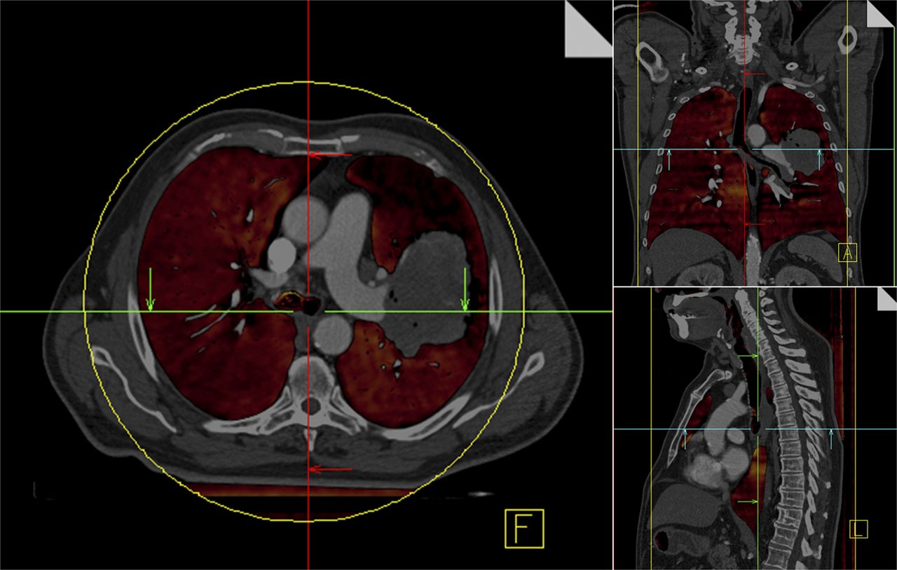
- FIGURE 1.
Perfused lung volume (color overlay) measured using contrast-enhanced dual-energy CT scan of patient with NSCLC tumor inside left lobe (shown in transverse, frontal, and sagittal views). Large perfusion defect surrounding tumor is clearly visible, most probably caused by obstruction of pulmonary vessels. (Courtesy of Wouter van Elmpt, Department of Radiation Oncology [MAASTRO Clinic], Maastricht University Medical Centre, Maastricht, The Netherlands, and Marco Das, Department of Radiology, Maastricht University Medical Centre, Maastricht, The Netherlands.)
- FIGURE 2.
18F-fluorothymidine PET/CT before therapy (A and D), in second week of therapy (B and E), and in fourth week (C and F). Top row shows slow decrease in 18F-fluorothymidine uptake in patient with cT4N2bM0 supraglottic laryngeal carcinoma treated with chemoradiotherapy. This patient developed local recurrence 7 mo after end of treatment and died of metastatic disease. Bottom row shows fast decrease of 18F-fluorothymidine uptake in patient with cT3N1M0 supraglottic laryngeal carcinoma treated with radiotherapy only. This patient developed no tumor-related event after 32 mo of follow-up. (Reprinted from (37).)
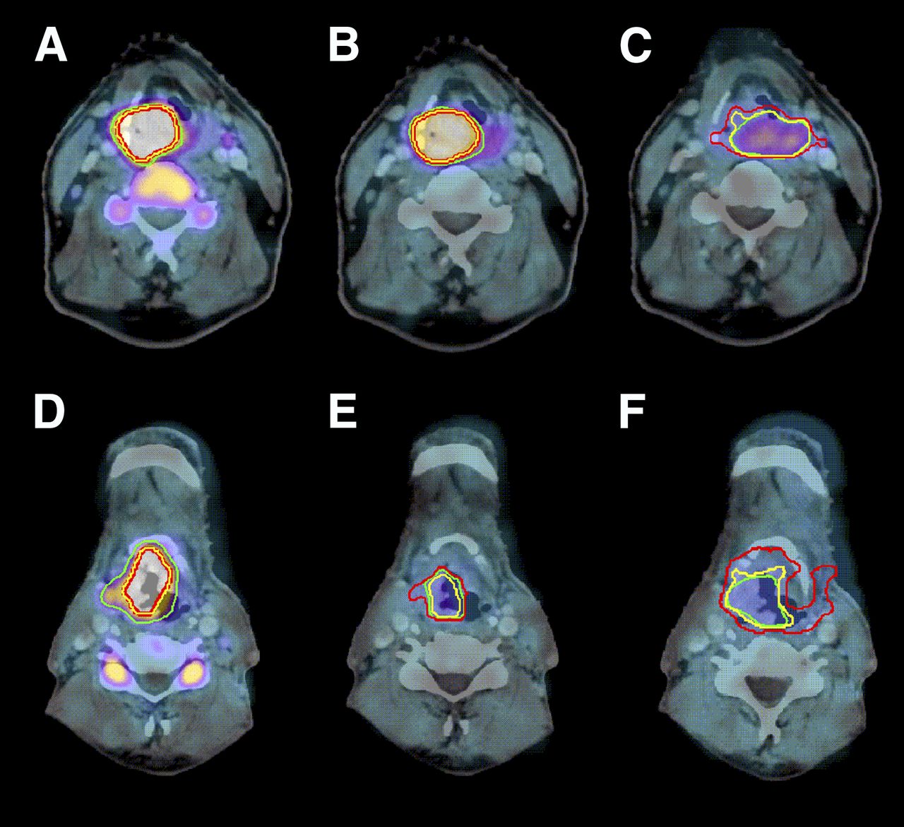
- FIGURE 3.
A 56-y-old woman with base-of-tongue HNSCC who underwent multiparametric functional PET/MRI before start of radiotherapy. (A) Combined 18F-fluoromisonidazole PET/MR image 3 h after injection (T2-weighted turbo inversion recovery magnitude). (B) 18F-fluoromisonidazole PET image registered to planning CT image showing different PTVs. (C) ADC map derived from DWI including PTV containing low ADCs inside PTV of first order. (D) Example radiotherapy plan with dose escalation of 20% prescribed to PTV containing low ADCs. (E) Probability map for radiation resistance of tumor derived from combination of 18F-fluoromisonidazole PET and ADC. (F) Dose-painting radiotherapy plan directly optimized on probability map shown in E. Radiation treatment plans were optimized using treatment-planning system Hyperion for VMAT treatment with 2 arcs and 6-MV photons. PTVs of interest are 70 Gy (red), PTV containing low ADCs (yellow), fluoromisonidazole (blue), 60 Gy (green), 54 Gy (orange), and spinal cord (purple). Isodose lines (from lowest to highest) are 45, 51, 57.5, 66.5, 73, 77, and 80 Gy.
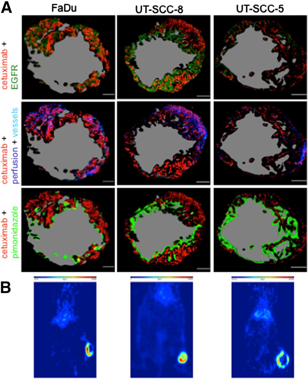
- FIGURE 4.
(A) Pseudocolored images of representative tumor sections of FaDu, UT-SCC-8, and UT-SCC-5 growing in NMRI (Naval Medical Research Institute) mice. Red = cetuximab; dark green = EGFR; dark blue = perfusion, Hoechst 33342; light blue = vascular endothelium, CD31; light green = hypoxia, pimonidazole; gray = necrotic area. (B) Representative PET images administered 86Y-cetuximab. (Reprinted with permission of (78).)


Jump to section
- Article
- Abstract
- FUNCTIONAL AND MOLECULAR IMAGING MODALITIES CURRENTLY AVAILABLE
- USE OF FUNCTIONAL MRI AND CT IN RADIOTHERAPY
- PET FOR RADIATION TREATMENT ADAPTATION AND NORMAL-TISSUE CHARACTERIZATION
- PET FOR IN VIVO TREATMENT VERIFICATION OF PROTON AND CARBON ION THERAPY
- MOLECULAR IMAGING IN SYSTEMIC RADIOTHERAPY AND TARGETED THERAPY
- CONCLUSION
- Footnotes
- REFERENCES
- Figures & Data
- Info & Metrics
Related Articles
Cited By...
- No citing articles found.