
Main menu

  • Home
  • Content
    • Current
    • Ahead of print
    • Past Issues
    • JNM Supplement
    • SNMMI Annual Meeting Abstracts
    • Continuing Education
    • JNM Podcasts
  • Subscriptions
    • Subscribers
    • Institutional and Non-member
    • Rates
    • Journal Claims
    • Corporate & Special Sales
  • Authors
    • Submit to JNM
    • Information for Authors
    • Assignment of Copyright
    • AQARA requirements
  • Info
    • Reviewers
    • Permissions
    • Advertisers
  • About
    • About Us
    • Editorial Board
    • Contact Information
  • More
    • Alerts
    • Feedback
    • Help
    • SNMMI Journals
  • SNMMI
    • JNM
    • JNMT
    • SNMMI Journals
    • SNMMI

User menu

  • Subscribe
  • My alerts
  • Log in
  • Log out
  • My Cart

Search

  • Advanced search
Journal of Nuclear Medicine
  • SNMMI
    • JNM
    • JNMT
    • SNMMI Journals
    • SNMMI
  • Subscribe
  • My alerts
  • Log in
  • Log out
  • My Cart
Journal of Nuclear Medicine

Advanced Search

  • Home
  • Content
    • Current
    • Ahead of print
    • Past Issues
    • JNM Supplement
    • SNMMI Annual Meeting Abstracts
    • Continuing Education
    • JNM Podcasts
  • Subscriptions
    • Subscribers
    • Institutional and Non-member
    • Rates
    • Journal Claims
    • Corporate & Special Sales
  • Authors
    • Submit to JNM
    • Information for Authors
    • Assignment of Copyright
    • AQARA requirements
  • Info
    • Reviewers
    • Permissions
    • Advertisers
  • About
    • About Us
    • Editorial Board
    • Contact Information
  • More
    • Alerts
    • Feedback
    • Help
    • SNMMI Journals
  • View or Listen to JNM Podcast
  • Visit JNM on Facebook
  • Join JNM on LinkedIn
  • Follow JNM on Twitter
  • Subscribe to our RSS feeds
Research ArticleClinical Investigations

Presence of Postsystolic Shortening Increases the Likelihood of Coronary Artery Disease: A Rest Electrocardiography-Gated Myocardial Perfusion SPECT Study

Yumiko Kanzaki, Yohei Yamauchi, Hideaki Morita, Masuo Hayashi, Tsuyoshi Komori, Akira Ukimura and Nobukazu Ishizaka
Journal of Nuclear Medicine December 2015, 56 (12) 1889-1894; DOI: https://doi.org/10.2967/jnumed.115.153791
Yumiko Kanzaki
1Department of Cardiology, Osaka Medical College, Takatsuki, Japan; and
  • Find this author on Google Scholar
  • Find this author on PubMed
  • Search for this author on this site
Yohei Yamauchi
1Department of Cardiology, Osaka Medical College, Takatsuki, Japan; and
  • Find this author on Google Scholar
  • Find this author on PubMed
  • Search for this author on this site
Hideaki Morita
1Department of Cardiology, Osaka Medical College, Takatsuki, Japan; and
  • Find this author on Google Scholar
  • Find this author on PubMed
  • Search for this author on this site
Masuo Hayashi
2Department of Radiology, Osaka Medical College, Takatsuki, Japan
  • Find this author on Google Scholar
  • Find this author on PubMed
  • Search for this author on this site
Tsuyoshi Komori
2Department of Radiology, Osaka Medical College, Takatsuki, Japan
  • Find this author on Google Scholar
  • Find this author on PubMed
  • Search for this author on this site
Akira Ukimura
1Department of Cardiology, Osaka Medical College, Takatsuki, Japan; and
  • Find this author on Google Scholar
  • Find this author on PubMed
  • Search for this author on this site
Nobukazu Ishizaka
1Department of Cardiology, Osaka Medical College, Takatsuki, Japan; and
  • Find this author on Google Scholar
  • Find this author on PubMed
  • Search for this author on this site
  • Article
  • Figures & Data
  • Info & Metrics
  • PDF
Loading

Article Figures & Data

Figures

  • Tables
  • FIGURE 1.
    • Download figure
    • Open in new tab
    • Download powerpoint
    FIGURE 1.

    Measurements of PSS index. (Upper) End-systole was defined on the basis of total average thickening curve. (Lower) PSS was determined from 17 segments using sum of difference between post–end-systolic max LV thickening (a) and end-systolic LV thickening (b) divided by post–end-systolic max LV thickening (a). If segment curve showed time to max LV thickening before end-systole, difference between max LV thickening and end-systolic LV thickening was assumed to be 0.
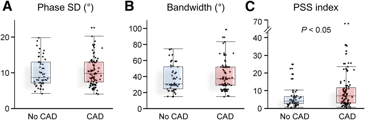
  • FIGURE 2.
    • Download figure
    • Open in new tab
    • Download powerpoint
    FIGURE 2.

    Box plots showing medians and 25th and 75th percentiles of phase SD, histogram bandwidth, and PSS index between patients with and without coronary artery disease. CAD = coronary artery disease.
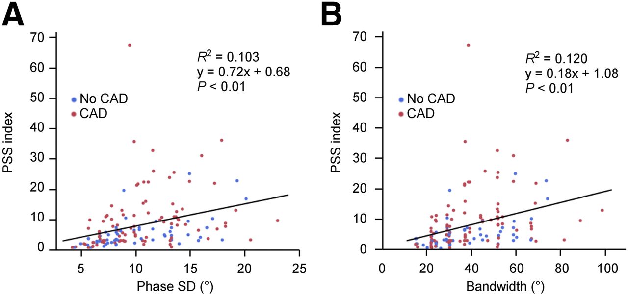
  • FIGURE 3.
    • Download figure
    • Open in new tab
    • Download powerpoint
    FIGURE 3.

    Comparisons of PSS index with phase SD and histogram bandwidth. CAD = coronary artery disease.

  • FIGURE 4.
    • Download figure
    • Open in new tab
    • Download powerpoint
    FIGURE 4.

    Representative cases of phase analysis and 17-segment thickening curves. Results from patient without coronary artery stenosis (A) and results from patient with significant coronary artery stenosis of right coronary artery and left circumflex artery (B). In A, phase SD = 10°, histogram bandwidth = 37°, and PSS index = 2.7, and in B, phase SD = 10°, histogram bandwidth = 39°, and PSS index = 29.6.
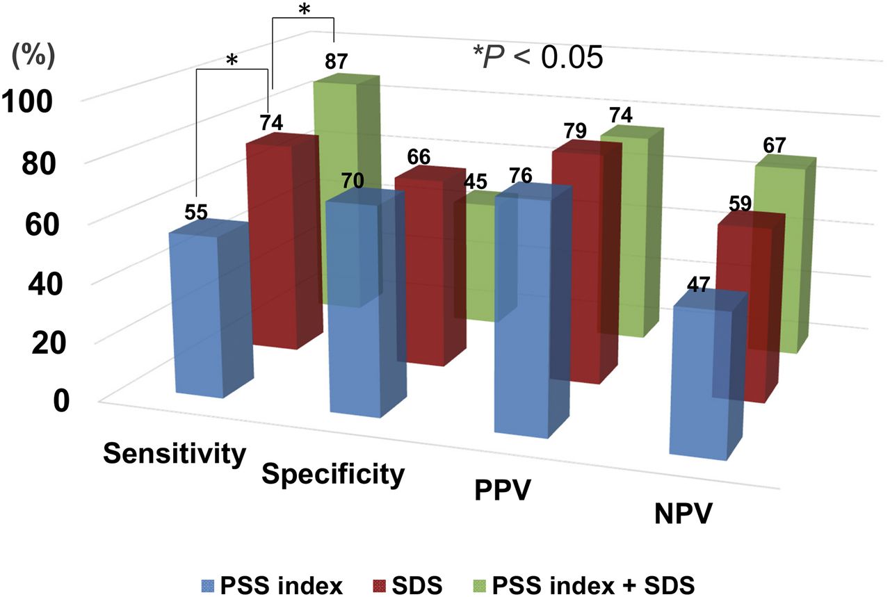
  • FIGURE 5.
    • Download figure
    • Open in new tab
    • Download powerpoint
    FIGURE 5.

    Diagnostic usefulness of PSS index, SDS, and combination of PSS index and SDS.

Tables

  • Figures
    • View popup
    TABLE 1

    Patient Characteristics

    CharacteristicNo coronary artery disease (n = 53)Coronary artery disease (n = 93)P
    Mean age ± SD (y)70.5 ± 8.370.6 ± 8.1NS
    Male sex (n)29 (55)69 (74)0.02
    Exercise21 (40)47 (51)NS
    Adenosine32 (60)46 (49)NS
    Body mass index23.7 ± 3.124.1 ± 3.0NS
    Hypertension (n)47 (89)83 (89)NS
    Dyslipidemia (n)33 (62)67 (72)NS
    Diabetes mellitus (n)14 (26)46 (49)<0.01
    Current smoker (n)7 (13)14 (15)NS
    Prior smoker (n)21 (40)37 (40)NS
    Family history of coronary artery disease (n)14 (26)23 (25)NS
    LV function at rest
     End-diastolic volume (mL)61.2 ± 17.263.6 ± 17.3NS
     End-systolic volume (mL)16.8 ± 8.119.8 ± 9.2NS
    Ejection fraction (%)74.1 ± 8.870.0 ± 8.30.01
    Transient ischemic dilatation1.0 ± 0.11.0 ± 0.1NS
    LV phase analysis
     Phase SD (°)10.2 ± 4.010.5 ± 3.9NS
     Histogram bandwidth (°)37.8 ± 16.240.5 ± 17.4NS
    PSS index5.6 ± 5.19.8 ± 10.2<0.01
    SSS3.8 ± 3.27.0 ± 4.8<0.01
    SRS1.7 ± 2.02.2 ± 3.0NS
    SDS2.1 ± 2.94.8 ± 3.8<0.01
    • Data are expressed as mean ± SD or as number, with percentage in parentheses.

    • NS = not significant.

    • View popup
    TABLE 2

    Diagnostic Accuracy for Detecting Coronary Artery Disease

    PredictorArea under curveSensitivity (%)Specificity (%)PPV (%)NPV (%)
    Phase SD0.5348576638
    Histogram bandwidth0.5351596840
    PSS index0.6455707647
    SSS0.7174557455
    SRS0.5245556436
    SDS0.7474667959
    • View popup
    TABLE 3

    Univariate and Multivariate Logistic Analysis for Detecting Coronary Artery Stenosis

    Univariate logistic analysisMultivariate logistic analysis
    PredictorOdds ratioSEE95% confidence intervalPOdds ratioSEE95% confidence intervalP
    Age1.000.020.96−1.04NS
    Men2.380.361.17−4.850.021.570.420.69−3.55NS
    Transient ischemic dilatation ≧ 1.10.760.360.38–1.53NS
    Phase SD ≧ 10°0.850.350.43−1.69NS
    Histogram bandwidth ≧ 37°1.440.350.73−2.85NS
    PSS index ≧ 62.810.371.37−5.74<0.012.460.401.11−5.430.03
    SSS ≧ 43.470.361.70−7.09<0.011.170.470.47–2.92NS
    SRS ≧ 21.000.350.51−1.96NS
    SDS ≧ 35.590.382.68−11.65<0.014.760.442.00−11.35<0.01
    • NS = not significant.

PreviousNext
Back to top

In this issue

Journal of Nuclear Medicine: 56 (12)
Journal of Nuclear Medicine
Vol. 56, Issue 12
December 1, 2015
  • Table of Contents
  • Table of Contents (PDF)
  • About the Cover
  • Index by author
Print
Download PDF
Article Alerts
Sign In to Email Alerts with your Email Address
Email Article

Thank you for your interest in spreading the word on Journal of Nuclear Medicine.

NOTE: We only request your email address so that the person you are recommending the page to knows that you wanted them to see it, and that it is not junk mail. We do not capture any email address.

Enter multiple addresses on separate lines or separate them with commas.
Presence of Postsystolic Shortening Increases the Likelihood of Coronary Artery Disease: A Rest Electrocardiography-Gated Myocardial Perfusion SPECT Study
(Your Name) has sent you a message from Journal of Nuclear Medicine
(Your Name) thought you would like to see the Journal of Nuclear Medicine web site.
Citation Tools
Presence of Postsystolic Shortening Increases the Likelihood of Coronary Artery Disease: A Rest Electrocardiography-Gated Myocardial Perfusion SPECT Study
Yumiko Kanzaki, Yohei Yamauchi, Hideaki Morita, Masuo Hayashi, Tsuyoshi Komori, Akira Ukimura, Nobukazu Ishizaka
Journal of Nuclear Medicine Dec 2015, 56 (12) 1889-1894; DOI: 10.2967/jnumed.115.153791

Citation Manager Formats

  • BibTeX
  • Bookends
  • EasyBib
  • EndNote (tagged)
  • EndNote 8 (xml)
  • Medlars
  • Mendeley
  • Papers
  • RefWorks Tagged
  • Ref Manager
  • RIS
  • Zotero
Share
Presence of Postsystolic Shortening Increases the Likelihood of Coronary Artery Disease: A Rest Electrocardiography-Gated Myocardial Perfusion SPECT Study
Yumiko Kanzaki, Yohei Yamauchi, Hideaki Morita, Masuo Hayashi, Tsuyoshi Komori, Akira Ukimura, Nobukazu Ishizaka
Journal of Nuclear Medicine Dec 2015, 56 (12) 1889-1894; DOI: 10.2967/jnumed.115.153791
Twitter logo Facebook logo LinkedIn logo Mendeley logo
  • Tweet Widget
  • Facebook Like
  • Google Plus One
Bookmark this article

Jump to section

  • Article
    • Abstract
    • MATERIALS AND METHODS
    • RESULTS
    • DISCUSSION
    • CONCLUSION
    • DISCLOSURE
    • Acknowledgments
    • Footnotes
    • REFERENCES
  • Figures & Data
  • Info & Metrics
  • PDF

Related Articles

  • This Month in JNM
  • PubMed
  • Google Scholar

Cited By...

  • No citing articles found.
  • Google Scholar

More in this TOC Section

  • Feasibility of Ultra-Low-Activity 18F-FDG PET/CT Imaging Using a Long–Axial-Field-of-View PET/CT System
  • Cardiac Presynaptic Sympathetic Nervous Function Evaluated by Cardiac PET in Patients with Chronotropic Incompetence Without Heart Failure
  • Validation and Evaluation of a Vendor-Provided Head Motion Correction Algorithm on the uMI Panorama PET/CT System
Show more Clinical Investigations

Similar Articles

Keywords

  • coronary artery disease
  • scintigraphy
  • postsystolic shortening
SNMMI

© 2025 SNMMI

Powered by HighWire