


Article Figures & Data
Figures
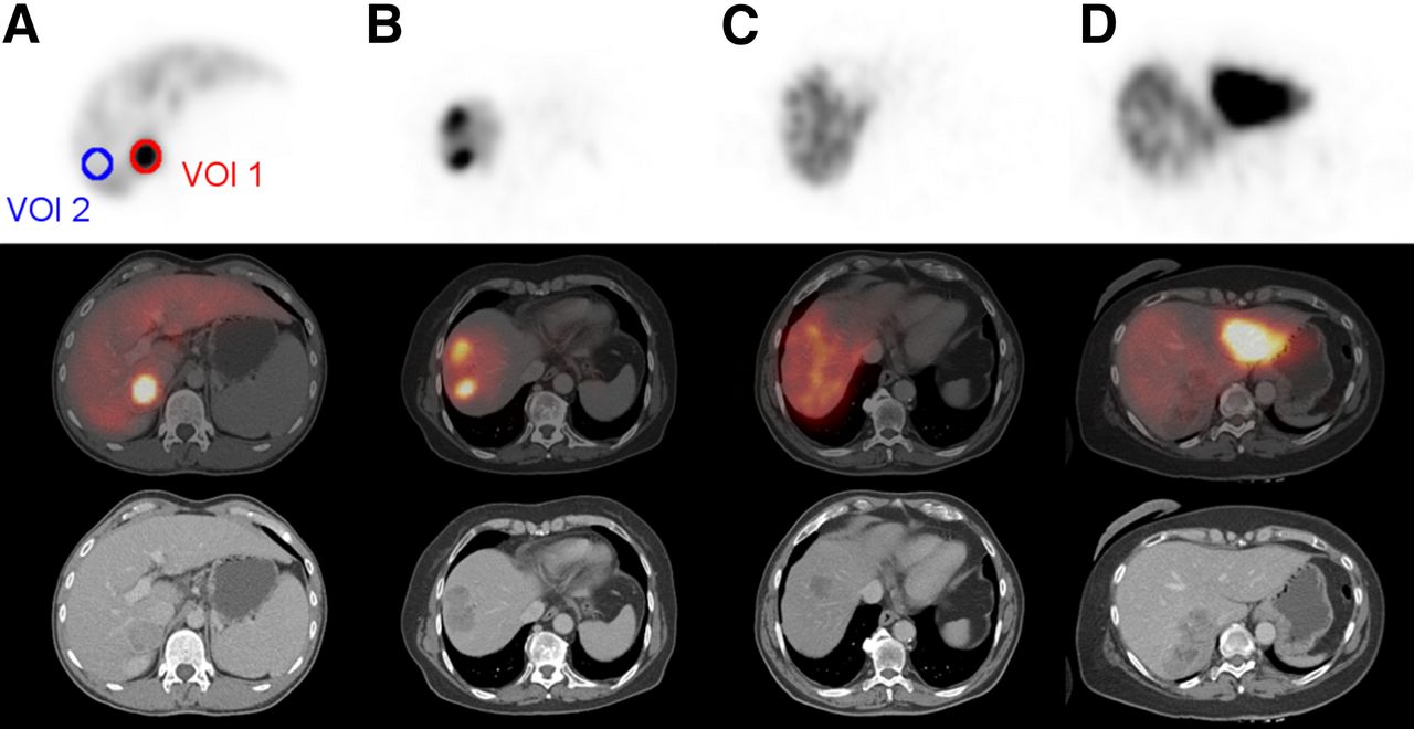
- FIGURE 1.
Grading of tracer accumulation in 99mTc-MAA SPECT. (A) Homogenously higher uptake than in normal liver tissue is grade 1. (B) Heterogeneously higher uptake than in normal liver tissue is grade 2. (C) Same uptake as in normal liver tissue is grade 3. (D) Lower uptake than in normal liver tissue is grade 4.
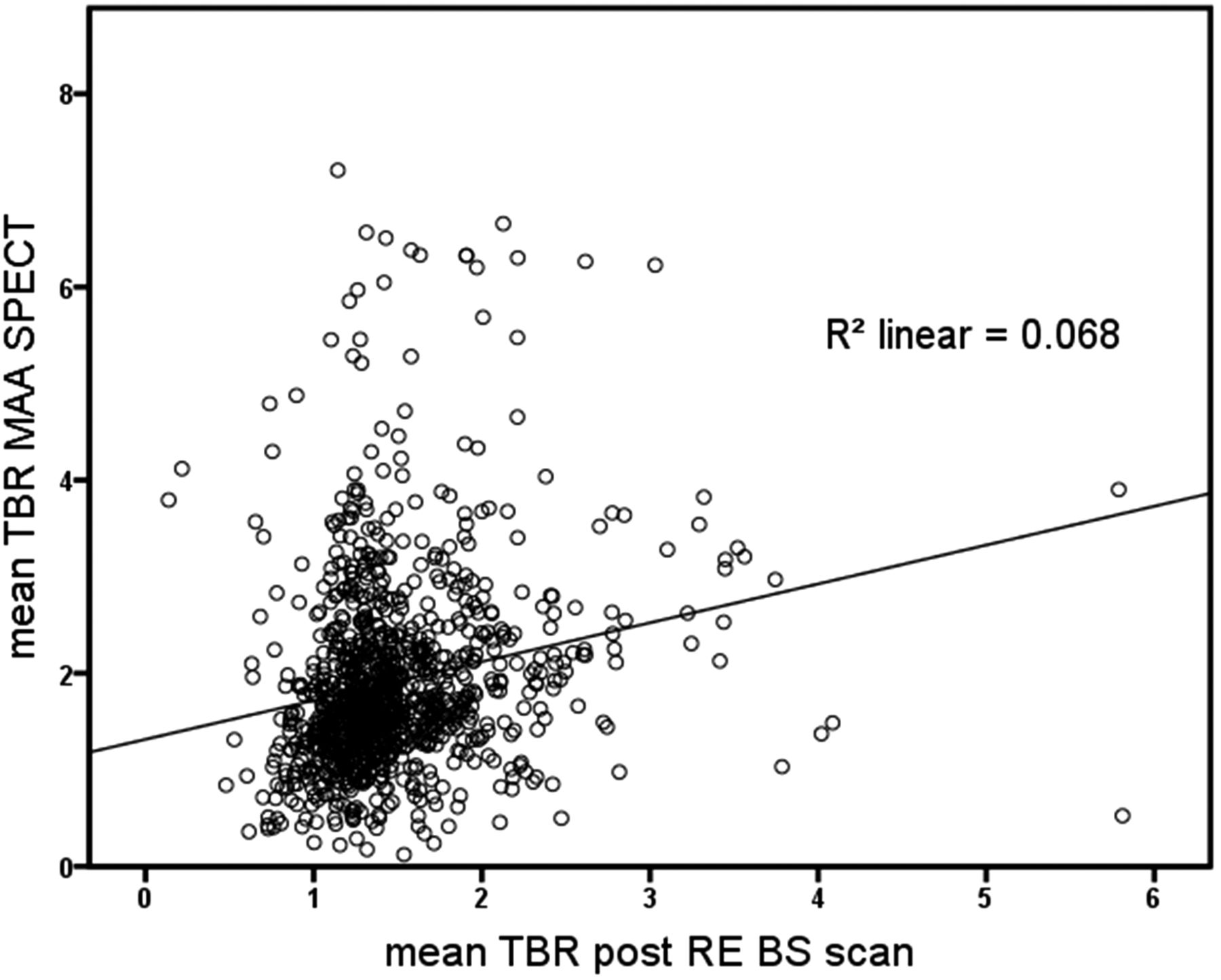
- FIGURE 2.
Scatterplot of mean TBR in 99mTc-MAA SPECT (x-axis) and mean TBR in 90Y-bremsstrahlung SPECT (y-axis).
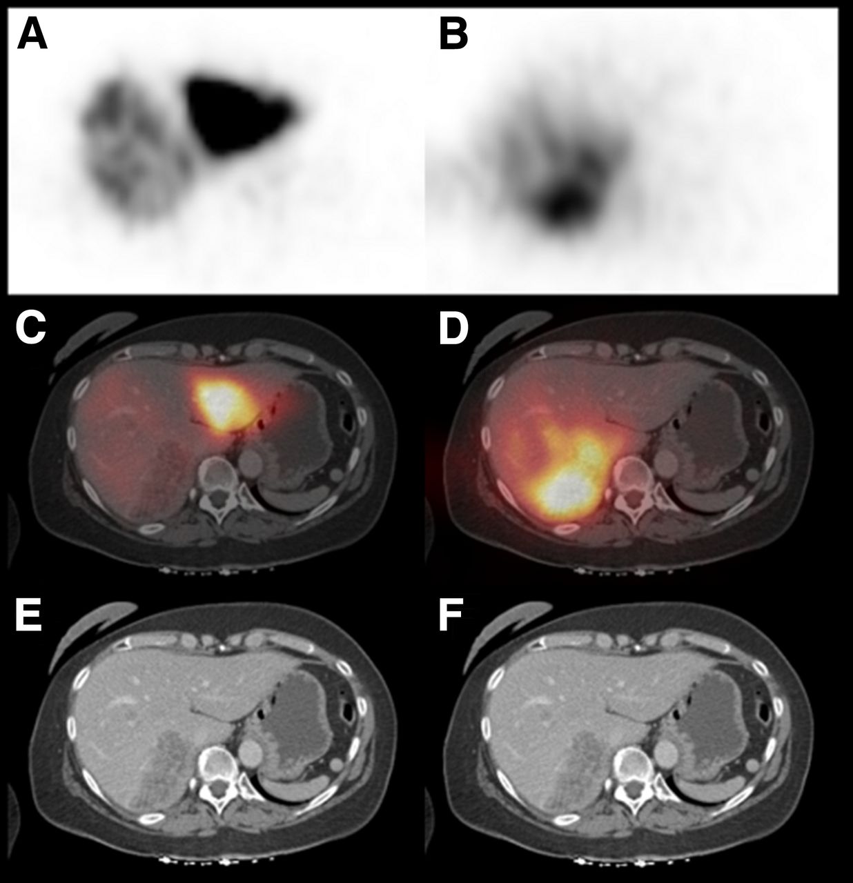
- FIGURE 3.
Coregistered diagnostic CT, 99mTc-MAA SPECT, and 90Y-bremsstrahlung SPECT scans of 60-y-old woman with hepatically metastasized breast cancer who had undergone sequential lobar radioembolization. Hepatic lesion in right lobe (E and F) does not show relevant uptake on 99mTc-MAA SPECT (A and C). 90Y-bremsstrahlung SPECT (B and D), however, reveals high uptake in tumor during radioembolization.
Tables
99mTc-MAA 90Y-bremsstrahlung Total Grade Total Primary Grade n % 1 2 3 4 Grade n % HCC 1 13 16.9 10 2 1 0 1 17 22.1 2 58 75.3 7 50 0 1 2 56 72.7 3 2 2.6 0 2 0 0 3 1 1.3 4 4 5.2 0 2 0 2 4 3 3.9 CCC 1 14 21.5 13 1 0 0 1 20 30.8 2 48 73.8 7 40 0 1 2 44 67.7 3 0 0.0 0 0 0 0 3 0 0.0 4 3 4.6 0 3 0 0 4 1 1.5 Breast cancer 1 45 36.6 42 2 1 0 1 65 52.8 2 66 53.7 18 48 0 0 2 53 43.1 3 2 1.6 2 0 0 0 3 1 0.8 4 10 8.1 3 3 0 4 4 4 3.3 CRC 1 76 17.2 57 17 1 1 1 102 23.1 2 338 76.5 37 290 6 5 2 319 72.2 3 7 1.6 1 4 2 0 3 9 2.0 4 21 4.8 7 8 0 6 4 12 2.7 NETs 1 57 31.7 49 5 2 1 1 83 46.1 2 108 60.0 31 76 1 0 2 87 48.3 3 4 2.2 1 1 2 0 3 7 3.9 4 11 6.1 2 5 2 2 4 3 1.7 Pancreatic cancer 1 6 25.0 3 3 0 0 1 5 20.8 2 14 58.3 2 12 0 0 2 16 66.7 3 0 0.0 0 0 0 0 3 1 4.2 4 4 16.7 0 1 1 2 4 2 8.3 Lung cancer 1 3 30.0 3 0 0 0 1 3 30.0 2 7 70.0 0 7 0 0 2 7 70.0 3 0 0.0 0 0 0 0 3 0 0.0 4 0 0.0 0 0 0 0 4 0 0.0 Malignant melanoma 1 2 11.1 0 2 0 0 1 2 11.1 2 15 83.3 2 12 0 1 2 15 83.3 3 0 0.0 0 0 0 0 3 0 0.0 4 1 5.6 0 1 0 0 4 1 5.6 Urologic tumor 1 5 16.7 2 3 0 0 1 3 10.0 2 25 83.3 1 24 0 0 2 27 90.0 3 0 0.0 0 0 0 0 3 0 0.0 4 0 0.0 0 0 0 0 4 0 0.0 Ear-nose-throat tumors 1 0 0.0 0 0 0 0 1 2 25.0 2 8 1000.0 2 4 0 2 2 4 50.0 3 0 0.0 0 0 0 0 3 0 0.0 4 0 0.0 0 0 0 0 4 2 25.0 Non-CRC gastrointestinal tumors 1 2 12.5 2 0 0 0 1 6 37.5 2 13 81.3 4 9 0 0 2 10 62.5 3 0 0.0 0 0 0 0 3 0 0.0 4 1 6.3 0 1 0 0 4 0 0.0 Sarcoma 1 7 46.7 5 2 0 0 1 6 40.0 2 6 40.0 1 4 0 1 2 8 53.3 3 0 0.0 0 0 0 0 3 0 0.0 4 2 13.3 0 2 0 0 4 1 6.7 - TABLE 2
Mean TBR on 99mTc-MAA and 90Y-Bremsstrahlung SPECT and Spearman Correlation Coefficient
Mean TBR ± SD Primary tumor n 99mTc-MAA 90Y-bremsstrahlung Spearman correlation coefficient P HCC 77 2.11 ± 1.25 1.56 ± 0.50 0.398 <0.001 CCC 65 2.05 ± 0.97 1.41 ± 0.37 0.279 0.024 Breast cancer 123 1.65 ± 0.76 1.41 ± 0.47 0.308 0.001 CRC 442 1.80 ± 0.92 1.43 ± 0.49 0.22 <0.001 NETs 180 2.16 ± 1.21 1.53 ± 0.47 0.197 0.008 PAN 24 1.92 ± 1.15 1.41 ± 0.52 0.323 0.123 PUL 10 1.99 ± 0.94 1.66 ± 0.76 0.442 0.2 MM 18 1.93 ± 1.13 1.53 ± 0.78 0.094 0.711 URO 30 1.97 ± 0.94 1.71 ± 0.31 0.05 0.795 ENT 8 2.61 ± 0.55 1.63 ± 0.90 0.143 0.736 GI 16 2.27 ± 1.05 1.81 ± 0.41 0.174 0.52 SAR 15 1.73 ± 1.10 1.64 ± 1.24 0.036 0.899 Total 1,008 1.90 ± 1.01 1.48 ± 0.51 0.261 <0.001 PAN = pancreatic cancer; PUL = lung cancer; MM = malignant carcinoma; URO = urologic tumor; ENT = ear-nose-throat tumor; GI = non-CRC gastrointestinal cancer; SAR = sarcoma.
- TABLE 3
Mean TBR and Grade in Comparison of Highly and Moderately Hypervascularized Tumors on 99mTc-MAA and 90Y-Bremsstrahlung SPECT
99mTc-MAA 90Y-bremsstrahlung Hypervascularized… Grade n % Mean TBR ± SD Grade n % Mean TBR ± SD Highly (n = 322; HCC, CCC, NETs) 1 84 26.1 2.32 ± 1.10 1 120 37.3 1.54 ± 0.41 2 214 66.5 2.20 ± 1.16 2 187 58.1 1.54 ± 0.47 3 6 1.9 1.24 ± 0.39 3 8 2.5 0.96 ± 0.21 4 18 5.6 0.61 ± 0.24 4 7 2.2 0.78 ± 0.17 Moderately (n = 565; CRC, breast cancer) 1 121 21.4 1.91 ± 0.88 1 167 29.6 1.50 ± 0.52 2 404 71.5 1.81 ± 0.87 2 372 65.8 1.42 ± 0.46 3 9 1.6 1.02 ± 0.28 3 10 1.8 1.13 ± 0.24 4 31 5.5 0.76 ± 0.23 4 16 2.8 0.86 ± 0.14


