


Article Figures & Data
Figures
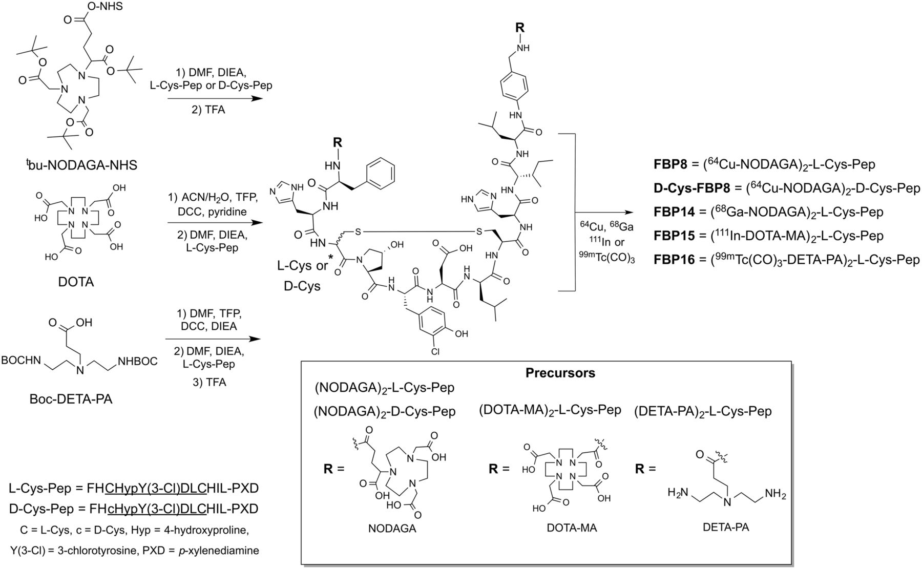
- FIGURE 1.
Synthesis of fibrin-binding probes.
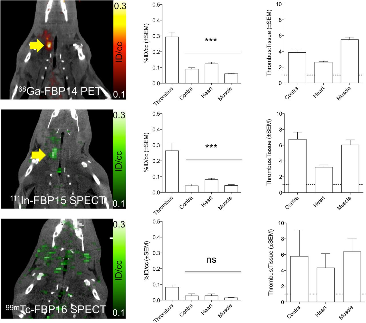
- FIGURE 2.
Representative PET and SPECT images (crush model, 30–90 min after injection) showing persistent thrombus signal (arrow) after injection of 68Ga-FBP14 (n = 5) and 111In-FBP15 (n = 4) but not for 99mTc-FP16 (n = 3). PET and SPECT quantification revealed high target radioactivity for 68Ga-FBP14 and 111In-FBP15 compared with background tissues, resulting in high thrombus-to-background radioactivity ratios. ***P < 0.001. ns = not significant.
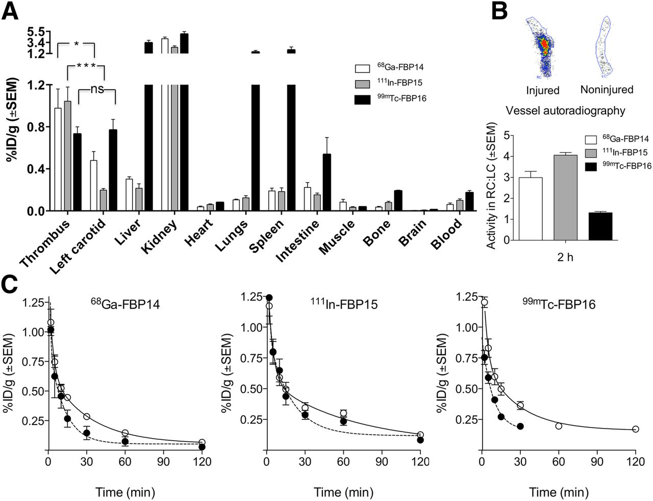
- FIGURE 3.
(A) Biodistribution at 120 min (crush model; 68Ga-FBP14, n = 5; 111In-FBP15, n = 8; 99mTc-FBP16, n = 6). (B) Representative autoradiographs of excised injured and contralateral carotid arteries after injection of 111In-FBP15 and ipsilateral-to-contralateral ratios for each probe (n = 5–6/probe). (C) Pharmacokinetic data from ex vivo blood analyses (68Ga-FBP14, n = 5; 111In-FBP15, n = 8; 99mTc-FBP16, n = 4). ○ = total radioactivity in plasma; ● = functional probe. *P < 0.05. ***P < 0.001. ns = not significant.
- FIGURE 4.
(A) CT, PET, and fused images from animal injected with 64Cu-d-Cys-FBP8 after carotid crush injury (n = 2). Contrast-enhanced CT angiography was used to detect common carotid artery (thin arrow). Thrombus location could not be detected by PET. (B) Time–radioactivity curves obtained from PET imaging, mean radioactivity values (30–90 min after injection), and representative photograph and autoradiograph of ipsilateral (ipsi) and contralateral (contra) carotid arteries showing comparable radioactivity levels between thrombus and background. (C) PET/CT imaging (n = 3) after sequential injection of 64Cu-d-Cys-FBP8, followed by injection of its active analog 64Cu-FBP8. Thrombus uptake was detected only after injection of 64Cu-FBP8 (blue arrow). (D) Statistically significant difference in thrombus versus contralateral artery uptake for 64Cu-FBP8 but not 64Cu-d-Cys-FBP8. In vitro binding studies of blood plasma incubated with immobilized fibrin showed significantly lower binding for 64Cu-d-Cys-FBP8 than 64Cu-FBP8 (n = 2). **P < 0.01. ***P < 0.001.
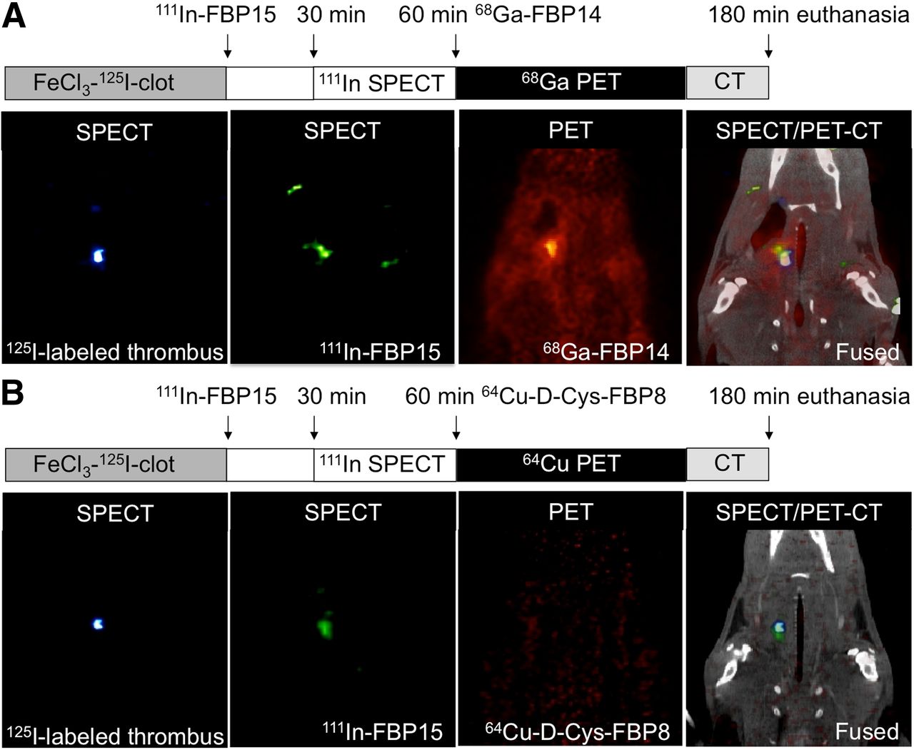
- FIGURE 5.
Triple-isotope SPECT/PET/CT studies in 125I-labeled thrombus–bearing rats. (A, left to right) A 35-keV SPECT image showing 125I-labeled thrombus; 171- to 245-keV SPECT image after 111In-FBP15 injection showing high uptake in region of thrombus; PET image after 68Ga-FBP14 injection showing focal signal intensity in region of carotid artery; fused SPECT/PET/CT images showing that 111In-FBP15 and 68Ga-FBP14 colocalize to 125I-labeled thrombus. (B, left to right) A 35-keV SPECT image showing 125I-labeled thrombus; 171- to 245-keV SPECT image after 111In-FBP15 injection showing high uptake in region of thrombus; PET image after 64Cu-d-Cys-FBP8 injection showing low signal intensity in field of view; fused SPECT/PET/CT images showing that fibrin-targeted 111In-FBP15 but not nonspecific 64Cu-d-Cys-FBP8 colocalizes to 125I-labeled thrombus.


Additional Files
Supplemental Data
Files in this Data Supplement:


Jump to section
Related Articles
Cited By...
- Brain-Penetrating Peptide and Antibody Radioligands for Proof-of-Concept PET Imaging of Fibrin in Alzheimers Disease
- Dosimetry of [64Cu]FBP8: a fibrin-binding PET probe
- Detection and Characterization of Thrombosis in Humans using Fibrin-Targeted Positron Emission Tomography and Magnetic Resonance
- Molecular Imaging of Atherothrombotic Diseases: Seeing Is Believing