


Article Figures & Data
Figures
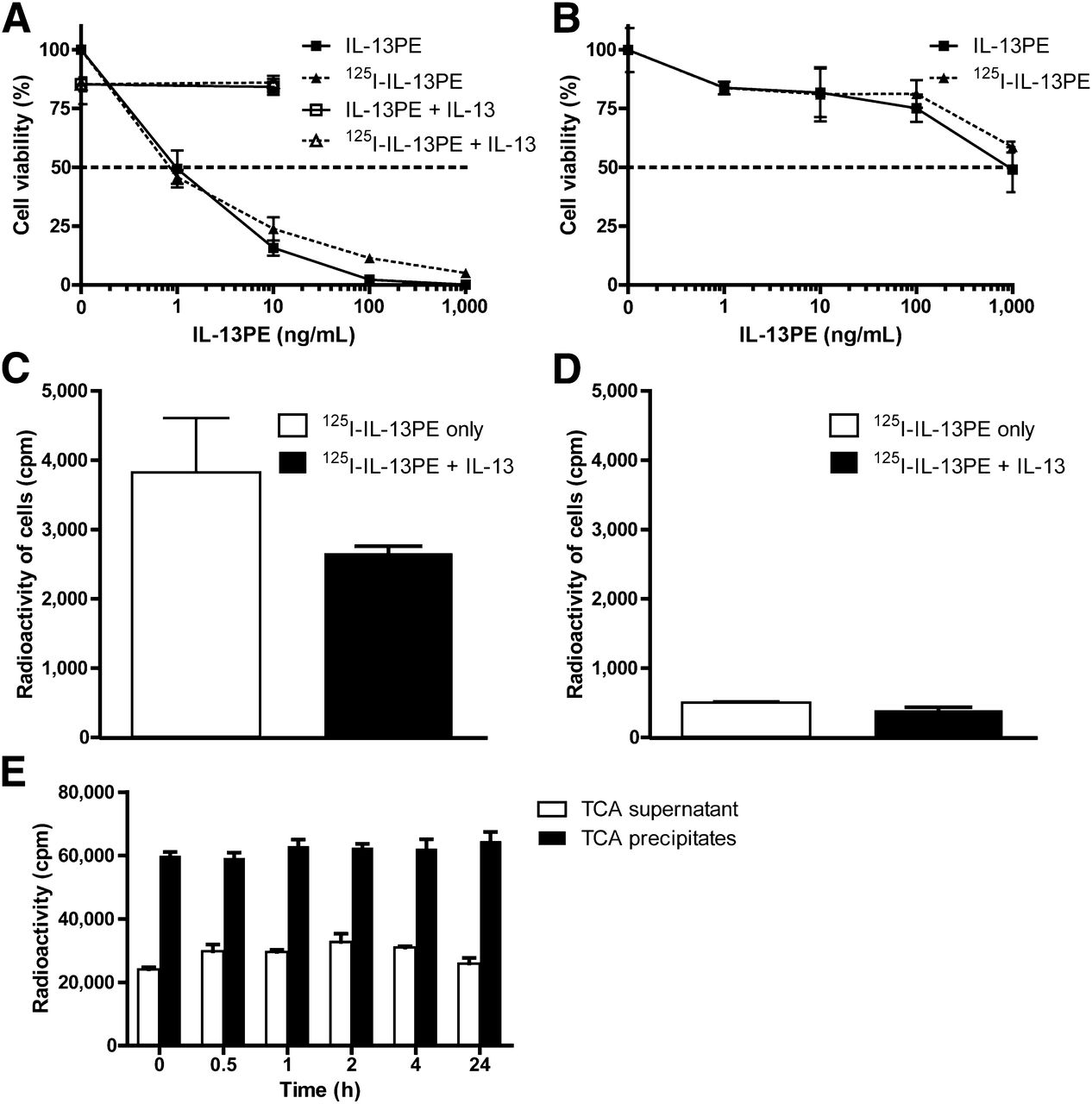
- FIGURE 1.
Radiolabeling and characterization of 125I-IL-13PE. Cytotoxicity of 125I-IL-13PE to glioma cell lines U251 (A) or T98G (B) was assessed by incubating cells with different concentrations of 125I-IL-13PE for 4 d. IL-13 (100 ng/mL) was used for blocking cytotoxicity of IL-13PE. Viable cells were counted by trypan blue exclusion. Binding of 125I-IL-13PE to U251 (C) or T98G (D) cells was assessed by incubating cells with 125I-IL-13PE (100 pM) at 4°C for 16 h with or without 100-fold excess of unlabeled IL-13. Cell-bound 125I-IL-13PE was separated by centrifugation through cushion of phthalate oil and counted by γ counter. Stability of 125I-IL-13PE in mouse serum (E) was determined by incubating with mouse serum at 37°C for 0.5–24 h. 125I-IL-13PE bound and free 125I were separated by trichloroacetic acid (TCA) precipitation. Radioactivity from protein precipitates and supernatant was measured by γ counter.
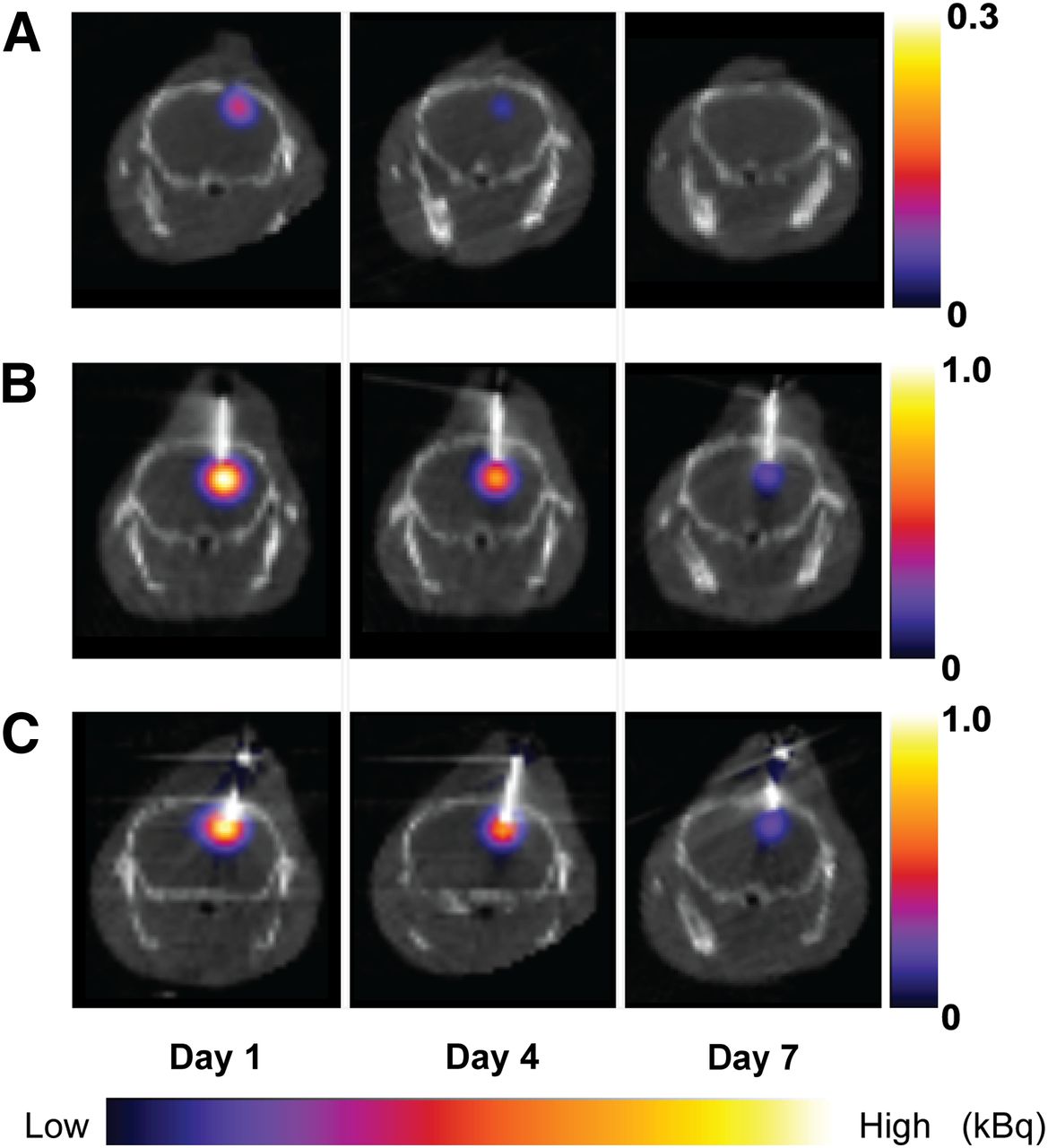
- FIGURE 2.
Coronal SPECT/CT images from mouse brain at days 1, 4, and 7 after 125I-IL-13PE drug infusion. Images after bolus injection showed focal accumulation of 125I-IL-13PE in brain tumor on day 1. Uptake decreased on day 4 and declined more on day 7 (A). Images after CED in both tumor-bearing and normal mouse showed accumulation of 125I-IL-13PE on day 1. Drug was found to be cleared by day 7 (B and C).
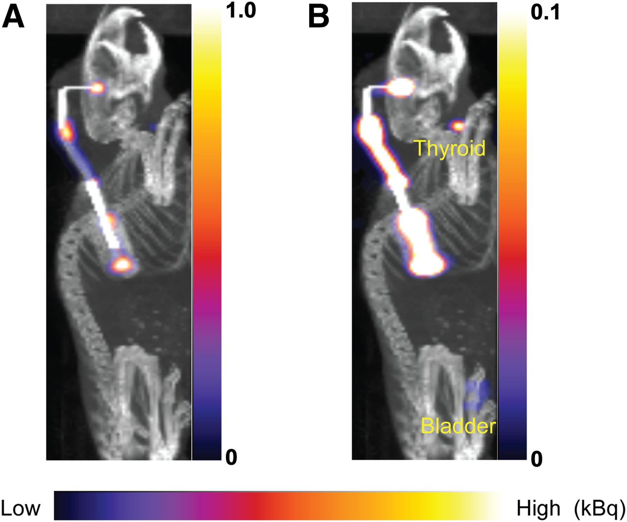
- FIGURE 3.
Maximum-intensity-projection SPECT/CT images from tumor-bearing mouse on day 1 after CED and shown on color scale. Images showed high uptake in brain, catheter tube, and osmotic pump (A). Mild uptake was seen in thyroid and bladder (B).
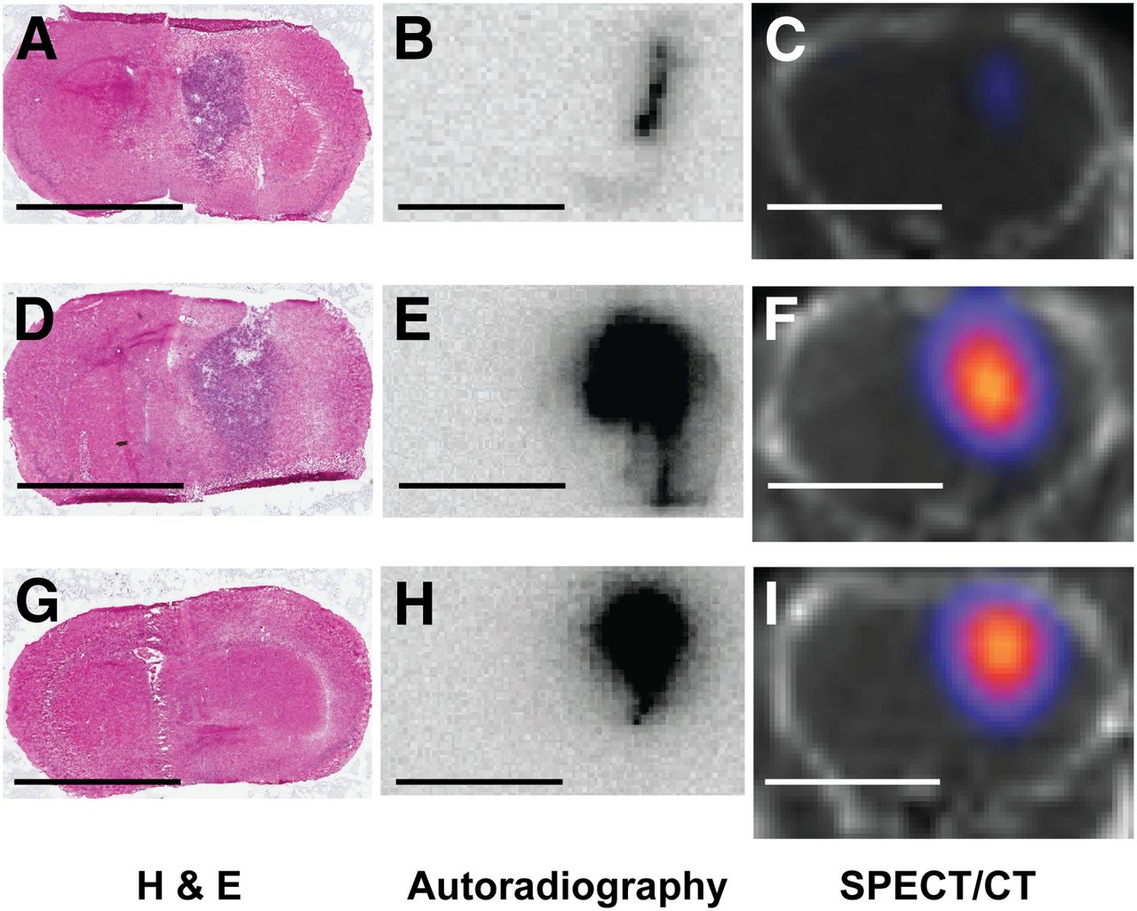
- FIGURE 4.
Hematoxylin and eosin–stained coronal sections, corresponding autoradiographs, and SPECT/CT images from center of injection site of U251 glioma–bearing mouse (A–F) and normal brain (G–I). Mice were euthanized on day 7 after either bolus (A–C) or CED (D–I) of 125I-IL-13PE injection, and frozen brains were sectioned coronally. U251 glioma tumors were seen in implanted site in cortex and spread into caudate (A and D). Autoradiography after bolus injection displayed weak levels of 125I-IL-13PE in tumor and around needle track (B), and after CED in tumor-bearing mouse showed that high concentrations of 125I-IL-13PE distributed throughout tumor and traveled along external capsule (E). Autoradiography after CED in normal brain showed high concentrations of 125I-IL-13PE distributed at injection site (H). Corresponding SPECT/CT images demonstrated similar anatomic epicenters of infusion (C, F, and I). Bar = 5 mm. H & E = hematoxylin and eosin.


Tables
Bolus injection in tumor CED in tumor CED in normal brain Organ day 1 (n = 3) day 7 (n = 3) day 1 (n = 3) day 7 (n = 3) day 1 (n = 1) day 7 (n = 2) Brain 2.787 ± 0.192* 0.549 ± 0.276† 4.155 ± 0.129* 1.057 ± 0.102† 3.985 1.016 ± 0.095† Thyroid 0.328 ± 0.169 0.780 ± 1.164 0.522 ± 0.277 1.021 ± 0.252 0.917 0.874 ± 0.392 Blood 0.818 ± 0.518‡ 0.021 ± 0.010‡ 0.260 ± 0.054 0.025 ± 0.007 0.615 0.037 ± 0.020 Kidney 0.252 ± 0.088§║ 0.048 ± 0.011§ 0.117 ± 0.017║ 0.065 ± 0.016 0.157 0.091 ± 0.030 Liver 0.808 ± 0.683 0.160 ± 0.032¶ 0.131 ± 0.054 0.026 ± 0.007¶ 0.198 0.064 ± 0.013¶ Spleen 0.048 ± 0.036 0.012 ± 0.004 0.010 ± 0.003 0.002 ± 0.001 0.014 0.006 ± 0.003 Heart 0.011 ± 0.001 0.002 ± 0.002 0.009 ± 0.003 0.001 ± 0.001 0.017 0.001 ± 0.001 Pump 14.03 ± 4.644 15.09 ± 0.335 14.74 12.22 ± 0.550 ↵*,†,‡,§,║,¶ Significant difference between groups (*P < 0.001, †P < 0.001, ‡P < 0.01, §P < 0.001, ║P < 0.01, and ¶P < 0.01).
Percentage of injected dose (%ID) of each organ is shown.
- TABLE 2
Tracking Accuracy of SPECT/CT for 125I-IL-13PE Distribution After Bolus Injection or CED into Glioma-Bearing Mouse Brain
SPECT Animal no. Injection method Time after injection Autoradiography, Vd (mm3) Vd (mm3) Percentage difference with autoradiography 1 Bolus Day 1 9.78 4.10 −41.9 2 Bolus Day 7 1.51 Not available Not available 3 Bolus Day 7 0.72 Not available Not available 4 Bolus Day 7 1.04 Not available Not available 5 CED Day 1 36.6 20.3 −55.4 6 CED Day 7 15.7 8.19 −52.2 7 CED Day 7 19.1 7.68 −40.3 8 CED Day 7 20.3 12.5 −61.7 Injection parameter SPECT Autoradiography Group Day Time Volume of injection (μL)* ID (kBq) Vd (mm3) No. of mice Vd (mm3) No. of mice Bolus injection in tumor 1 10 min 5 925 3.11 ± 0.70* 4 9.78 1 7 10 min 5 925 Not available 6 1.09 ± 0.40† 3 CED in tumor 1 3 d 100 3,700 18.3 ± 1.37* 4 36.6 1 7 3 d 100 3,700 12.0 ± 3.43 6 18.4 ± 2.41† 3 CED in normal brain 1 3 d 100 3,700 11.7 1 Not available 0 7 3 d 100 3,700 10.9 ± 4.97 4 14.3 ± 5.42† 2


Jump to section
Related Articles
Cited By...
- No citing articles found.