


Abstract
The somatostatin receptor, which is overexpressed by many neuroendocrine tumors, is a well-known target for molecular imaging and peptide receptor radionuclide therapy. Recently, 57Co-labeled DOTATOC, an octreotide analog, was shown to have the highest affinity yet found for somatostatin receptor subtype 2. The aim of this study was to evaluate the biologic effects of novel cobalt-labeled octreotide analogs targeting the somatostatin receptor to identify promising candidates for molecular imaging and Auger electron–based radionuclide therapy. Methods: Cobalt-labeled DOTATATE, DOTATOC, and DOTANOC were prepared with 57Co or 58mCo for SPECT or Auger electron–based therapy, respectively. The cellular uptake and intracellular distribution of the radioligands were characterized with the pancreatic tumor cell line AR42J in vitro, including assessment of the therapeutic effects of 58mCo-DOTATATE via DNA double-strand break and proliferation assays. Comparisons with the therapeutic effects of 111In- and 177Lu-DOTATATE were also performed. Tumor uptake and normal tissue uptake were characterized in a subcutaneous pancreatic tumor mouse model. Results: All 3 cobalt-conjugated peptides resulted in time-dependent and receptor-specific uptake, with a high level (≥88%) of cellular internalization in vitro of the total cell-associated radioactivity. The DNA double-strand break yield showed a dose-dependent increase with activity, whereas cell survival showed a dose-dependent decrease. 58mCo-DOTATATE was significantly more efficient in cell killing per cumulated decay than 111In- and 177Lu-DOTATATE. The in vivo pharmacokinetic studies showed a high level of receptor-specific tumor uptake. Conclusion: All cobalt-labeled radioligands showed a high level of receptor-specific uptake both in vitro and in vivo in tumor-bearing mice. Furthermore, 58mCo-DOTATATE showed considerable therapeutic effects in vitro and, thus, could be an effective agent for eradicating disseminated tumor cells and micrometastases.
The somatostatin receptor, which is overexpressed by many neuroendocrine tumors, is a well-known target for molecular imaging (i.e., PET and SPECT) and for peptide receptor radionuclide therapy (1,2). One of the most widely used radiopharmaceuticals for SPECT imaging of these tumors is 111In-diethylenetriaminepentaacetic acid (DTPA)-d-Phe1-octreotide (OctreoScan; Mallinckrodt), which was developed from the somatostatin analog octreotide. Several modifications of this peptide have been made over time, leading to new generations of the ligand with improved binding to somatostatin receptor–positive tissues—for example, DOTATOC (DOTA-[Tyr3]-octreotide) (3), DOTATATE (DOTA-[Tyr3, Thr8]-octreotide) (4), and DOTANOC (DOTA-[1-Nal3]-octreotide) (5).
Recently, 57Co-labeled DOTATOC was shown to have the highest affinity yet found for the somatostatin receptor subtype 2 (SSTR2) (6). Furthermore, the rate of internalization of this compound into the pancreatic tumor cell line AR42J was the highest found for any somatostatin-based radioligand. In a study by Uusijärvi et al. (7) of 59 therapeutic radionuclides (α, β, and Auger electron emitters), the Auger electron emitter 58mCo was the radionuclide that resulted in the second highest theoretic tumor-to-normal tissue dose rate ratio. Therefore, the therapeutic potency of the 58mCo radionuclide and the remarkable obtainable internalization rates and affinities for SSTR2-expressing tumor cells make 58mCo-labeled octreotide analogs attractive candidates for targeted Auger electron–based therapy of small somatostatin receptor–positive tumors and micrometastases.
The purpose of this study was to evaluate the biologic effects of selected cobalt-labeled octreotide analogs targeting the somatostatin receptor to identify promising candidates for molecular imaging and Auger electron–based radionuclide therapy.
MATERIALS AND METHODS
Cell Culture
The rat pancreatic tumor cell line AR42J (CLS Cell Lines Service GmbH), which expresses SSTR2, was grown in complete medium (Nutrients Mixture F-12 Ham [Sigma-Aldrich] supplemented with 10% fetal bovine serum [Life Technologies], 1% penicillin/streptomycin [Sigma-Aldrich], and 2 mM l-glutamine [Sigma-Aldrich]). The cells were maintained in a humidified 5% CO2 atmosphere at 37°C.
Chemicals and Syntheses
All solvents and reagents were used as received unless otherwise noted. Water was deionized with a Millipore Direct-Q 3 ultraviolet water purification system. DOTATOC was obtained from JPT Peptide Technologies and exhibited a purity of greater than 99.5%. DOTATATE and DOTANOC were obtained from ABX and exhibited a purity of greater than 99%. Sodium acetate buffer for complexometry (pH 4.6; Sigma-Aldrich) was applied for labeling. The γ spectra were measured by use of a calibrated broad-energy germanium detector (BEGE2020-7600SL; Canberra) with detector software Genie 2000 (version 3.2.1; Canberra) and the nuclear data shown in Table 1.
Nuclear Data for Radioisotopes Used in This Work (21)
Cobalt-labeled DOTATATE, DOTATOC, and DOTANOC were prepared with 57Co for assessment of the biokinetics of the radioligands in vitro and in vivo, and 58mCo-labeled DOTATATE was used for Auger electron–based radionuclide therapy. 57Co was purchased as 57CoCl2 from Perkin Elmer. 58mCo activity was produced and the activity was measured from the coproduced 58gCo as previously described (8) with irradiation times of up to 6 h. An additional purification step was added to increase the specific activity of the labeled peptide. In brief, the additional purification step was performed with a Chromafix 30-PS-HCO3 cartridge (45 mg; Macherey-Nagel) that had been conditioned with 5 mL of 4 M HCl. The radiocobalt solution was eluted through the cartridge, and then the cartridge was washed with 0.2 mL of 4 M HCl to completely remove the cobalt. The solution (0.5 mL) was evaporated to dryness and redissolved in 0.3 mL of 0.04 M HCl for labeling. Sodium acetate buffer (30–50 μL; pH 4.6), water (for injection; 75 μL; Braun), DOTA-peptide (5–250 μg), and the radionuclide in 0.04–0.05 M HCl were loaded into a small glass vial and heated at 85°C in a water bath for 30 min.
For comparison purposes, 111In-DOTATATE was prepared with the same labeling conditions as those used for cobalt, but 177Lu-DOTATATE was prepared as described by Kwekkeboom et al. with minor deviations (4). In brief, a kit consisting of DOTATATE (0.25 mg), sodium ascorbate (125 mg), gentisic acid (25 mg), and water (for injection; 0.80 mL) was added to 177Lu (in 0.04 M HCl; 0.200 mL; 7.6 GBq). The solution was heated in a water bath at 85°C for 30 min, and 1 mL of DTPA (2 mg/mL in isotonic saline) was added. The labeling yield and the radiochemical purity were always greater than 99%. 111In was purchased as 111InCl3 from Dupharma, and 177Lu was purchased as 177LuCl3 from ITG Isotope Technologies.
Radiochemical purity was assessed by use of analytic reverse-phase high-performance liquid chromatography with a Hitachi LaChrom Elite system and a Phenomenex Jupiter C18 300A column (150 × 4.60 mm; 5 μm). The following eluents were applied: H2O with 0.1% trifluoroacetic acid (eluent A) and acetonitrile (eluent B). The following program was used: 86% eluent A and 14% eluent B for 0–2 min, 86% eluent A and 14% eluent B to 37% eluent A and 63% eluent B for 2–10 min, and 37% eluent A and 63% eluent B to 86% eluent A and 14% eluent B for 10–12 min at a flow of 1 mL/min. The retention times were approximately 2.2 min for unbound radiometal and approximately 8.2 min for radiometal-DOTA-peptide.
Subcellular Distribution and Efflux
AR42J cells were seeded in 6-well plates (400,000 cells per well) 48 h before the experiment. On the day of the experiment, the cells were washed twice with incubation medium (1% bovine serum albumin in serum-free Nutrients Mixture F-12 Ham). The cells were incubated with 57Co-labeled peptide or 111In-DOTATATE at 0.02 MBq/mL (specific activity, 3–4 MBq/nmol) in incubation medium with or without an excess of octreotide (Sequoia Research Products) at 37°C for various times. Cellular uptake was stopped by removal of the incubation medium, and the cells were washed 3 times with cold phosphate-buffered saline. For removal of receptor-bound radioligand, the cells were incubated for 5 min in ice-cold acid wash buffer (0.2 M sodium acetate, 0.5 M NaCl; pH 2.5) on ice (9). The cells were separated into cytoplasmic and nuclear fractions with a Nuclei EZ Prep nuclei isolation kit (Sigma-Aldrich) in accordance with the manufacturer’s instructions. The radioactivity of the incubation medium and the receptor-bound, cytoplasmic, and nuclear fractions was measured with a 2470 Wizard Automatic γ Counter (Perkin Elmer).
For efflux studies, cellular uptake was stopped after 2 h by removal of the incubation medium. Unbound radioactivity was removed by washing the cells 3 times with cold phosphate-buffered saline, and receptor-bound activity was removed as described earlier. The cells were washed with incubation medium and returned to the incubator with fresh incubation medium. At various times, the medium was collected and replaced with fresh incubation medium. Thereafter, the cells were incubated with 1 M NaOH to extract the remaining cell-associated radioligand. The radioactivity of the incubation medium and the receptor-bound, external, and internal fractions was measured with the 2470 Wizard Automatic γ Counter.
Assessment of Yield of DNA Double-Strand Breaks from 58mCo-, 111In-, or 177Lu-DOTATATE
AR42J cells were washed 3 times with incubation medium and incubated with radioligand at 0–50 MBq/mL (specific activity, 44–84 MBq/nmol) for 1 h at 37°C with or without an excess of octreotide. The cells were subsequently fixed with 4% paraformaldehyde and permeabilized with 0.2% Triton X-100 (Dow Chemical Co.). DNA double-strand breaks (DSBs) were visualized by incubating the cells with rabbit polyclonal anti–phospho-H2AX (P-Ser139) antibody (Sigma-Aldrich) and then with AlexaFluor-conjugated goat antirabbit antibody (Life Technologies). The cells were counterstained with 4′,6-diamidino-2-phenylindole (Sigma-Aldrich) and analyzed with an Olympus FV1000 confocal laser scanning microscope. γH2AX foci (from >30 cells for each activity concentration) were analyzed with FociCounter analysis software (10).
Antiproliferative Effects of 58mCo-, 111In-, or 177Lu-DOTATATE
AR42J cells were seeded in 96-well plates (5,000 cells per well); 24 h later, the cells were washed 3 times with incubation medium and then incubated with radioligand at 0–50 MBq/mL (specific activity, 44–84 MBq/nmol) with or without an excess of octreotide for 24 h. On the next day, the cells were washed 3 times with phosphate-buffered saline and then incubated in complete medium for 7 d. The WST-1 (Roche Applied Science) assay was performed in accordance with the manufacturer’s instructions.
In Vivo Animal Studies
Female Swiss nude mice (NTac:NIHS-Foxn1nu; Taconic) and C.B-17 SCID mice (C.B-Igh-1b/IcrTac-Prkdcscid; Taconic), 6–10 wk old, were anesthetized with isoflurane in 100% oxygen. Next, the mice were inoculated in the right flank with 106 AR42J cells that had been prepared in 50 μL of medium containing extracellular matrix gel (Sigma-Aldrich; ratio, 1:1). Tumors were allowed to grow for 10–14 d, reaching a weight of 67–422 mg. All animal experiments were approved by The Animal Experiments Inspectorate in Denmark.
Studies of the biodistribution of 57Co-DOTATATE in tumor-bearing C.B-17 SCID mice were performed with 105 ± 18 kBq (mean ± SD) of the tracer (32 ± 5 pmol of peptide in 0.1% bovine serum albumin) injected retroorbitally into the venous sinus of anesthetized mice (11). At various times after injection of the tracer, the mice (n = 3 in each group) were euthanized and dissected. Organs were collected and weighed, and activities were measured with the 2470 Wizard Automatic γ Counter. The γ counter was cross-calibrated with the Ge detector used to determine the injected activities. Organ tracer activity distributions were determined as the percentage injected activity per gram of tissue.
SPECT/CT scans of AR42J tumor–bearing Swiss nude mice (inoculated as described earlier) were performed with a Siemens Inveon small-animal scanner. The animals were anesthetized with isoflurane in 100% oxygen and then injected intravenously with 57Co-DOTATATE or 57Co-DOTATOC (5.5–7.8 MBq; 1–4 nmol). At various times after injection, the animals were scanned. Before the last scan, at 4 h after injection, the animals were euthanized to prevent further redistribution of the radioligand during the rather long SPECT scan (up to 280 min) at this time point. A receptor-blocking experiment was performed by coinjecting 0.93 μmol of octreotide intraperitoneally with the radioligand to visualize nonspecific binding. The SPECT scans were performed with pinhole collimators (two 5 × 1.0 mm) and acquisition times of 10–280 min. The images were reconstructed with the 3-dimensional ordered-subset maximization expectation algorithm. The CT scans were performed with 80-kVp x-rays (500 μA), a rotation of 360°, and 360 projections.
Statistical Analysis
Data were analyzed with unpaired, 2-tailed Student t tests. The F test was used to compare best-fit curves of the survival data as a function of cumulated decays with GraphPad Prism (GraphPad Software, Inc.). A P value of less than 0.05 was considered statistically significant.
RESULTS
Up to 0.58 GBq of therapeutic 58mCo-DOTATATE was produced with a specific activity of up to 84 MBq/nmol. The radionuclide and radiochemical purities of 58mCo-DOTATATE were always greater than 98% and 99%, respectively, at the end of synthesis—the former due to the inevitable coproduction of the ground state of 58Co.
The subcellular distributions of the cobalt-labeled peptides and, for comparison purposes, of the Auger electron–emitting compound 111In-DOTATATE in the pancreatic cancer cell line AR42J as a function of increasing incubation times are shown in Figure 1. All 3 cobalt-labeled peptides resulted in time-dependent uptake, with a high level (≥88%) of cellular internalization in vitro of the total cell-associated radioactivity. Similar internalization was found for 111In-DOTATATE. Moreover, the uptake was receptor-specific, because an excess of octreotide could block the internalization. The overall level of radioligand uptake was significantly higher (49%–123% higher) for the cobalt-labeled peptides than for 111In-DOTATATE at all time points. However, only 2%–4% of the cell-associated cobalt activity was transferred to the cell nuclei, where Auger electrons are most effective for cell killing regardless of the peptide used, whereas 4%–7% nucleus translocation was seen with 111In-DOTATATE.
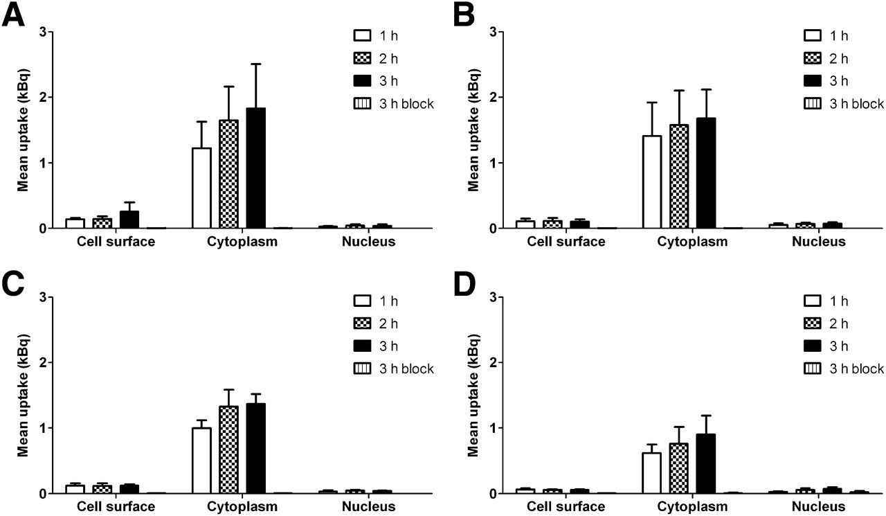
Subcellular distributions of cobalt-labeled peptides 57Co-DOTATOC (A), 57Co-DOTANOC (B), 57Co-DOTATATE (C), and 111In-DOTATATE (D) in pancreatic cancer cell line AR42J as function of increasing incubation times (1–3 h). For each radiopharmaceutical, receptor-blocking experiment was performed with excess octreotide (“3 h block”). Data are mean ± SD from triplicate experiments.
For determination of the retention of the cobalt-labeled peptides in the cells, the rates of externalization of the intracellular radioactivity (efflux) after 2 h of preloading were measured for the 3 cobalt-labeled peptides and for 111In-DOTATATE (results not shown). The externalization rates were similar for the 4 radioligands tested. Of the cobalt-labeled peptides, 57Co-DOTATATE showed the highest level of retention in the cells (41% ± 5% at 24 h), 57Co-DOTANOC showed the lowest level of retention (33% ± 7% at 24 h), and 57Co-DOTATOC retained 37% ± 3% at 24 h. For comparison 111In-DOTATATE retained 45% ± 5% at 24 h (decay corrected). Between 64% and 73% of the internalized 57Co or 111In activities were still retained in the cells after 4 h.
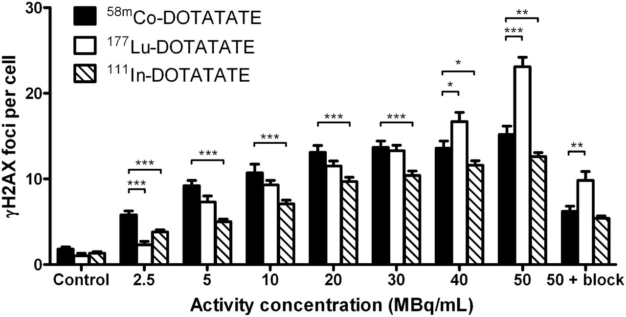
The formation of DNA DSBs showed a clear dose-dependent increase with activity for both 58mCo-DOTATATE and 111In-DOTATATE (Fig. 2). The effect could be strongly diminished by blocking the somatostatin receptors with excess octreotide. For comparison, the DSB yield was also measured for the β−-emitting 177Lu-DOTATATE, which is currently among the best radioligands for peptide receptor radionuclide therapy in patients (2). At the lowest activity concentration used, 58mCo-DOTATATE resulted in the highest DSB yield of the tested compounds (P < 0.001). At the highest activity concentration, 177Lu-DOTATATE resulted in the highest DSB yield, but this radioligand also resulted in the highest nonspecific toxicity with blocked receptors (P < 0.01). 111In-DOTATATE showed low nonspecific toxicity comparable to that of 58mCo-DOTATATE but was significantly less efficient in causing DSBs at all activity concentrations (Fig. 2).

Formation of DNA DSBs in AR42J cells exposed for 1 h to increasing amounts of 58mCo-, 111In-, and 177Lu-DOTATATE activities (mean ± SEM). Nonspecific toxicity at 50 MBq/mL was assessed by blocking somatostatin receptors with excess octreotide (“50 + block”). P values were determined with 2-tailed Student t test. *P < 0.05. **P < 0.01. ***P < 0.001.
The antiproliferative effects of 58mCo-, 111In-, and 177Lu-DOTATATE are shown in Figure 3. All radioligands resulted in a dose-dependent decrease in cell survival that could be strongly diminished by blocking the receptors with excess octreotide. At activity concentrations of less than or equal to 30 MBq/mL, 58mCo-DOTATATE showed a general trend to be more effective. Because the half-lives of the 3 therapeutic isotopes are different (58mCo, 9.04 h; 111In, 2.80 d; 177Lu, 6.73 d), the numbers of cumulated decays from the radiopharmaceuticals during the cell survival experiments also were different. Therefore, a calculation was performed to determine the cumulated decays during the 24-h period of incubation with the radioligands. The cell survival as a function of the cumulated decays from the radiopharmaceuticals during this period is shown in Figure 4. The data indicated that 58mCo-DOTATATE was significantly more efficient in cell killing per cumulated decay than the other radioligands.

Antiproliferative effects in AR42J cells exposed for 24 h to increasing amounts of 58mCo-, 111In-, and 177Lu-DOTATATE activities. Nonspecific toxicity at 50 MBq/mL was assessed by blocking somatostatin receptors with excess octreotide (“50 + block”). Data are mean ± SEM from 5 replicate experiments. P values were determined with 2-tailed Student t test. *P < 0.05. **P < 0.01. ***P < 0.001.
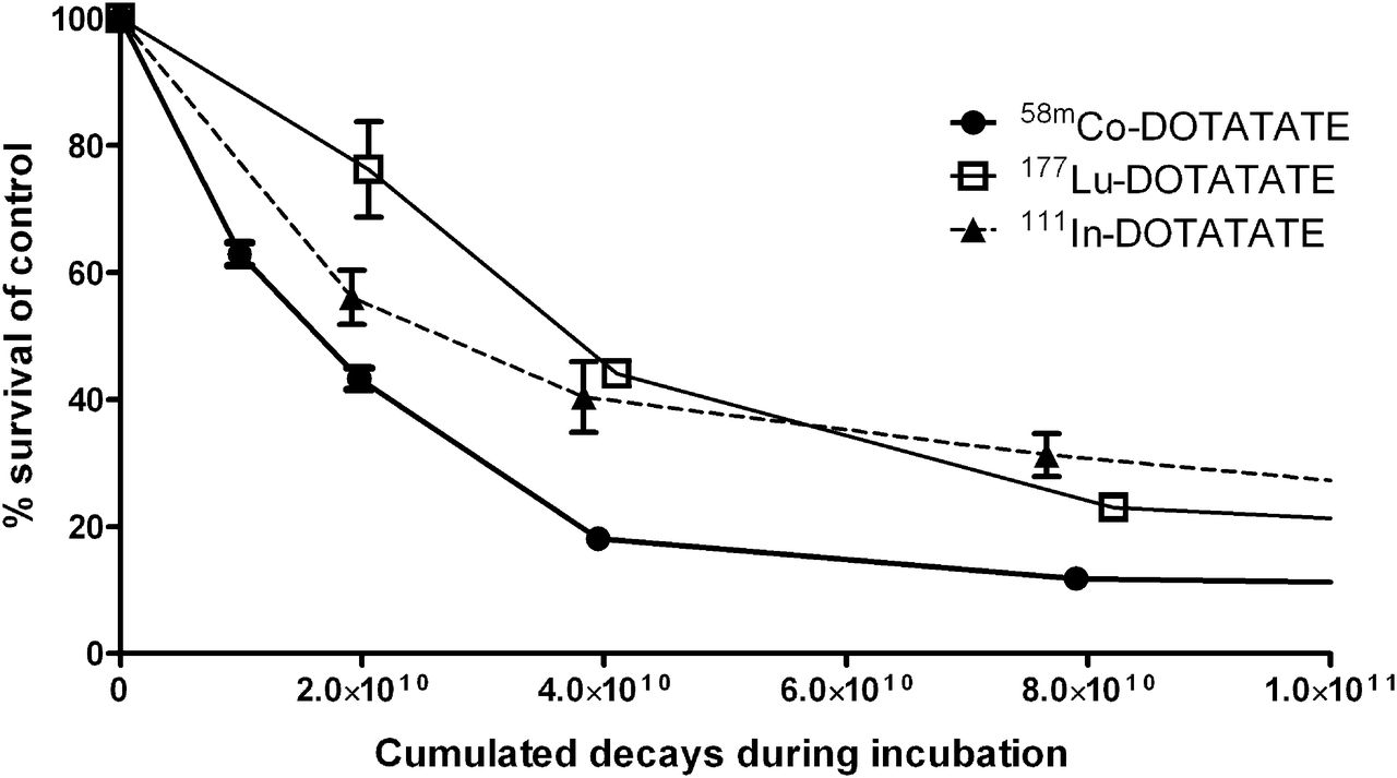
Antiproliferative effects in AR42J cells as function of number of cumulated decays during 24-h incubation with increasing amounts of 58mCo-, 111In-, and 177Lu-DOTATATE.
For determination of the in vivo pharmacokinetics of cobalt-labeled DOTATATE, biodistribution and SPECT/CT studies of 57Co-DOTATATE (57Co was used as a surrogate for 55Co for PET) in a pancreatic tumor–bearing mouse model were performed. 57Co-DOTATATE showed a high level of receptor-specific uptake in the tumors (Fig. 5A). Figure 5B shows the nonspecific uptake achieved by blocking the somatostatin receptors with a coinjection of excess octreotide; the radioligand was found mainly in the kidneys, the main route of excretion for somatostatin analogs.
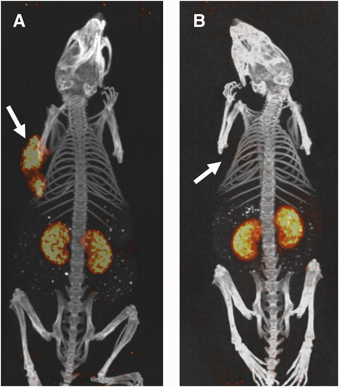
Maximum-intensity-projection images from SPECT/CT scans of AR42J tumor–bearing mice injected with 5.5–7.8 MBq of 57Co-DOTATATE (1–4 nmol of peptide) and without (A) or with (B) coinjection of 0.93 μmol of octreotide to visualize nonspecific binding or localization. Scans were performed at 4 h after injection. Arrows indicate locations of tumors.
The biodistribution of 57Co-DOTATATE is shown in Figure 6 (as percentage injected activity per gram of tissue). A rapid, high level of uptake of 57Co-DOTATATE was observed in the tumors, but a moderate to high level of uptake (as percentage injected activity per gram of tissue) also was found in the somatostatin receptor–positive organs (pancreas, stomach, lungs, and adrenal glands) and in the kidneys because of excretion. However, when measured as the percentage injected activity, the highest overall tissue activity at 4 h and 24 h after injection was found in the tumors. Tumor uptake between 4 h and 24 h decreased only to about 43%, in good agreement with our in vitro efflux results. Clearance from the blood and the somatostatin receptor–negative tissues also was fast. The tumor-to-kidney, tumor-to-liver, and tumor-to-blood ratios at 4 h and 24 h after injection were 1.3–1.4, 33–52, and 121–137, respectively.

Biodistribution of 57Co-DOTATATE (105 ± 18 kBq; 32 ± 5 pmol of peptide) in AR42J tumor–bearing mice at 1, 4, and 24 h after injection (mean ± SD; n = 3 in each group).
DISCUSSION
The development of novel radiopharmaceuticals for targeted radionuclide therapy of cancer is a rapidly growing research area (12). Most emerging radiopharmaceuticals consist of commercially available radionuclides bound to new targeting ligands. Fewer studies develop and test novel therapeutic radionuclides and address the consequences of changing the radionuclide to a putative therapeutically more potent one. As shown in Figure 1, it was possible to obtain a considerably higher level of receptor-mediated uptake in the pancreatic cancer cell line AR42J, which exclusively expresses SSTR2, by labeling DOTATATE with cobalt instead of indium at the same specific activity. This finding was in good agreement with the measured affinities for DOTATOC labeled with cobalt, gallium, or yttrium, as reported by Heppeler et al. (6); in that study, 57Co-DOTATOC resulted in the highest affinity for SSTR2. The authors concluded that 57Co-DOTATOC had the highest SSTR2 affinity of any somatostatin-based radioligand found so far. However, in the present study, the relative subcellular distributions of the cell-associated radioactivity in the pancreatic cancer cell line were similar for the 2 radiometals (Fig. 1).
As mentioned earlier, the retention and externalization (efflux) of the radiometals from AR42J tumor cells after incubation with the 3 cobalt-labeled peptides and 111In-DOTATATE were rather similar. The measured retention of 57Co-DOTATOC (∼67%) at 4 h was in good agreement with that in the study of Heppeler et al. (6), who found that about 60% of 57Co-DOTATOC was retained after 4 h. However, they also found rapid tumor clearance in vivo in an AR42J pancreatic tumor mouse model of 57Co-DOTATOC, with a decrease in tumor uptake between 4 h and 24 h of more than 90%. These results were not observed in our in vivo pharmacokinetic study with 57Co-DOTATATE and a similar AR42J tumor mouse model (Fig. 6). Tumor uptake between 4 h and 24 h decreased only to about 43%, in good agreement with our in vitro efflux results.
The high level of lung uptake was somewhat surprising, although it has been seen in other studies with metal-labeled DOTATATE in mice (13). However, mouse lung tissue is known to express the SST4 receptor subtype (14), and DOTATATE has some affinity for this subtype (15). Furthermore, labeling of DOTATOC with Co rather than Ga or Y increases its SSTR4 affinity strongly, leading to lung uptake of the ligand (6). Hence, one could speculate that labeling of DOTATATE with Co2+ instead of typical trivalent radiometals could further increase the SSTR4 affinity, leading to increased lung uptake, although this notion was not investigated in the present study. Except for the lung uptake, the measured biodistribution of 57Co-DOTATATE was comparable to that found for 111In-DOTATATE by Froidevaux et al. (16).
As mentioned earlier, only a small fraction of the internalized cobalt or indium activity was translocated to the nuclei of the pancreatic tumor cells; the majority was found in the cytoplasm. It is generally well established that Auger electron emitters should be positioned in close vicinity to the genomic DNA to be maximally effective in cell killing, with high–linear energy transfer–like effects (17). Such was not the case for the peptides used in the present study; for those peptides, no DNA binding was assumed to take place. Hence, the observed effects were not expected to be due to high–linear energy transfer “Auger effects” and, thus, may have been inferior to the effects of peptide receptor radionuclide therapy with α emitters. Nevertheless, both 58mCo-DOTATATE and 111In-DOTATATE resulted in high DSB yields, with the former producing the largest number of DSBs (P < 0.05), and both compounds could effectively reduce the proliferation of AR42J cancer cells compared with the results for untreated controls. However, per cumulated decay from the radiopharmaceuticals during the incubation period, 58mCo-DOTATATE was significantly more effective in cell killing than 111In-DOTATATE.
From the published theoretic radiation doses to the cell nucleus (and, thus, the DNA) per decay from the radionuclides used in the present study (i.e., the cellular S values (absorbed dose per unit cumulated activity) (18,19)), it can be calculated that 58mCo delivers approximately 2 and 6 times higher doses to the nucleus for decays taking place in the nucleus and the cytoplasm, respectively, than 111In. (The corresponding doses are approximately 2 and 11 times higher, respectively, for 58mCo than for 58gCo. Hence, the dose contribution from this impurity, constituting <2% at the end of synthesis, is negligible.) This calculation is in good agreement with the experimental findings. Compared with 177Lu, 58mCo delivers approximately 3 times higher doses to the nucleus for decays taking place in the nucleus and the cytoplasm, respectively.
Furthermore, given the theoretic tumor-to-normal tissue dose rate ratios calculated by Uusijärvi et al. (7), 58mCo has tumor-to-normal tissue dose rate ratios that are approximately 15 and 11 times higher than those of 111In for activity located in the nucleus and the cytoplasm, respectively, of tumor cells. Hence, 58mCo-DOTATATE theoretically should exhibit much less nonspecific toxicity in vivo in patients than 111In-DOTATATE; therefore, it should be possible to increase the injected activity considerably to achieve a higher radiation dose to the tumor without increasing the dose to the normal tissue.
At the lowest activity concentration, 58mCo-DOTATATE was more effective in DSB formation and cell killing than β−-emitting 177Lu-DOTATATE, but the opposite was found at the highest activity concentration. However, when the number of cumulated decays was considered, 58mCo-DOTATATE was found to be significantly more effective in cell killing (Fig. 4). This finding is in good agreement with the previously mentioned theoretic radiation doses per decay: the long range of the β−-particles from 177Lu results in “wasted” energy deposited outside the cell (considered as single cells in the cellular S value formalism). On the other hand, radionuclide therapy of macroscopic tumors with β−-emitting radionuclides relies mainly on cross-fire effects; in contrast, therapy with Auger electron emitters is based on localized energy depositions. Because almost no cross-fire takes place for cells growing in monolayers in vitro, as in the proliferation assay in the present study, a general prediction of the in vivo efficacy from the in vitro comparison described earlier is meaningless. However, for small micrometastases or single cells, for which cross-fire effects are insignificant, the comparison is valid. That is, in a future clinical setting, 58mCo-DOTATATE could be an effective agent for eradicating circulating and disseminated tumor cells and micrometastases when used in combination with a long-range β−-emitting radionuclide or another treatment modality for eradicating macroscopic tumor tissue. However, further studies of the therapeutic efficacy in vivo of 58mCo-DOTATATE are required to determine the full potential of this promising radiopharmaceutical. If such studies become successful, then translation to clinical applications should be feasible. We recently calculated that it will be possible to produce more than 120 GBq of 58mCo in 3 h with a small PET cyclotron (19). Hence, clinical applications with, for example, the administration of 18.5 GBq per patient—as in a study using 111In-DTPA-octreotide (20)—should be achievable with 58mCo-DOTATATE.
CONCLUSION
We evaluated the biologic effects of cobalt-labeled octreotide analogs targeting the somatostatin receptor in molecular imaging and Auger electron–based radionuclide therapy. All cobalt-labeled radioligands showed a high level of receptor-specific uptake both in vitro and in vivo in tumor-bearing mice. Overall, 58mCo-DOTATATE showed better therapeutic effects in vitro than 111In-DOTATATE and 177Lu-DOTATATE, especially when the number of cumulated decays from the radiopharmaceuticals was considered. Hence, further in vivo studies of the therapeutic efficacy of this promising Auger electron–emitting radiopharmaceutical are warranted.
DISCLOSURE
The costs of publication of this article were defrayed in part by the payment of page charges. Therefore, and solely to indicate this fact, this article is hereby marked “advertisement” in accordance with 18 USC section 1734. The bioimaging experiments reported in this article were performed at DaMBIC, a bioimaging research core facility, at the University of Southern Denmark. DaMBIC was established by an equipment grant from the Danish Agency for Science Technology and Innovation and by internal funding from the University of Southern Denmark. This work was supported by the Lundbeckfonden Center of Excellence NanoCAN grant, the Region Syddanmarks Forskningspulje 2012 (j. nr.: 12/6433) and the Odense Universitetshospitals Forskningspulje 2010 (j. nr.: 2-41-4-00066-201). No other potential conflict of interest relevant to this article was reported.
Footnotes
↵* Contributed equally to this work.
Published online May 29, 2014.
- © 2014 by the Society of Nuclear Medicine and Molecular Imaging, Inc.
REFERENCES
- Received for publication January 6, 2014.
- Accepted for publication April 9, 2014.


