
Main menu

  • Home
  • Content
    • Current
    • Ahead of print
    • Past Issues
    • JNM Supplement
    • SNMMI Annual Meeting Abstracts
    • Continuing Education
    • JNM Podcasts
  • Subscriptions
    • Subscribers
    • Institutional and Non-member
    • Rates
    • Journal Claims
    • Corporate & Special Sales
  • Authors
    • Submit to JNM
    • Information for Authors
    • Assignment of Copyright
    • AQARA requirements
  • Info
    • Reviewers
    • Permissions
    • Advertisers
  • About
    • About Us
    • Editorial Board
    • Contact Information
  • More
    • Alerts
    • Feedback
    • Help
    • SNMMI Journals
  • SNMMI
    • JNM
    • JNMT
    • SNMMI Journals
    • SNMMI

User menu

  • Subscribe
  • My alerts
  • Log in
  • My Cart

Search

  • Advanced search
Journal of Nuclear Medicine
  • SNMMI
    • JNM
    • JNMT
    • SNMMI Journals
    • SNMMI
  • Subscribe
  • My alerts
  • Log in
  • My Cart
Journal of Nuclear Medicine

Advanced Search

  • Home
  • Content
    • Current
    • Ahead of print
    • Past Issues
    • JNM Supplement
    • SNMMI Annual Meeting Abstracts
    • Continuing Education
    • JNM Podcasts
  • Subscriptions
    • Subscribers
    • Institutional and Non-member
    • Rates
    • Journal Claims
    • Corporate & Special Sales
  • Authors
    • Submit to JNM
    • Information for Authors
    • Assignment of Copyright
    • AQARA requirements
  • Info
    • Reviewers
    • Permissions
    • Advertisers
  • About
    • About Us
    • Editorial Board
    • Contact Information
  • More
    • Alerts
    • Feedback
    • Help
    • SNMMI Journals
  • View or Listen to JNM Podcast
  • Visit JNM on Facebook
  • Join JNM on LinkedIn
  • Follow JNM on Twitter
  • Subscribe to our RSS feeds
Research ArticleBasic Science Investigations

Optical Imaging of Renal Cell Carcinoma with Anti–Carbonic Anhydrase IX Monoclonal Antibody Girentuximab

Constantijn H.J. Muselaers, Alexander B. Stillebroer, Mark Rijpkema, Gerben M. Franssen, Egbert Oosterwijk, Peter F.A. Mulders, Wim J.G. Oyen and Otto C. Boerman
Journal of Nuclear Medicine June 2014, 55 (6) 1035-1040; DOI: https://doi.org/10.2967/jnumed.114.137356
Constantijn H.J. Muselaers
1Department of Urology, Radboud University Medical Center, Nijmegen, The Netherlands; and
2Department of Nuclear Medicine, Radboud University Medical Center, Nijmegen, The Netherlands
  • Find this author on Google Scholar
  • Find this author on PubMed
  • Search for this author on this site
Alexander B. Stillebroer
1Department of Urology, Radboud University Medical Center, Nijmegen, The Netherlands; and
2Department of Nuclear Medicine, Radboud University Medical Center, Nijmegen, The Netherlands
  • Find this author on Google Scholar
  • Find this author on PubMed
  • Search for this author on this site
Mark Rijpkema
2Department of Nuclear Medicine, Radboud University Medical Center, Nijmegen, The Netherlands
  • Find this author on Google Scholar
  • Find this author on PubMed
  • Search for this author on this site
Gerben M. Franssen
2Department of Nuclear Medicine, Radboud University Medical Center, Nijmegen, The Netherlands
  • Find this author on Google Scholar
  • Find this author on PubMed
  • Search for this author on this site
Egbert Oosterwijk
1Department of Urology, Radboud University Medical Center, Nijmegen, The Netherlands; and
  • Find this author on Google Scholar
  • Find this author on PubMed
  • Search for this author on this site
Peter F.A. Mulders
1Department of Urology, Radboud University Medical Center, Nijmegen, The Netherlands; and
  • Find this author on Google Scholar
  • Find this author on PubMed
  • Search for this author on this site
Wim J.G. Oyen
2Department of Nuclear Medicine, Radboud University Medical Center, Nijmegen, The Netherlands
  • Find this author on Google Scholar
  • Find this author on PubMed
  • Search for this author on this site
Otto C. Boerman
2Department of Nuclear Medicine, Radboud University Medical Center, Nijmegen, The Netherlands
  • Find this author on Google Scholar
  • Find this author on PubMed
  • Search for this author on this site
  • Article
  • Figures & Data
  • Info & Metrics
  • PDF
Loading

Article Figures & Data

Figures

  • FIGURE 1.
    • Download figure
    • Open in new tab
    • Download powerpoint
    FIGURE 1.

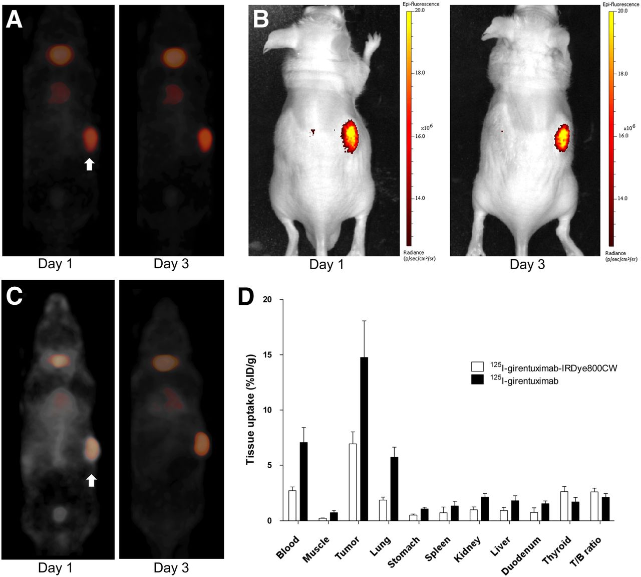
    (A) Micro-SPECT images of mouse bearing SK-RC-52 tumor on right flank (arrow) at 1 and 3 d after injection of 125I-girentuximab-IRDye800CW. In addition to tumor uptake, minimal uptake in heart and thyroid was observed. (B) Optical images of same mouse at 1 and 3 d after injection (same image settings were used for days 1 and 3). Some reflectance-induced image artifacts on backs of mice were observed. (C) Micro-SPECT images of mouse bearing SK-RC-52 tumor on right flank (arrow) at 1 and 3 d after injection of 125I-girentuximab. (D) Biodistribution of 125I-girentuximab-IRDye800CW and 125I-girentuximab in mice with subcutaneous SK-RC-52 tumor at 3 d after injection. Values are expressed as mean ± SD. T/B ratio = tumor-to-blood ratio.

  • FIGURE 2.
    • Download figure
    • Open in new tab
    • Download powerpoint
    FIGURE 2.

    (A) Optical images of mouse bearing CAIX-positive SK-RC-52 tumor on left flank and CAIX-negative SK-RC-59 tumor on right flank (arrows) at 1 and 3 d after injection of 125I-girentuximab-IRDye800CW (same image settings were used for days 1 and 3). Some reflectance-induced image artifacts on backs of mice were observed. (B) Biodistribution at 3 d after injection of 125I-girentuximab-IRDye800CW or 125I-MOPC21-IRDye800CW in mice with subcutaneous SK-RC-52 tumor and subcutaneous SK-RC-59 tumor. Values are expressed as mean ± SD.

  • FIGURE 3.
    • Download figure
    • Open in new tab
    • Download powerpoint
    FIGURE 3.

    Ex vivo biodistribution of 125I-girentuximab-IRDye800CW and excess unlabeled girentuximab. T/B ratio = tumor-to-blood ratio.

  • FIGURE 4.
    • Download figure
    • Open in new tab
    • Download powerpoint
    FIGURE 4.

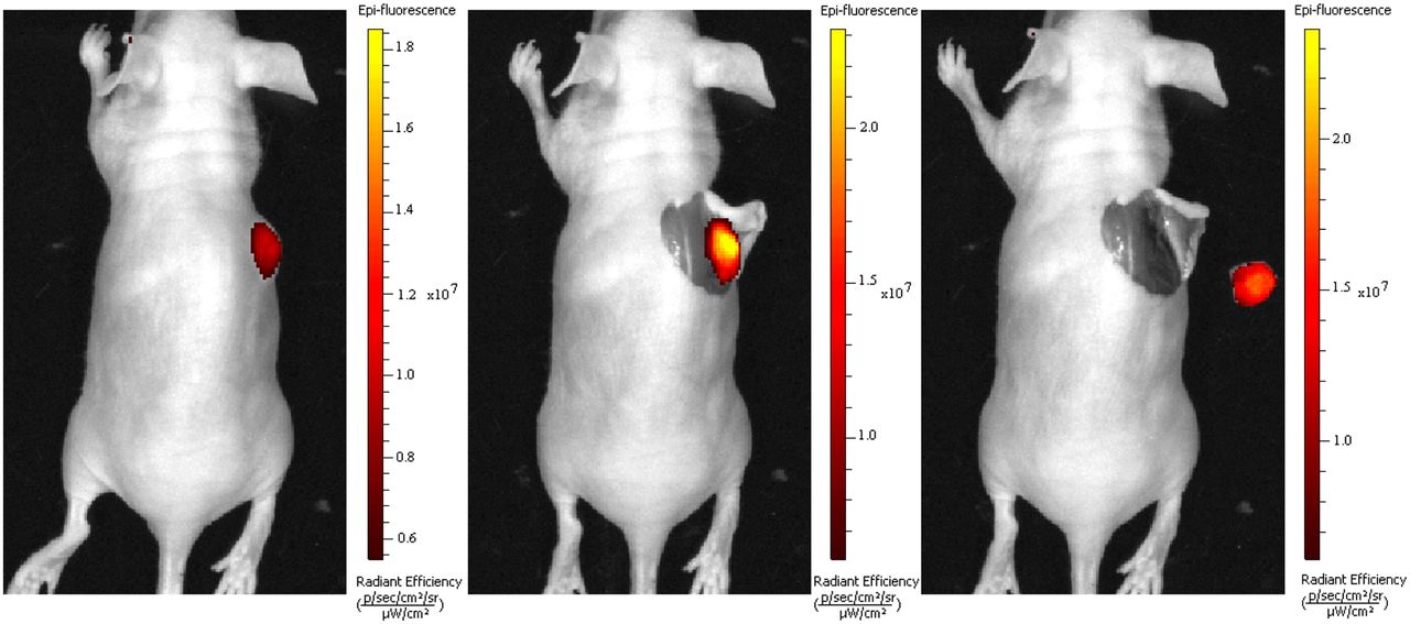
    (Left) Preoperative fluorescence image of mouse with subcutaneous growing ccRCC tumor lesion at 3 d after injection with 125I-girentuximab-IRDye800CW. (Middle) Tumor lesion was subsequently removed by fluorescence image–guided surgery. (Right) After resection, no residual tumor was detected by fluorescence imaging or macroscopically.

PreviousNext
Back to top

In this issue

Journal of Nuclear Medicine: 55 (6)
Journal of Nuclear Medicine
Vol. 55, Issue 6
June 1, 2014
  • Table of Contents
  • Table of Contents (PDF)
  • About the Cover
  • Index by author
Print
Download PDF
Article Alerts
Sign In to Email Alerts with your Email Address
Email Article

Thank you for your interest in spreading the word on Journal of Nuclear Medicine.

NOTE: We only request your email address so that the person you are recommending the page to knows that you wanted them to see it, and that it is not junk mail. We do not capture any email address.

Enter multiple addresses on separate lines or separate them with commas.
Optical Imaging of Renal Cell Carcinoma with Anti–Carbonic Anhydrase IX Monoclonal Antibody Girentuximab
(Your Name) has sent you a message from Journal of Nuclear Medicine
(Your Name) thought you would like to see the Journal of Nuclear Medicine web site.
Citation Tools
Optical Imaging of Renal Cell Carcinoma with Anti–Carbonic Anhydrase IX Monoclonal Antibody Girentuximab
Constantijn H.J. Muselaers, Alexander B. Stillebroer, Mark Rijpkema, Gerben M. Franssen, Egbert Oosterwijk, Peter F.A. Mulders, Wim J.G. Oyen, Otto C. Boerman
Journal of Nuclear Medicine Jun 2014, 55 (6) 1035-1040; DOI: 10.2967/jnumed.114.137356

Citation Manager Formats

  • BibTeX
  • Bookends
  • EasyBib
  • EndNote (tagged)
  • EndNote 8 (xml)
  • Medlars
  • Mendeley
  • Papers
  • RefWorks Tagged
  • Ref Manager
  • RIS
  • Zotero
Share
Optical Imaging of Renal Cell Carcinoma with Anti–Carbonic Anhydrase IX Monoclonal Antibody Girentuximab
Constantijn H.J. Muselaers, Alexander B. Stillebroer, Mark Rijpkema, Gerben M. Franssen, Egbert Oosterwijk, Peter F.A. Mulders, Wim J.G. Oyen, Otto C. Boerman
Journal of Nuclear Medicine Jun 2014, 55 (6) 1035-1040; DOI: 10.2967/jnumed.114.137356
Twitter logo Facebook logo LinkedIn logo Mendeley logo
  • Tweet Widget
  • Facebook Like
  • Google Plus One
Bookmark this article

Jump to section

  • Article
    • Abstract
    • MATERIALS AND METHODS
    • RESULTS
    • DISCUSSION
    • CONCLUSION
    • DISCLOSURE
    • Footnotes
    • REFERENCES
  • Figures & Data
  • Info & Metrics
  • PDF

Related Articles

  • This Month in JNM
  • PubMed
  • Google Scholar

Cited By...

  • New Developments in Dual-Labeled Molecular Imaging Agents
  • Roadmap for the Development and Clinical Translation of Optical Tracers Cetuximab-800CW and Trastuzumab-800CW
  • Evaluation of Nonpeptidic Ligand Conjugates for the Treatment of Hypoxic and Carbonic Anhydrase IX-Expressing Cancers
  • Targeted Dual-Modality Imaging in Renal Cell Carcinoma: An Ex Vivo Kidney Perfusion Study
  • Noninvasive brain cancer imaging with a bispecific antibody fragment, generated via click chemistry
  • Google Scholar

More in this TOC Section

  • PET/MRI of Hypoxic Atherosclerosis Using 64Cu-ATSM in a Rabbit Model
  • Tumor Uptake of Anti-CD20 Fabs Depends on Tumor Perfusion
  • How Sensitive Is the Upper Gastrointestinal Tract to 90Y Radioembolization? A Histologic and Dosimetric Analysis in a Porcine Model
Show more Basic Science Investigations

Similar Articles

Keywords

  • renal cell carcinoma
  • optical imaging
  • girentuximab
  • monoclonal antibody
  • CAIX
SNMMI

© 2025 SNMMI

Powered by HighWire