
Main menu

  • Home
  • Content
    • Current
    • Ahead of print
    • Past Issues
    • JNM Supplement
    • SNMMI Annual Meeting Abstracts
    • Continuing Education
    • JNM Podcasts
  • Subscriptions
    • Subscribers
    • Institutional and Non-member
    • Rates
    • Journal Claims
    • Corporate & Special Sales
  • Authors
    • Submit to JNM
    • Information for Authors
    • Assignment of Copyright
    • AQARA requirements
  • Info
    • Reviewers
    • Permissions
    • Advertisers
  • About
    • About Us
    • Editorial Board
    • Contact Information
  • More
    • Alerts
    • Feedback
    • Help
    • SNMMI Journals
  • SNMMI
    • JNM
    • JNMT
    • SNMMI Journals
    • SNMMI

User menu

  • Subscribe
  • My alerts
  • Log in
  • Log out
  • My Cart

Search

  • Advanced search
Journal of Nuclear Medicine
  • SNMMI
    • JNM
    • JNMT
    • SNMMI Journals
    • SNMMI
  • Subscribe
  • My alerts
  • Log in
  • Log out
  • My Cart
Journal of Nuclear Medicine

Advanced Search

  • Home
  • Content
    • Current
    • Ahead of print
    • Past Issues
    • JNM Supplement
    • SNMMI Annual Meeting Abstracts
    • Continuing Education
    • JNM Podcasts
  • Subscriptions
    • Subscribers
    • Institutional and Non-member
    • Rates
    • Journal Claims
    • Corporate & Special Sales
  • Authors
    • Submit to JNM
    • Information for Authors
    • Assignment of Copyright
    • AQARA requirements
  • Info
    • Reviewers
    • Permissions
    • Advertisers
  • About
    • About Us
    • Editorial Board
    • Contact Information
  • More
    • Alerts
    • Feedback
    • Help
    • SNMMI Journals
  • View or Listen to JNM Podcast
  • Visit JNM on Facebook
  • Join JNM on LinkedIn
  • Follow JNM on Twitter
  • Subscribe to our RSS feeds
Research ArticleBasic Science Investigations

Evaluation of Prostate-Specific Membrane Antigen as an Imaging Reporter

Mark A. Castanares, Amarnath Mukherjee, Wasim H. Chowdhury, Minzhi Liu, Ying Chen, Ronnie C. Mease, Yuchuan Wang, Ronald Rodriguez, Shawn E. Lupold and Martin G. Pomper
Journal of Nuclear Medicine May 2014, 55 (5) 805-811; DOI: https://doi.org/10.2967/jnumed.113.134031
Mark A. Castanares
1Department of Pharmacology and Molecular Sciences, Johns Hopkins University School of Medicine, Baltimore, Maryland
  • Find this author on Google Scholar
  • Find this author on PubMed
  • Search for this author on this site
Amarnath Mukherjee
2James Buchanan Brady Urological Institute and Department of Urology, Johns Hopkins University School of Medicine, Baltimore, Maryland; and
  • Find this author on Google Scholar
  • Find this author on PubMed
  • Search for this author on this site
Wasim H. Chowdhury
2James Buchanan Brady Urological Institute and Department of Urology, Johns Hopkins University School of Medicine, Baltimore, Maryland; and
  • Find this author on Google Scholar
  • Find this author on PubMed
  • Search for this author on this site
Minzhi Liu
2James Buchanan Brady Urological Institute and Department of Urology, Johns Hopkins University School of Medicine, Baltimore, Maryland; and
  • Find this author on Google Scholar
  • Find this author on PubMed
  • Search for this author on this site
Ying Chen
3Russell H. Morgan Department of Radiology and Radiological Science, Johns Hopkins University School of Medicine, Baltimore, Maryland
  • Find this author on Google Scholar
  • Find this author on PubMed
  • Search for this author on this site
Ronnie C. Mease
3Russell H. Morgan Department of Radiology and Radiological Science, Johns Hopkins University School of Medicine, Baltimore, Maryland
  • Find this author on Google Scholar
  • Find this author on PubMed
  • Search for this author on this site
Yuchuan Wang
3Russell H. Morgan Department of Radiology and Radiological Science, Johns Hopkins University School of Medicine, Baltimore, Maryland
  • Find this author on Google Scholar
  • Find this author on PubMed
  • Search for this author on this site
Ronald Rodriguez
2James Buchanan Brady Urological Institute and Department of Urology, Johns Hopkins University School of Medicine, Baltimore, Maryland; and
  • Find this author on Google Scholar
  • Find this author on PubMed
  • Search for this author on this site
Shawn E. Lupold
2James Buchanan Brady Urological Institute and Department of Urology, Johns Hopkins University School of Medicine, Baltimore, Maryland; and
  • Find this author on Google Scholar
  • Find this author on PubMed
  • Search for this author on this site
Martin G. Pomper
1Department of Pharmacology and Molecular Sciences, Johns Hopkins University School of Medicine, Baltimore, Maryland
3Russell H. Morgan Department of Radiology and Radiological Science, Johns Hopkins University School of Medicine, Baltimore, Maryland
  • Find this author on Google Scholar
  • Find this author on PubMed
  • Search for this author on this site
  • Article
  • Figures & Data
  • Supplemental
  • Info & Metrics
  • PDF
Loading

Article Figures & Data

Figures

  • Tables
  • Additional Files
  • FIGURE 1.
    • Download figure
    • Open in new tab
    • Download powerpoint
    FIGURE 1.

    In vitro characterization of reporter adenovirus. (A) Reporter expression was validated by Western blot in HCT116 cells and by fluorescent cellular immunoassay in PC3-CAR cells 48 h after infection of reporter adenovirus. Supplemental data provide experimental details. (B–D) Functionality of each reporter was verified in HCT116 and PC3-CAR cells by 3H-ganciclovir (HGCV) uptake (B), 125I uptake (C), and YC-I-27 binding (D). Data were normalized to cellular GFP expression and plotted as mean ± SE of mean of 3 replicates. *P < 0.01.

  • FIGURE 2.
    • Download figure
    • Open in new tab
    • Download powerpoint
    FIGURE 2.

    In vivo comparison of imaging reporters. Mice were contralaterally implanted with 2 Matrigel suspensions of cells expressing either reporter or negative control gene (luciferase) in upper flanks. Luciferase was not used for imaging but as a control gene. After 48 h, each radiotracer was injected and uptake was quantified from regions of interest drawn around the reporter, luciferase control, and muscle. Graphs illustrate time–activity curves for respective reporter-probe systems in the Matrigel suspension model for HSV-sr39tk:18F-FHBG (A), hNIS:125I (C), and PSMA:18F-DCFPyL (E) and reporter-derived signal-to-control and signal-to-muscle ratios for HSV-sr39tk (B), hNIS (D), and PSMA (F) systems, as well as comparison of peak signal-to-noise ratios of each reporter-probe pair (G). Data are mean ± SE of mean of 4 animals. *P < 0.05.

  • FIGURE 3.
    • Download figure
    • Open in new tab
    • Download powerpoint
    FIGURE 3.

    PET/CT imaging of reporter-probe systems. Representative maximum-intensity projections of reporter-probe systems of data collected from 60 to 90 min after radiotracer injection. (A) Images cropped above liver and below thyroid display the ability of each reporter to sequester its respective probe. (B) Uncropped images demonstrate background associated with each reporter-probe system. All images were decay-corrected and scaled to the same maximum value.
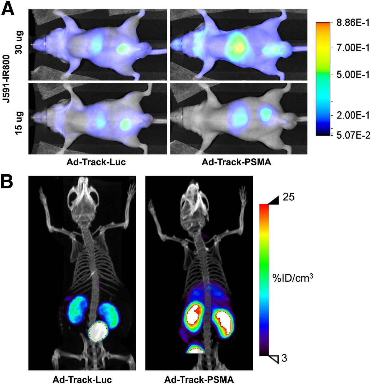
  • FIGURE 4.
    • Download figure
    • Open in new tab
    • Download powerpoint
    FIGURE 4.

    Monitoring hepatic infection and transgene expression by imaging PSMA in a murine model. Athymic nude mice were intravenously administered adenovirus expressing PSMA or negative control gene (luciferase [Luc]). Seventy-two hours after injection of adenovirus, animals were imaged and specific uptake was observed in liver of animals by optical (A) and SPECT (B) imaging. SPECT images were obtained 15 min after injection of radiotracer, with similar levels of radioactivity in liver at 4 h after injection (Supplemental Fig. 4).

Tables

  • Figures
  • Additional Files
    • View popup
    TABLE 1

    Description of Reporter Adenoviruses and Respective Imaging Modalities Used for Detection of Reporter Genes

    VirusReporter geneImaging modality
    Ad-Track-PSMAProstate-specific membrane antigenOptical, PET, SPECT
    Ad-Track-hNISHuman sodium iodide symporterPET, SPECT
    Ad-HSV-sr39tkHerpes simplex virus mutant thymidine kinasePET

Additional Files

  • Figures
  • Tables
  • Supplemental Data

    Files in this Data Supplement:

    • Supplemental Data
PreviousNext
Back to top

In this issue

Journal of Nuclear Medicine: 55 (5)
Journal of Nuclear Medicine
Vol. 55, Issue 5
May 1, 2014
  • Table of Contents
  • Table of Contents (PDF)
  • About the Cover
  • Index by author
Print
Download PDF
Article Alerts
Sign In to Email Alerts with your Email Address
Email Article

Thank you for your interest in spreading the word on Journal of Nuclear Medicine.

NOTE: We only request your email address so that the person you are recommending the page to knows that you wanted them to see it, and that it is not junk mail. We do not capture any email address.

Enter multiple addresses on separate lines or separate them with commas.
Evaluation of Prostate-Specific Membrane Antigen as an Imaging Reporter
(Your Name) has sent you a message from Journal of Nuclear Medicine
(Your Name) thought you would like to see the Journal of Nuclear Medicine web site.
Citation Tools
Evaluation of Prostate-Specific Membrane Antigen as an Imaging Reporter
Mark A. Castanares, Amarnath Mukherjee, Wasim H. Chowdhury, Minzhi Liu, Ying Chen, Ronnie C. Mease, Yuchuan Wang, Ronald Rodriguez, Shawn E. Lupold, Martin G. Pomper
Journal of Nuclear Medicine May 2014, 55 (5) 805-811; DOI: 10.2967/jnumed.113.134031

Citation Manager Formats

  • BibTeX
  • Bookends
  • EasyBib
  • EndNote (tagged)
  • EndNote 8 (xml)
  • Medlars
  • Mendeley
  • Papers
  • RefWorks Tagged
  • Ref Manager
  • RIS
  • Zotero
Share
Evaluation of Prostate-Specific Membrane Antigen as an Imaging Reporter
Mark A. Castanares, Amarnath Mukherjee, Wasim H. Chowdhury, Minzhi Liu, Ying Chen, Ronnie C. Mease, Yuchuan Wang, Ronald Rodriguez, Shawn E. Lupold, Martin G. Pomper
Journal of Nuclear Medicine May 2014, 55 (5) 805-811; DOI: 10.2967/jnumed.113.134031
Twitter logo Facebook logo LinkedIn logo Mendeley logo
  • Tweet Widget
  • Facebook Like
  • Google Plus One
Bookmark this article

Jump to section

  • Article
    • Abstract
    • MATERIALS AND METHODS
    • RESULTS
    • DISCUSSION
    • CONCLUSION
    • DISCLOSURE
    • Acknowledgments
    • Footnotes
    • REFERENCES
  • Figures & Data
  • Supplemental
  • Info & Metrics
  • PDF

Related Articles

  • This Month in JNM
  • PubMed
  • Google Scholar

Cited By...

  • Approaches to Imaging Immune Activation Using PET
  • Intracellular Vesicle Entrapment of Nanobubble Ultrasound Contrast Agents Targeted to PSMA Promotes Prolonged Enhancement and Stability In Vivo and In Vitro
  • Imaging of T-cell Responses in the Context of Cancer Immunotherapy
  • Molecular Imaging with Reporter Genes: Has Its Promise Been Delivered?
  • Imaging CAR T cell therapy with PSMA-targeted positron emission tomography
  • Real Time Ultrasound Molecular Imaging of Prostate Cancer with PSMA-targeted Nanobubbles
  • AEG-1 Promoter-Mediated Imaging of Prostate Cancer
  • Google Scholar

More in this TOC Section

  • PET/MRI of Hypoxic Atherosclerosis Using 64Cu-ATSM in a Rabbit Model
  • Tumor Uptake of Anti-CD20 Fabs Depends on Tumor Perfusion
  • How Sensitive Is the Upper Gastrointestinal Tract to 90Y Radioembolization? A Histologic and Dosimetric Analysis in a Porcine Model
Show more Basic Science Investigations

Similar Articles

Keywords

  • PSMA
  • molecular-genetic imaging
  • DCFPyL
  • Positron Emission Tomography
  • optical imaging
SNMMI

© 2025 SNMMI

Powered by HighWire