


Article Figures & Data
Figures
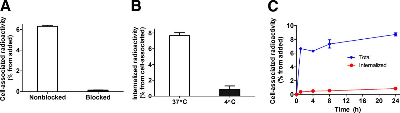
- FIGURE 1.
(A) In vitro binding specificity of 111In-DOTA-Z09591 to glioma U-87 MG cell line. In blocking group, receptors were presaturated by 200-fold excess of nonlabeled DOTA-Z09591. (B) Internalization of 111In-DOTA-Z09591 by glioma U-87 MG cells after 4-h incubation at 37°C and 4°C. (C) Internalization of 111In-DOTA-Z09591 by glioma U-87 MG cells during continuous incubation. Cells were incubated with conjugate (0.5 nM) at 37°C. Data presented as mean values from 3 cell dishes ± SD. Error bars might be not seen because they are smaller than symbols.
- FIGURE 2.
Imaging of PDFGRβ expression in U-87 MG xenograft (arrow) in BALB/C nu/nu mouse using small-animal SPECT/CT (maximum-intensity projection). Image was acquired at 3 h after injection. SPECT scale, 0.02–0.04; CT scale, 400–2000 HU.

Tables
- TABLE 1
Biodistribution of 111In-DOTA-Z09591 at 2 Hours After Injection in BALB/C nu/nu Mice Bearing U-87 MG Xenografts
Total injected dose (μg) Organ 0.1 0.5 1 70 Blood 0.34 ± 0.05 0.35 ± 0.07 0.26 ± 0.04 0.14 ± 0.02 Lung 0.8 ± 0.2 0.72 ± 0.08 0.7 ± 0.2 0.21 ± 0.02 Liver 0.99 ± 0.07 *† 0.84 ± 0.07 0.8 ± 0.1 0.39 ± 0.05 Spleen 1.8 ± 0.4 2.0 ± 0.3 1.6 ± 0.2 0.25 ± 0.06 Stomach 1.1 ± 0.3 1.0 ± 0.2 0.8 ± 0.2 0.19 ± 0.02 Colon 1.3 ± 0.3 1.5 ± 0.2 1.1 ± 0.4 0.23 ± 0.05 Kidney 288 ± 15 267 ± 40 256 ± 19 310 ± 32 Tumor 5.6 ± 1.1 6.5 ± 0.9 7.2 ± 2.4 0.89 ± 0.04 Muscle 0.4 ± 0.1 0.4 ± 0.1 0.3 ± 0.1 0.07 ± 0.02 Bone 0.6 ± 0.1 0.8 ± 0.2 0.6 ± 0.1 0.14 ± 0.05 GI tract‡ 1.2 ± 0.2 1.7 ± 0.7 1.2 ± 0.2 0.50 ± 0.13 ↵* Significant difference (P < 0.05) between doses 0.1 and 0.5 μg.
↵† Significant difference (P < 0.05) between doses 0.1 and 1 μg.
↵‡ Data for GI tract are presented as %ID per whole sample.
Uptake in all organs and tissues, except kidneys, after injection of 70 μg of 111In-DOTA-Z09591 was significantly lower than after injection of any other dose. Data are presented as average %ID/g and SD for 4 mice.
- TABLE 2
Influence of Coinjection of DOTA-Z09591 on Biodistribution of Anti-HER2 111In-DOTA-Z02395 at 2 Hours After Injection in BALB/C nu/nu Mice
Organ 111In-DOTA-Z02395 (1 μg) 111In-DOTA-Z02395 (1 μg) + 70 μg of DOTA-Z09591 111In-DOTA-Z02395 (70 μg) Blood 1.5 ± 0.4 1.2 ± 0.1 1.0 ± 0.2 Lung 1.8 ± 0.3 2.1 ± 0.5 1.8 ± 0.2 Liver 1.5 ± 0.3 1.7 ± 0.1 1.2 ± 0.1 Spleen 0.7 ± 0.1 0.8 ± 0.2 0.7 ± 0.1 Stomach 0.9 ± 0.1 1.3 ± 0.2* 0.9 ± 0.2 Colon 0.9 ± 0.1 1.4 ± 0.3* 0.83 ± 0.06 Kidney 238 ± 32 271 ± 21 215 ± 35 Muscle 0.47 ± 0.08 0.49 ± 0.08 0.43 ± 0.06 Bone 0.65 ± 0.08 0.77 ± 0.09 0.6 ± 0.1 ↵* Significant difference (P < 0.05) between control and coinjection groups.
Data are presented as average %ID/g and SD for 4 mice.
- TABLE 3
Tumor-to-Organ Ratios of 111In-DOTA-Z09591 Affibody Molecule (2 Hours After Injection) in BALB/C nu/nu Mice Bearing U-87 MG Xenografts
Dose (μg) Organ 0.1 0.5 1 Blood 17 ± 3 19 ± 5 28 ± 14 Lung 7 ± 2 9 ± 2 12 ± 4 Liver 5.6 ± 0.7 8 ± 2 9 ± 4 Spleen 3.2 ± 0.7 3.3 ± 0.5 5 ± 2 Stomach 5 ± 1* 7 ± 2 10 ± 3 Colon 4.2 ± 0.7* 4 ± 1 6 ± 1 Muscle 13 ± 3* 17 ± 7 28 ± 7 Bone 8.8 ± 0.8 9 ± 3 13 ± 4 ↵* Significant difference (P < 0.05) between doses 0.1 and 1 μg.
Data are presented as average of 4 animals and SD.
- TABLE 4
Biodistribution of 111In-DOTA-Z09591 (Injected Dose 1 μg) at 1, 2, and 4 Hours After Injection in BALB/C nu/nu Mice Bearing U-87 MG Xenografts
Organ 1 h 2 h 4 h Blood 0.53 ± 0.04*† 0.35 ± 0.06‡ 0.20 ± 0.07 Lung 1.2 ± 0.2† 0.8 ± 0.3‡ 0.3 ± 0.1 Liver 0.90 ± 0.04* 0.80 ± 0.07 0.6 ± 0.2 Spleen 2.3 ± 0.4† 1.7 ± 0.3‡ 1.0 ± 0.2 Stomach 1.3 ± 0.1*† 1.0 ± 0.2‡ 0.5 ± 0.1 Colon 1.8 ± 0.4† 1.3 ± 0.2‡ 0.6 ± 0.2 Kidney 210 ± 26* 249 ± 15 232 ± 66 Tumor 5.7 ± 0.6† 5.9 ± 1.7 4.0 ± 0.8 Muscle 0.61 ± 0.09*† 0.34 ± 0.03‡ 0.15 ± 0.05 Bone 0.79 ± 0.03*† 0.56 ± 0.06‡ 0.25 ± 0.03 GI tract§ 1.9 ± 0.2*† 1.2 ± 0.2‡ 0.8 ± 0.2 ↵* Significant difference (P < 0.05) between 1 and 2 h after injection.
↵† Significant difference (P < 0.05) between 1 and 4 h after injection.
↵‡ Significant difference (P < 0.05) between 2 and 4 h after injection.
↵§ Data for GI tract are presented as %ID per whole sample.
There was no significant difference between data for 2-h time point and data reported for injected dose of 1 μg in Table 1. Data are presented as an average % ID/g and SD for 4 mice.
- TABLE 5
Tumor-to-Organ Ratios for 111In-DOTA-Z09591 Affibody Molecule (Injected Dose 1 μg) at 1, 2, and 4 Hours After Injection in BALB/C nu/nu Mice Bearing U-87 MG Xenografts
Organ 1 h 2 h 4 h Blood 11 ± 1*† 17 ± 3 21 ± 6 Lung 5 ± 1† 8 ± 3 12 ± 2 Liver 6 ± 1† 8 ± 3 7 ± 2 Spleen 2.6 ± 0.6† 3.5 ± 0.7 4.2 ± 0.4 Stomach 4.4 ± 0.5*† 6.0 ± 0.7 8 ± 1 Colon 3.3 ± 0.7*† 4.6 ± 0.7‡ 6.6 ± 0.5 Muscle 10 ± 2*† 18 ± 6 30 ± 14 Bone 7 ± 1† 11 ± 3 16 ± 3
Supplemental Data
Files in this Data Supplement:


