


Article Figures & Data
Figures
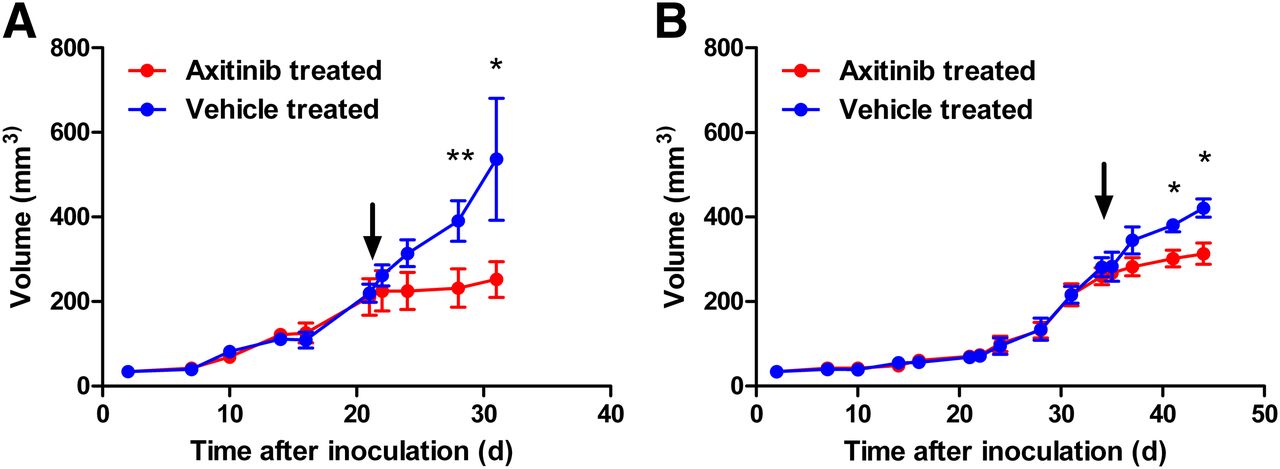
- FIGURE 1.
Growth kinetics of U87-MG tumors (A) and MDA-MB-231 tumors (B) in vehicle-treated animals and axitinib-treated animals (volume is reported in mm3 [mean ± SD; n = 4]). Arrows denote day 0, 1 d before initiation of therapy regimen. *P < 0.05. **P < 0.01.
- FIGURE 2.
Uptake of 18F-FDG in U87-MG tumors (A; n = 7) and MDA-MB-231 tumors (B; n = 4) in vehicle-treated animals and axitinib-treated animals, normalized to day 0 (% normalized uptake from day 0 [mean ± SD]). **P < 0.01.
- FIGURE 3.
Uptake of 18F-FLT in U87-MG tumors (A; n = 7) and MDA-MB-231 tumors (B; n = 4) in vehicle-treated animals and axitinib-treated animals, normalized to day 0 (% normalized uptake from day 0 [mean ± SD]). *P < 0.05. **P < 0.01.
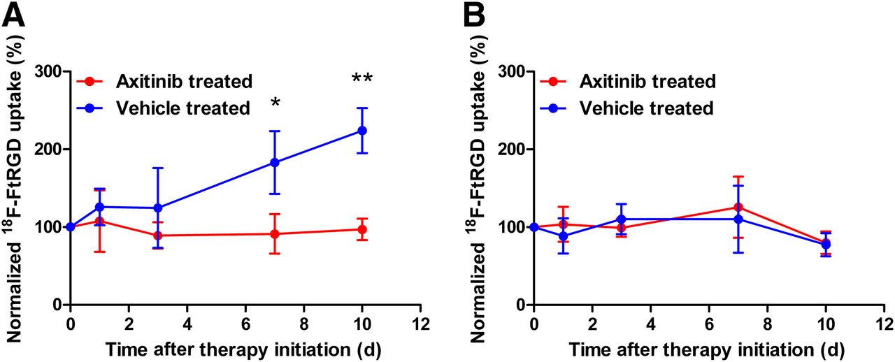
- FIGURE 4.
Uptake of 18F-FtRGD in U87-MG tumors (A; n = 4) and MDA-MB-231 tumors (B; n = 7) in vehicle-treated animals and axitinib-treated animals, normalized to day 0 (% normalized uptake from day 0 [mean ± SD]). *P < 0.05. **P < 0.01.
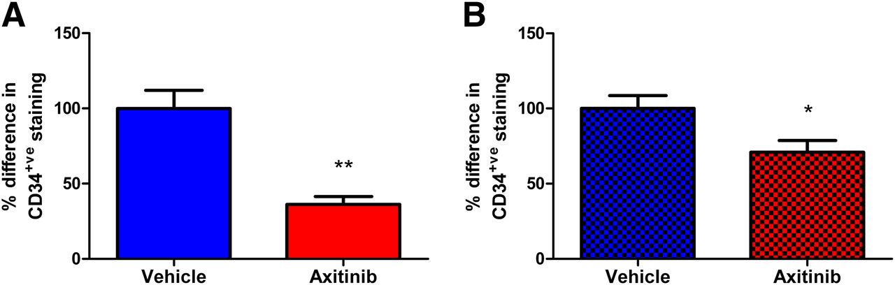
- FIGURE 5.
Angiogenesis (represented by MVD) in U87-MG and MDA-MB-231 tumors. MVD was significantly lower in both U87-MG tumors (A; n = 10) and MDA-MB-231 tumors (B; n = 10) from axitinib-treated mice than in tumors from vehicle-treated mice 10 d after treatment initiation. Data are reported as percentage difference in CD34-positive (CD34+ve) staining per square millimeter, normalized to average for vehicle-treated specimens. *P < 0.05. **P < 0.01.
- FIGURE 6.
Representative sections were taken from both tumor types 10 d after initiation of therapy regimen. Representative sections from axitinib-treated U87-MG tumors (A) and MDA-MB-231 tumors (C) were analyzed for MVD after staining for endothelial cells with CD34 (bars = 100 μm) and compared with sections from vehicle-treated U87-MG tumors (B) and MDA-MB-231 tumors (D).
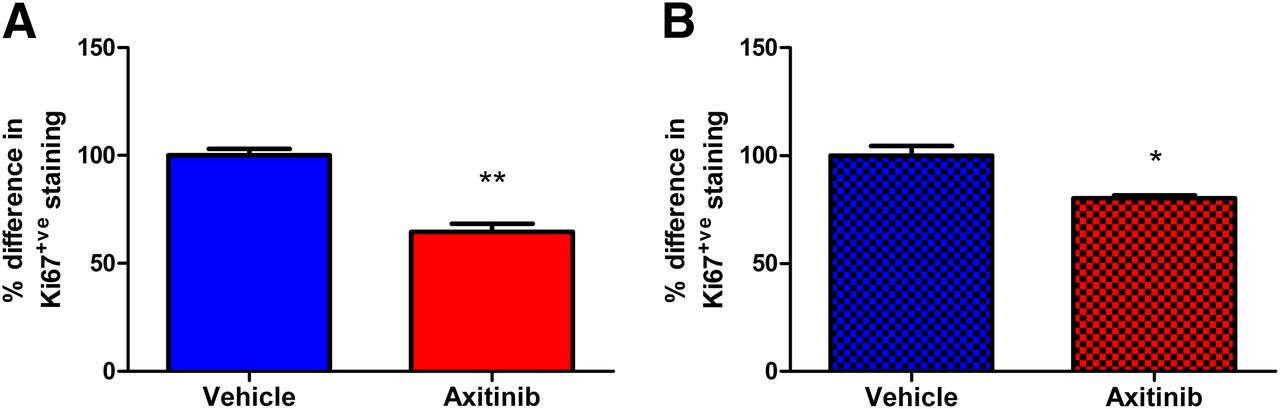
- FIGURE 7.
Rate of proliferation (Ki67) of U87-MG and MDA-MB-231 tumors. Ki67 levels were significantly lower in both U87-MG tumors (A; n = 6) and MDA-MB-231 tumors (B; n = 6) from axitinib-treated mice than in tumors from vehicle-treated mice 10 d after treatment initiation. Data are reported as percentage difference in Ki67-positive (Ki67+ve) cells per square millimeter, normalized to average for vehicle-treated specimens. *P < 0.05. **P < 0.01.


Tables
Uptake (mean ± SD %ID/g) of 18F-FDG in: U87-MG tumors MDA-MB-231 tumors Days after treatment Axitinib treated Vehicle treated Axitinib treated Vehicle treated 0 3.03 ± 0.46 2.99 ± 0.31 1.84 ± 0.10 2.05 ± 0.16 1 3.07 ± 0.62 3.24 ± 0.33 1.93 ± 0.19 2.09 ± 0.17 3 3.76 ± 1.10 4.29 ± 0.60 1.98 ± 0.12 2.25 ± 0.19 7 3.66 ± 0.83 4.94 ± 1.20 1.79 ± 0.05 2.25 ± 0.20 10 3.68 ± 0.41 5.26 ± 0.48 1.54 ± 0.07 2.22 ± 0.21 Uptake (mean ± SD %ID/g) of 18F-FLT in: U87-MG tumors MDA-MB-231 tumors Days after treatment Axitinib treated Vehicle treated Axitinib treated Vehicle treated 0 3.02 ± 1.18 2.82 ± 0.69 3.37 ± 0.33 2.90 ± 0.46 1 3.47 ± 1.23 4.3 ± 0.14 3.28 ± 0.29 3.08 ± 0.53 3 3.37 ± 0.84 4.9 ± 0.68 2.54 ± 0.24 2.89 ± 0.83 7 2.65 ± 0.60 4.14 ± 0.56 1.99 ± 0.68 3.29 ± 0.53 10 3.34 ± 0.60 4.68 ± 0.89 1.84 ± 0.50 3.30 ± 0.55 Uptake (mean ± SD %ID/g) of 18F-FtRGD in: U87-MG tumors MDA-MB-231 tumors Days after treatment Axitinib treated Vehicle treated Axitinib treated Vehicle treated 0 1.62 ± 0.32 1.50 ± 0.25 1.59 ± 0.72 1.58 ± 0.41 1 1.66 ± 0.23 1.74 ± 0.29 1.69 ± 0.17 1.71 ± 0.14 3 1.37 ± 0.29 1.61 ± 0.43 1.07 ± 0.28 1.36 ± 0.06 7 1.43 ± 0.22 2.40 ± 0.56 2.01 ± 0.54 1.90 ± 0.59 10 1.65 ± 0.25 3.33 ± 0.63 1.28 ± 0.06 1.50 ± 0.03


