


Article Figures & Data
Figures
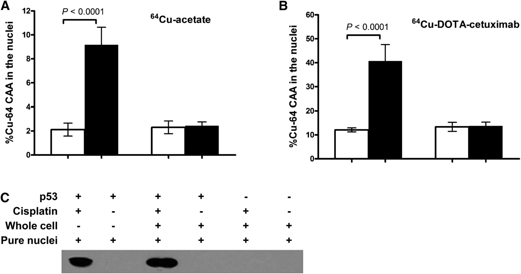
- FIGURE 1.
Effect of cisplatin on nuclear uptake of 64Cu in p53 wild-type and null HCT116 cell lines. HCT116 cells were incubated with or without 40 μM cisplatin before incubation with 64Cu-acetate (A) or 64Cu-DOTA-cetuximab (B) for another 20 h. (C) Expression level of p53 in HCT116 cells cultured with 64Cu-DOTA-cetuximab, either with or without prior incubation with cisplatin, was detected by Western blot. Amount of p53 detected in both whole cells and pure nuclei are shown.
- FIGURE 2.
(A) Biodistribution of 64Cu-DOTA-cetuximab in HCT116 +/+ tumor–bearing nude mice. Data are presented as percentage injected dose per gram (%ID/g) ± SD (n = 5 for each time point). (B) One milligram of unlabeled cetuximab was injected to each mouse 24 h before administration of radiotracer to block specific uptake of 64Cu-DOTA-cetuximab (P < 0.001). (C) Mice bilaterally implanted with both HCT116 +/+ and HCT116 −/− tumors showed tumor uptake to be similar (P = 0.07 at 24 h after injection, P = 0.8 at 72 h after injection).
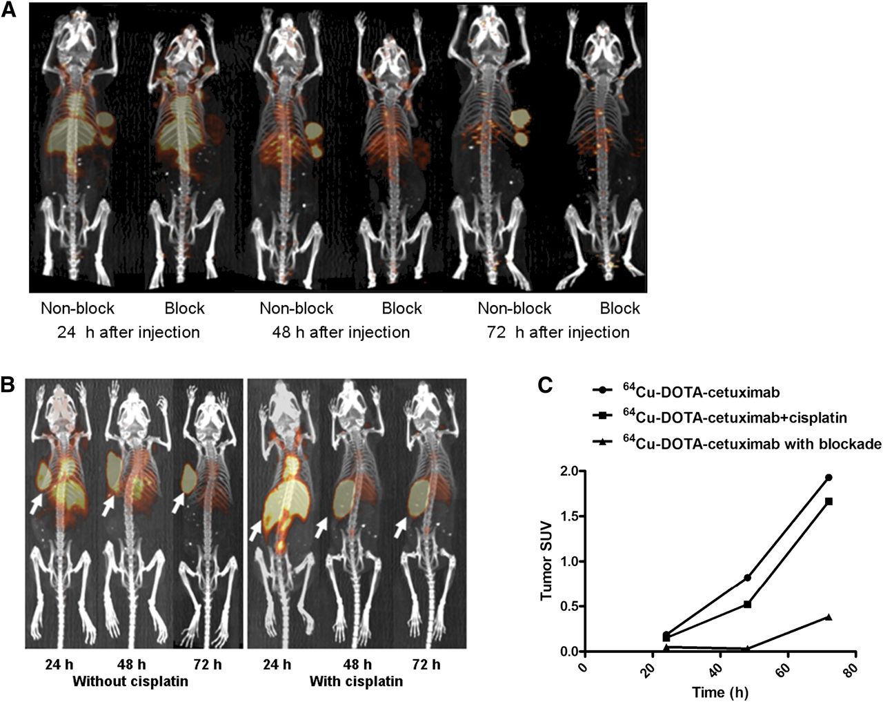
- FIGURE 3.
(A) Small-animal PET/CT projection images of HCT116 +/+ tumor–bearing nude mice at 24, 48, and 72 h after injection of 64Cu-DOTA-cetuximab, with or without 24 h preinjection of excess amount of unlabeled cetuximab. (B) Small-animal PET/CT projection images of HCT116 +/+ tumor–bearing nude mice at 24, 48, and 72 h after injection of 64Cu-DOTA-cetuximab, with or without 24 h preinjection of cisplatin (5 mg/kg). (C) SUVs were determined from quantifying activity in regions of interest from PET images of HCT116 +/+ tumor–bearing nude mice.
- FIGURE 4.
Radioimmunotherapy experiments in HCT116 +/+ (A) and HCT116 −/− (B) tumor–bearing nude mice. Comparison of average tumor growth in treated and control groups was shown. Tumor growth in individual mice of treated and control group was included in supplemental figures. Dash arrows = days when cisplatin was given; solid arrows = days when radioimmunotherapy was given.
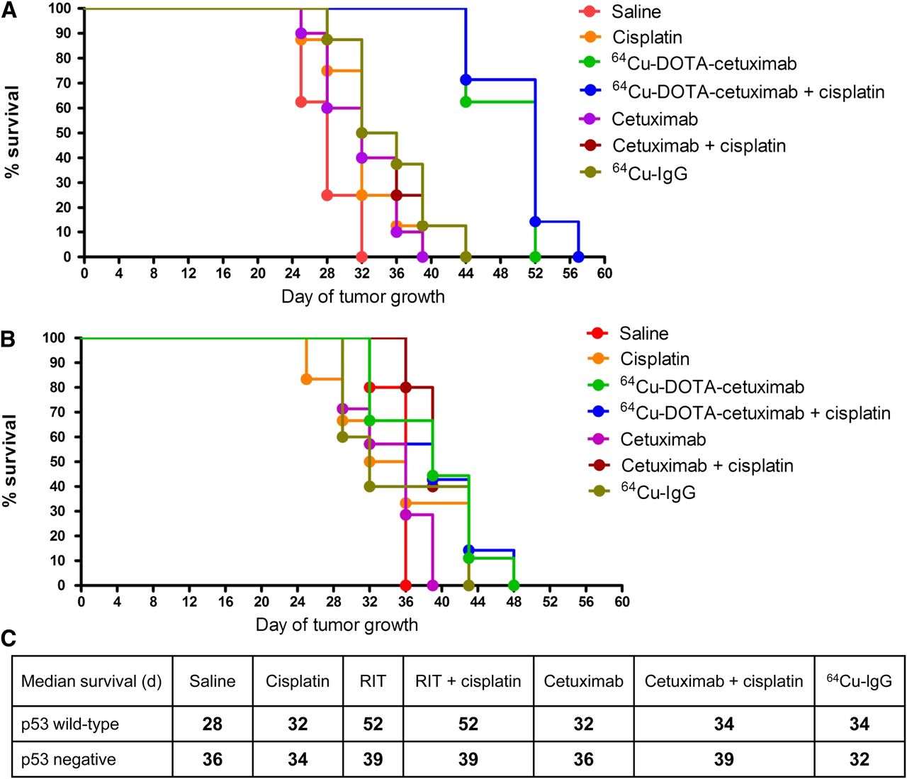
- FIGURE 5.
Effect of p53 status in p53 wild-type (A) and p53-null (B) tumors on survival of HCT116 tumor–bearing nude mice in treated and control groups. Estimated survival distribution function was generated using Kaplan–Meier time-to-death analysis. Median survivals of all treated and control groups with HCT116 +/+ and HCT116 −/− tumors are summarized in C. ***P < 0.001 vs. radioimmunotherapy group. **P < 0.01 vs. radioimmunotherapy group. RIT = radioimmunotherapy.


Tables
Group Injection 1 Intraperitoneal: 150 μL of saline 2 Intraperitoneal: cisplatin (5 mg/kg) in 150 μL of saline 3 Intravenous: 22.2 MBq (600 μCi) of 64Cu-DOTA-cetuximab in 150 μL of saline 4 Intraperitoneal: cisplatin (5 mg/kg ) in 150 μL of saline, followed by intravenous injection of 22.2 MBq (600 μCi) of 64Cu-DOTA-cetuximab in 150 μL of saline after 24 h 5 Intravenous: 50 μg of cetuximab in 150 μL of saline 6 Intraperitoneal: cisplatin (5 mg/kg) in 150 μL of saline, followed by intravenous injection of 50 μg of cetuximab in 150 μL of saline after 24 h 7 Intravenous: 22.2 MBq (600 μCi) of 64Cu-IgG in 150 μL of saline Mice received 2 treatments 1 wk apart for each group.
Dose Organ mGy/MBq rad/mCi Lower large intestine wall 0.14 0.525 Small intestine 0.030 0.111 Stomach wall 0.044 0.163 Upper large intestine wall 0.083 0.308 Kidneys 0.043 0.160 Liver 0.12 0.434 Lungs 0.028 0.103 Pancreas 0.030 0.111 Red marrow 0.046 0.171 Spleen 0.066 0.244 Osteogenic cells 0.11 0.414 Heart wall 0.085 0.315 Urinary bladder wall 0.026 0.096 Total body 0.032 0.118 Effective dose was 0.050 mSv/MBq (0.183 [rem/mCi]).
Supplemental Data
Files in this Data Supplement:


