
Main menu

  • Home
  • Content
    • Current
    • Ahead of print
    • Past Issues
    • JNM Supplement
    • SNMMI Annual Meeting Abstracts
    • Continuing Education
    • JNM Podcasts
  • Subscriptions
    • Subscribers
    • Institutional and Non-member
    • Rates
    • Journal Claims
    • Corporate & Special Sales
  • Authors
    • Submit to JNM
    • Information for Authors
    • Assignment of Copyright
    • AQARA requirements
  • Info
    • Reviewers
    • Permissions
    • Advertisers
  • About
    • About Us
    • Editorial Board
    • Contact Information
  • More
    • Alerts
    • Feedback
    • Help
    • SNMMI Journals
  • SNMMI
    • JNM
    • JNMT
    • SNMMI Journals
    • SNMMI

User menu

  • Subscribe
  • My alerts
  • Log in
  • Log out
  • My Cart

Search

  • Advanced search
Journal of Nuclear Medicine
  • SNMMI
    • JNM
    • JNMT
    • SNMMI Journals
    • SNMMI
  • Subscribe
  • My alerts
  • Log in
  • Log out
  • My Cart
Journal of Nuclear Medicine

Advanced Search

  • Home
  • Content
    • Current
    • Ahead of print
    • Past Issues
    • JNM Supplement
    • SNMMI Annual Meeting Abstracts
    • Continuing Education
    • JNM Podcasts
  • Subscriptions
    • Subscribers
    • Institutional and Non-member
    • Rates
    • Journal Claims
    • Corporate & Special Sales
  • Authors
    • Submit to JNM
    • Information for Authors
    • Assignment of Copyright
    • AQARA requirements
  • Info
    • Reviewers
    • Permissions
    • Advertisers
  • About
    • About Us
    • Editorial Board
    • Contact Information
  • More
    • Alerts
    • Feedback
    • Help
    • SNMMI Journals
  • View or Listen to JNM Podcast
  • Visit JNM on Facebook
  • Join JNM on LinkedIn
  • Follow JNM on Twitter
  • Subscribe to our RSS feeds
Research ArticleClinical Investigations

Longitudinal Progression of Cognitive Decline Correlates with Changes in the Spatial Pattern of Brain 18F-FDG PET

Sepideh Shokouhi, Daniel Claassen, Hakmook Kang, Zhaohua Ding, Baxter Rogers, Arabinda Mishra, William R. Riddle and for The Alzheimer’s Disease Neuroimaging Initiative
Journal of Nuclear Medicine September 2013, 54 (9) 1564-1569; DOI: https://doi.org/10.2967/jnumed.112.116137
Sepideh Shokouhi
1Department of Radiology and Radiological Sciences, Vanderbilt University Institute of Imaging Science, Nashville, Tennessee
  • Find this author on Google Scholar
  • Find this author on PubMed
  • Search for this author on this site
Daniel Claassen
2Department of Neurology, Vanderbilt University, Nashville, Tennessee; and
  • Find this author on Google Scholar
  • Find this author on PubMed
  • Search for this author on this site
Hakmook Kang
3Department of Biostatistics, Vanderbilt University, , Nashville, Tennessee
  • Find this author on Google Scholar
  • Find this author on PubMed
  • Search for this author on this site
Zhaohua Ding
1Department of Radiology and Radiological Sciences, Vanderbilt University Institute of Imaging Science, Nashville, Tennessee
  • Find this author on Google Scholar
  • Find this author on PubMed
  • Search for this author on this site
Baxter Rogers
1Department of Radiology and Radiological Sciences, Vanderbilt University Institute of Imaging Science, Nashville, Tennessee
  • Find this author on Google Scholar
  • Find this author on PubMed
  • Search for this author on this site
Arabinda Mishra
1Department of Radiology and Radiological Sciences, Vanderbilt University Institute of Imaging Science, Nashville, Tennessee
  • Find this author on Google Scholar
  • Find this author on PubMed
  • Search for this author on this site
William R. Riddle
1Department of Radiology and Radiological Sciences, Vanderbilt University Institute of Imaging Science, Nashville, Tennessee
  • Find this author on Google Scholar
  • Find this author on PubMed
  • Search for this author on this site
  • Article
  • Figures & Data
  • Info & Metrics
  • PDF
Loading

Article Figures & Data

Figures

  • Tables
  • FIGURE 1.
    • Download figure
    • Open in new tab
    • Download powerpoint
    FIGURE 1.

    Illustration of regional binary masks warped from reference brain to ADNI image volume with nonrigid deformation field. Regions include cerebellum (1); midbrain (2); pons (3); occipital lobe (4); cingulate cortex (5); sublobar region (6) including corpus callosum, caudate, putamen, globus pallidus, thalamus, and right and left frontal lobes (7, 8); right and left parietal lobes (9, 10); and right and left temporal lobes (11, 12).
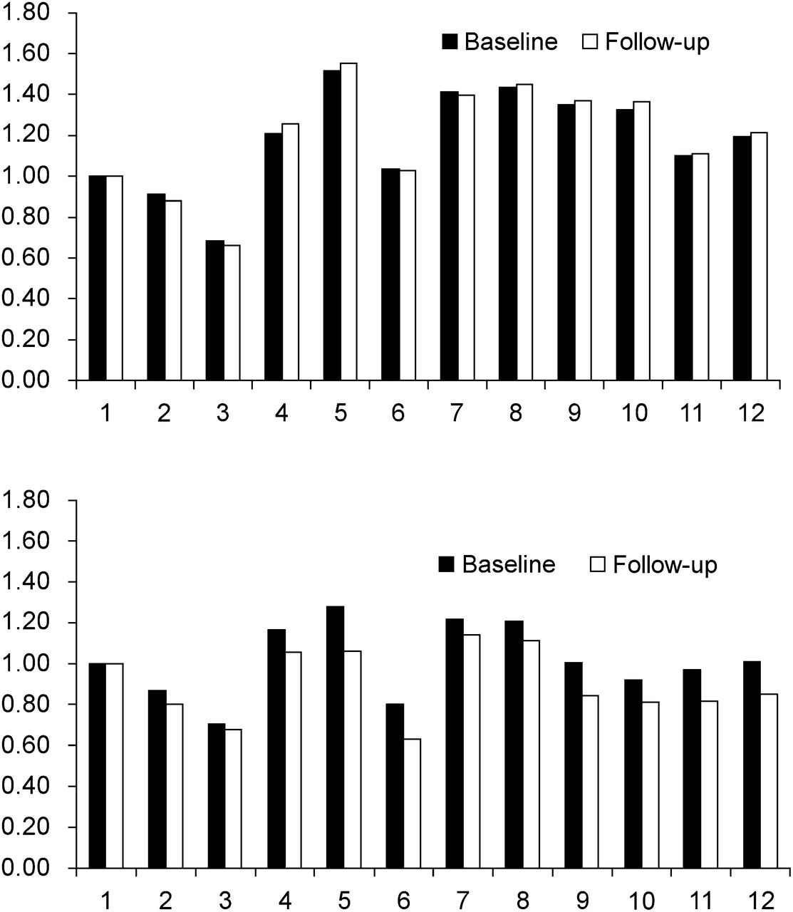
  • FIGURE 2.
    • Download figure
    • Open in new tab
    • Download powerpoint
    FIGURE 2.

    Comparison between 18F-FDG vectors at baseline and follow-up in 2 subjects. Lower figure shows subject with low correlation, and upper figure shows subject with high correlation.
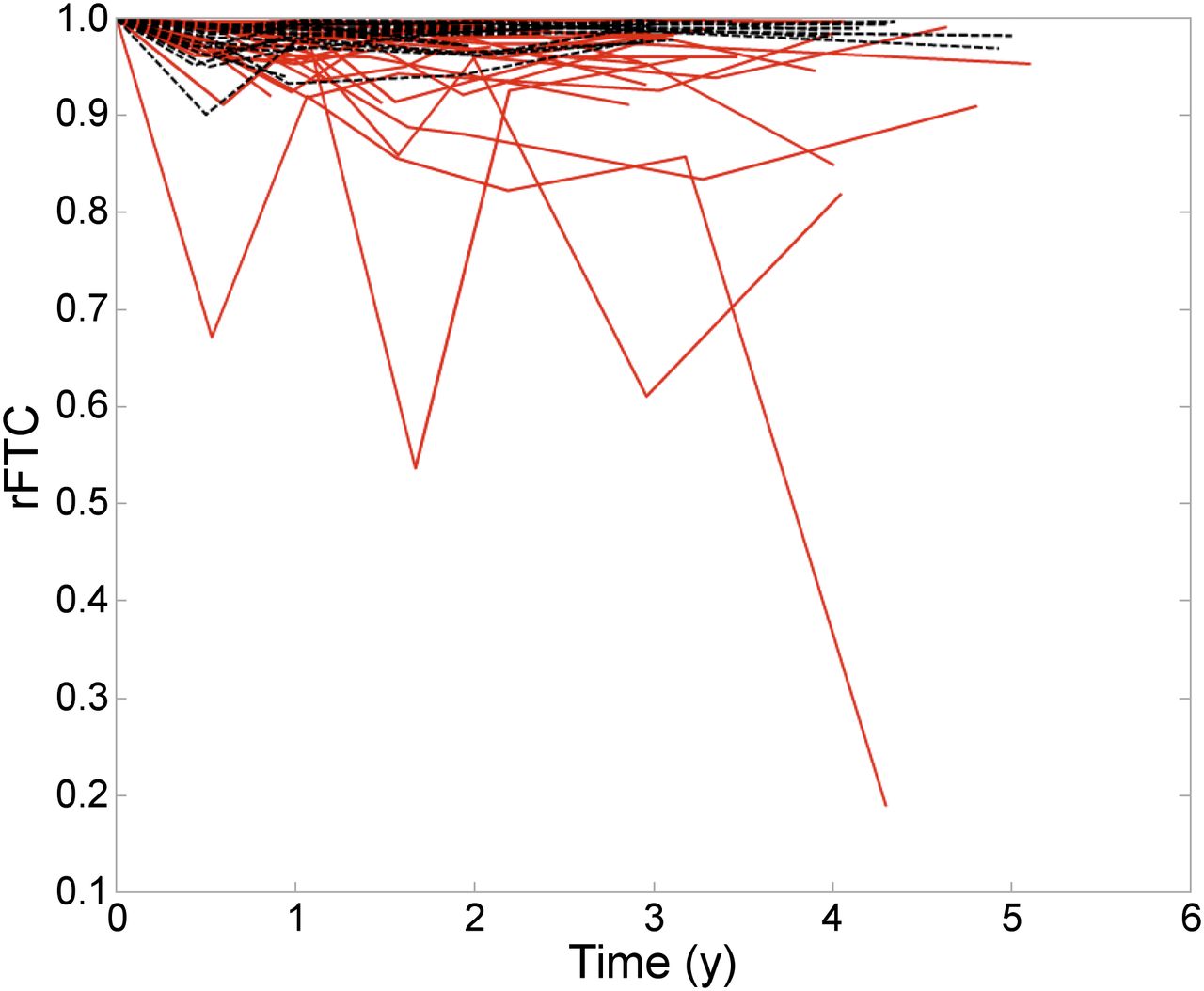
  • FIGURE 3.
    • Download figure
    • Open in new tab
    • Download powerpoint
    FIGURE 3.

    Comparison between normal (black dashed line) and MCI (red solid line) subjects with respect to their rFTC(t).
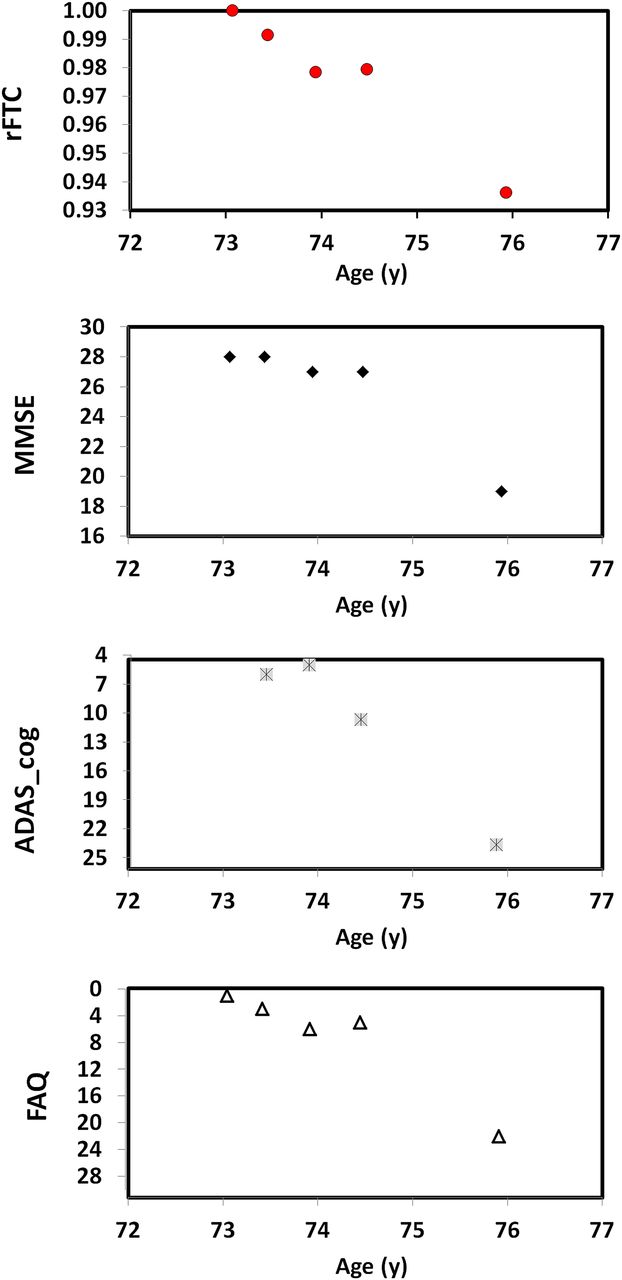
  • FIGURE 4.
    • Download figure
    • Open in new tab
    • Download powerpoint
    FIGURE 4.

    Changes in rFTC, MMSE, FAQ, and ADAS_cog values with age in MCI subject who has large decline in cognitive scores.
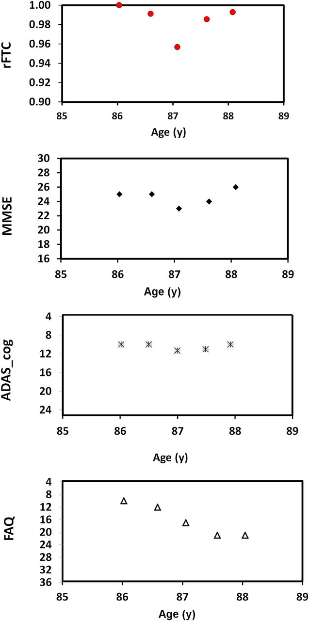
  • FIGURE 5.
    • Download figure
    • Open in new tab
    • Download powerpoint
    FIGURE 5.

    Changes in rFTC, MMSE, FAQ, and ADAS_cog values with age in MCI subject who has moderate decline in cognitive scores.

Tables

  • Figures
    • View popup
    TABLE 1

    Clinical Data of ADNI Subjects Used for This Study

    CharacteristicMCINormal
    No. of subjects2724
     Female1311
     Male1413
    Average age at baseline74.6 ± 7.274.53 ± 5.7
    APOE-ε4176
    Baseline ADAS_cog11 ± 4.76 ± 2.6
    Baseline FAQ3.2 ± 2.80.1 ± 0.6
    Baseline MMSE27.4 ± 1.429.2 ± 0.9
    • View popup
    TABLE 2

    Outcome Results of 2 Mixed-Effects Models

    ModelFixed effectEstimateP
    1
     ADAS_cogtime:MCI1.60.0001
     rFTCtime:MCI−0.020.0001
     MMSEtime:MCI−0.820.0002
     FAQtime:MCI2.50.0001
    2
     ADAS_cogtime:APOE1.140.001
     rFTCtime:APOE−0.0140.008
     MMSEtime:APOE−0.440.007
     FAQtime:APOE1.320.0001
    • Both models were performed for rFTC(t), MMSE(t), ADAS-cog(t), and FAQ(t) as outcome variables. With the first model (time:MCI), we see how changes in these variables over time differed between MCI and normal subjects. Second model (time:APOE) shows how changes in these variables over time differed between the group with positive APOE-ε4 and control group.

    • View popup
    TABLE 3

    Correlation Between rFTC and Cognitive Tests

    Method AMethod B
    VariableMedianPMedianP
    ADAS-cog/rFTC0.50.0110.810.0005
    MMSE/rFTC0.50.00390.640.00004
    FAQ/rFTC0.60.00160.40.0002
    • Wilcoxon rank-sum tests of Spearman rank correlation between rFTC and each of 3 cognitive tests (method A) and correlation between residuals of linear regressions on rFTC(t) and 3 cognitive tests (method B).

PreviousNext
Back to top

In this issue

Journal of Nuclear Medicine: 54 (9)
Journal of Nuclear Medicine
Vol. 54, Issue 9
September 1, 2013
  • Table of Contents
  • Table of Contents (PDF)
  • About the Cover
  • Index by author
Print
Download PDF
Article Alerts
Sign In to Email Alerts with your Email Address
Email Article

Thank you for your interest in spreading the word on Journal of Nuclear Medicine.

NOTE: We only request your email address so that the person you are recommending the page to knows that you wanted them to see it, and that it is not junk mail. We do not capture any email address.

Enter multiple addresses on separate lines or separate them with commas.
Longitudinal Progression of Cognitive Decline Correlates with Changes in the Spatial Pattern of Brain 18F-FDG PET
(Your Name) has sent you a message from Journal of Nuclear Medicine
(Your Name) thought you would like to see the Journal of Nuclear Medicine web site.
Citation Tools
Longitudinal Progression of Cognitive Decline Correlates with Changes in the Spatial Pattern of Brain 18F-FDG PET
Sepideh Shokouhi, Daniel Claassen, Hakmook Kang, Zhaohua Ding, Baxter Rogers, Arabinda Mishra, William R. Riddle, for The Alzheimer’s Disease Neuroimaging Initiative
Journal of Nuclear Medicine Sep 2013, 54 (9) 1564-1569; DOI: 10.2967/jnumed.112.116137

Citation Manager Formats

  • BibTeX
  • Bookends
  • EasyBib
  • EndNote (tagged)
  • EndNote 8 (xml)
  • Medlars
  • Mendeley
  • Papers
  • RefWorks Tagged
  • Ref Manager
  • RIS
  • Zotero
Share
Longitudinal Progression of Cognitive Decline Correlates with Changes in the Spatial Pattern of Brain 18F-FDG PET
Sepideh Shokouhi, Daniel Claassen, Hakmook Kang, Zhaohua Ding, Baxter Rogers, Arabinda Mishra, William R. Riddle, for The Alzheimer’s Disease Neuroimaging Initiative
Journal of Nuclear Medicine Sep 2013, 54 (9) 1564-1569; DOI: 10.2967/jnumed.112.116137
Twitter logo Facebook logo LinkedIn logo Mendeley logo
  • Tweet Widget
  • Facebook Like
  • Google Plus One
Bookmark this article

Jump to section

  • Article
    • Abstract
    • MATERIALS AND METHODS
    • RESULTS
    • DISCUSSION
    • CONCLUSION
    • DISCLOSURE
    • Acknowledgments
    • Footnotes
    • REFERENCES
  • Figures & Data
  • Info & Metrics
  • PDF

Related Articles

  • This Month in JNM
  • PubMed
  • Google Scholar

Cited By...

  • Insulin Resistance Predicts Medial Temporal Hypermetabolism in Mild Cognitive Impairment Conversion to Alzheimer Disease
  • Google Scholar

More in this TOC Section

  • Feasibility of Ultra-Low-Activity 18F-FDG PET/CT Imaging Using a Long–Axial-Field-of-View PET/CT System
  • Cardiac Presynaptic Sympathetic Nervous Function Evaluated by Cardiac PET in Patients with Chronotropic Incompetence Without Heart Failure
  • Validation and Evaluation of a Vendor-Provided Head Motion Correction Algorithm on the uMI Panorama PET/CT System
Show more Clinical Investigations

Similar Articles

Keywords

  • Alzheimer imaging
  • 18F-FDG PET
  • 18F-FDG time correlation
  • Mild cognitive impairment
  • longitudinal 18F-FDG imaging
SNMMI

© 2025 SNMMI

Powered by HighWire