


Article Figures & Data
Figures
- FIGURE 1.
Histologic (hematoxylin, erythrosine, and safran) and immunohistologic staining of VCAM-1 and Mac-2 expression in ApoE−/− mouse carotid vessels. Histochemistry and immunohistochemistry were performed on adjacent transverse sections of atherosclerotic lesions from ligated left carotid arteries (A–E), with magnifications centered on intimal and adventitial areas (F–J and K–O, respectively) and on nonligated left carotid arteries from sham-operated ApoE−/− animals (P–T) and on contralateral, right, carotid arteries (U–Y). Positive VCAM-1 and Mac-2 immunostaining was observed in atherosclerotic lesions developing at site of left carotid artery ligation but not in vessels from sham-operated animals and right carotid arteries. Specificity of immunostaining was assessed by performing control experiments in absence of VCAM-1– and Mac-2–specific primary antibodies (without VCAM-1 Ab and without MAC-2 Ab). Ab = antibody; HES = hematoxylin, erythrosine, and safran; w/o = without.*Vessel lumen.
- FIGURE 2.
Left-to-right carotid (A) and left carotid-to-blood (B) activity ratios of 99mTc-labeled peptidic sequences as determined by γ-well counting at 180 min after intravenous tracer injection. mis. = mismatch. *P < 0.05 vs. 99mTc-B2702p. †P < 0.05 vs. 99mTc-B2702p1 mismatch.
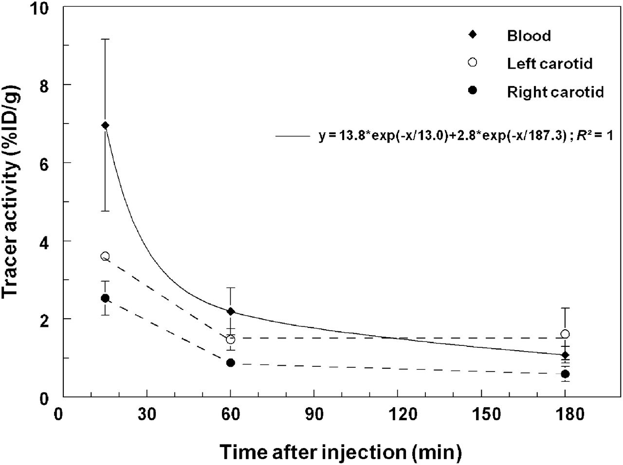
- FIGURE 3.
Time–activity curves of 99mTc-B2702p1 in blood and left and right carotid arteries at 15, 60, and 180 min after intravenous injection of tracer.
- FIGURE 4.
(A) Representative planar images of ApoE−/− mouse with left carotid artery ligation at 180 min after injection of 99mTc-B2702p1, 99mTc-B2702p1 mismatch, and 99mTc-B2702p; corresponding left-to-right carotid tracer activity ratios are indicated in parentheses. (B) 99mTc-B2702p1, 99mTc-B2702p1 mismatch, and 99mTc-B2702p left-to-right carotid activity ratios from in vivo planar image quantification. *P < 0.05 vs. 99mTc-B2702p. †P < 0.05 vs. 99mTc-B2702p1 mismatch. mis = mismatch.
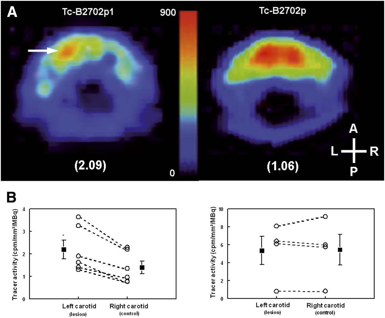
- FIGURE 5.
(A) Representative pinhole SPECT image of 99mTc-B2702p1 and 99mTc-B2702p activity at level of atherosclerotic lesion developing at site of left carotid artery ligation in ApoE−/− mouse. Corresponding left-to-right carotid tracer activity ratios are indicated in parentheses. (B) Left and right 99mTc-B2702p1 (left) and 99mTc-B2702p (right) carotid activities from in vivo pinhole SPECT image quantification. *P < 0.05 vs. right carotid tracer activity.
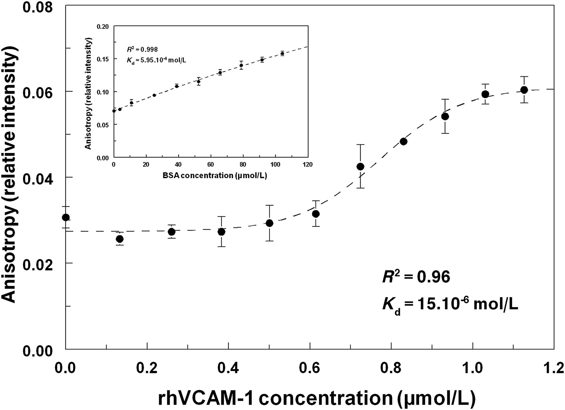
- FIGURE 6.
Anisotropy values from [F]-B2702p1 in presence of increasing concentrations of recombinant human VCAM-1. Results from control experiments performed in absence of VCAM-1 and in presence of nonspecific target bovine serum albumin are shown in inset.


Tables
- TABLE 1
Peptidic Sequence, Radiochemical Purity, and Urinary Stability of 99mTc-B2702p1-20 Peptides
Tracer Peptidic sequence RCP (%) Stability (%) 99mTc-B2702p H2N-HGR ENL RIA LRY-COOH 90 93 99mTc-B2702p1 H2N-HGR ANL RIL ARY-COOH 92 94 99mTc-B2702p1 mismatch H2N- HGL RAY IRA RNL-COOH 90 89 99mTc-B2702p2 H2N-HGR ENL AIL ARY-COOH 95 96 99mTc-B2702p3 H2N-HGR ENL RIL ARA-COOH 97 83 99mTc-B2702p4 H2N-HGR ENL RIL AAY-COOH 95 92 99mTc-B2702p5 H2N-HGR ENL RIL ARY-COOH 50 N/D 99mTc-B2702p6 H2N-HGR ENA RIL ARY-COOH 93 95 99mTc-B2702p7 H2N-HGA ENL RIL ARY-COOH 95 50 99mTc-B2702p8 H2N-HGR ENL RIA ARY-COOH 80 84 99mTc-B2702p9 H2N-HGR EAL RIL ARY-COOH 95 97 99mTc-B2702p10 H2N-HGR ENL RIL ARY-COOH 95 99 99mTc-B2702p11 H2N-HGA ENL RIA LRY-COOH 97 82 99mTc-B2702p12 H2N-HGR ANL RIA LRY-COOH 88 80 99mTc-B2702p13 H2N-HGR EAL RIA LRY-COOH 96 79 99mTc-B2702p14 H2N-HGR ENA RIA LRY-COOH 96 76 99mTc-B2702p15 H2N-HGR ENL AIA LRY-COOH 96 85 99mTc-B2702p16 H2N-HGR ENL RAA LRY-COOH 91 70 99mTc-B2702p17 H2N-HGR ENL RIA LAY-COOH 90 80 99mTc-B2702p18 H2N-HGR ENL RIA LRA-COOH 88 81 99mTc-B2702p19 H2N-HGR ANL RIL ARA-COOH 88 79 99mTc-B2702p20 H2N-HGR ANL RIL AAY-COOH 92 78 N/D = not determined; RCP = radiochemical purity.
- TABLE 2
Biodistribution of 99mTc-B2702p1-20 at 180 Minutes After Injection in ApoE−/− Mice with Left Carotid Artery Ligation
Tracer Heart Aorta Lung Liver Spleen Kidney Fat Muscle Blood Thyroid L. carotid R. carotid 99mTc-B2702p 1.5 ± 0.1 2.7 ± 0.4 3.9 ± 0.4 10.6 ± 2.6 2.2 ± 0.3 69.0 ± 13.0 1.0 ± 0.2 1.2 ± 0.2 5.7 ± 0.6 9.7 ± 3.0 2.4 ± 0.4 1.8 ± 0.3 99mTc-B2702p1 0.3 ± 0.1 0.5 ± 0.1 0.8 ± 0.1 3.1 ± 0.6 1.6 ± 0.7 4.6 ± 0.5 0.2 ± 0.0 0.1 ± 0.0 0.9 ± 0.2† 0.7 ± 0.2 1.3 ± 0.4* 0.5 ± 0.1† 99mTc-B2702p1 mismatch 0.7 ± 0.2 1.7 ± 0.7 1.7 ± 0.4 22.1 ± 5.9 0.8 ± 0.1 15.5 ± 4.2 0.5 ± 0.2 0.5 ± 0.1 2.4 ± 0.8 1.1 ± 0.4 1.2 ± 0.6 1.2 ± 0.6 99mTc-B2702p2 0.3 ± 0.1 0.9 ± 0.6 0.8 ± 0.3 2.7 ± 0.3 1.4 ± 0.4 13.4 ± 1.9 0.3 ± 0.2 0.2 ± 0.1 0.9 ± 0.3† 1.3 ± 0.1 0.5 ± 0.2† 0.3 ± 0.1† 99mTc-B2702p3 0.4 ± 0.1 0.7 ± 0.2 0.9 ± 0.2 4.2 ± 0.6 0.8 ± 0.2 33.1 ± 0.7 0.3 ± 0.1 0.2 ± 0.1 1.3 ± 0.3† 0.6 ± 0.1 0.5 ± 0.1† 0.4 ± 0.1† 99mTc-B2702p4 0.3 ± 0.1 0.6 ± 0.3 0.6 ± 0.2 4.7 ± 0.9 0.6 ± 0.2 26.2 ± 5.1 0.2 ± 0.1 0.2 ± 0.1 0.9 ± 0.4† 0.6 ± 0.2 0.7 ± 0.4† 0.2 ± 0.1† 99mTc-B2702p6 0.3 ± 0.0 0.6 ± 0.0 0.7 ± 0.0 5.9 ± 1.0 0.5 ± 0.0 8.2 ± 0.7 0.2 ± 0.0 0.1 ± 0.0 1.1 ± 0.1† 1.5 ± 0.3 0.5 ± 0.1† 0.4 ± 0.2† 99mTc-B2702p7 0.4 ± 0.0 0.7 ± 0.1 0.9 ± 0.1 8.2 ± 0.4 1.0 ± 0.3 8.0 ± 1.3 0.2 ± 0.0 0.2 ± 0.0 1.3 ± 0.1† 1.9 ± 0.7 0.4 ± 0.2† 0.4 ± 0.2† 99mTc-B2702p8 0.3 ± 0.1 0.7 ± 0.1 0.9 ± 0.1 6.9 ± 1.6 0.8 ± 0.2 20.6 ± 10.8 0.2 ± 0.1 0.2 ± 0.1 1.2 ± 0.3† 1.6 ± 0.6 0.4 ± 0.1† 0.2 ± 0.1† 99mTc-B2702p9 0.1 ± 0.0 0.3 ± 0.0 0.4 ± 0.0 2.1 ± 0.1 0.4 ± 0.1 2.2 ± 0.2 0.1 ± 0.0 0.1 ± 0.0 0.4 ± 0.0† 0.4 ± 0.0 0.1 ± 0.0† 0.1 ± 0.0† 99mTc-B2702P10 0.5 ± 0.3 0.3 ± 0.0 0.4 ± 0.1 2.8 ± 0.5 0.4 ± 0.1 23.6 ± 3.7 0.1 ± 0.0 0.1 ± 0.0 0.4 ± 0.0† 0.3 ± 0.0 0.1 ± 0.0† 0.4 ± 0.2† 99mTc-B2702p11 1.8 ± 0.1 2.7 ± 0.1 3.6 ± 0.3 10.8 ± 0.6 2.9 ± 0.1 19.2 ± 0.8 1.5 ± 0.3 0.6 ± 0.1 6.7 ± 0.4 14.2 ± 6.9 2.1 ± 0.3 1.9 ± 0.7 99mTc-B2702p12 0.4 ± 0.1 2.8 ± 0.9 4.2 ± 1.5 5.5 ± 0.6 3.1 ± 0.2 36.9 ± 11.4 0.8 ± 0.1 0.8 ± 0.4 4.4 ± 1.2 8.8 ± 2.8 2.1 ± 0.9 1.4 ± 0.4 99mTc-B2702p13 1.6 ± 0.3 2.3 ± 0.3 3.3 ± 0.4 8.0 ± 0.6 2.9 ± 0.4 22.7 ± 7.5 0.8 ± 0.3 0.5 ± 0.1 5.9 ± 0.5 11.0 ± 3.2 1.7 ± 0.3 1.8 ± 0.4 99mTc-B2702p14 2.5 ± 0.7 3.2 ± 0.6 5.7 ± 1.4 12.7 ± 2.4 3.7 ± 1.0 34.6 ± 1.9 0.8 ± 0.1 0.7 ± 0.2 10.7 ± 2.2 15.9 ± 2.6 2.2 ± 0.2 2.2 ± 0.2 99mTc-B2702p15 4.5 ± 0.3 5.2 ± 0.1 11.2 ± 0.7 18.1 ± 0.7 6.4 ± 0.2 34.0 ± 4.4 1.6 ± 0.7 1.2 ± 0.1 16.4 ± 0.9 6.3 ± 1.1 3.3 ± 0.7 1.9 ± 0.5 99mTc-B2702p16 0.3 ± 0.0 0.6 ± 0.1 0.9 ± 0.1 3.4 ± 0.4 1.2 ± 0.1 98.9 ± 18.1 0.4 ± 0.1 0.2 ± 0.0 1.0 ± 0.1† 1.7 ± 0.2 0.4 ± 0.0† 0.4 ± 0.2† 99mTc-B2702p17 0.4 ± 0.1 0.6 ± 0.2 1.2 ± 0.5 3.5 ± 1.2 1.6 ± 0.7 85.3 ± 25.7 0.3 ± 0.1 0.3 ± 0.2 1.3 ± 0.4† 2.8 ± 0.4 0.4 ± 0.1† 0.2 ± 0.1† 99mTc-B2702p18 0.5 ± 0.1 2.5 ± 0.4 3.2 ± 0.5 6.3 ± 1.5 2.3 ± 0.7 63.9 ± 17.2 1.2 ± 0.5 0.7 ± 0.0 5.9 ± 1.0 18.8 ± 2.7 2.0 ± 0.2* 1.0 ± 0.1† 99mTc-B2702p19 0.5 ± 0.1 0.9 ± 0.3 1.3 ± 0.2 5.5 ± 0.5 1.5 ± 0.1 15.2 ± 6.0 0.2 ± 0.0 0.3 ± 0.0 2.0 ± 0.5† 1.9 ± 1.5 0.7 ± 0.1† 1.1 ± 0.5 99mTc-B2702p20 0.6 ± 0.2 1.4 ± 0.4 1.2 ± 0.3 6.1 ± 1.1 1.0 ± 0.4 11.1 ± 2.0 0.2 ± 0.1 0.2 ± 0.1 2.2 ± 0.6† 0.5 ± 0.2 0.9 ± 0.1† 1.2 ± 0.8 Data are mean ± SE %ID/g. *P < 0.05 vs. right carotid. †P < 0.05 vs. 99mTc-B2702p.


Jump to section
Related Articles
Cited By...
- Contrast-Enhanced, Molecular Imaging of Vascular Inflammation in the Mouse Model by Simultaneous PET/MRI
- In Vivo Translation of the CIRPI System: Revealing Molecular Pathology of Rabbit Aortic Atherosclerotic Plaques
- Molecular imaging of atherosclerosis: spotlight on Raman spectroscopy and surface-enhanced Raman scattering
- Scintillating Balloon-Enabled Fiber-Optic System for Radionuclide Imaging of Atherosclerotic Plaques
- PET/CT Imaging of Chemokine Receptor CCR5 in Vascular Injury Model Using Targeted Nanoparticle