


Article Figures & Data
Figures
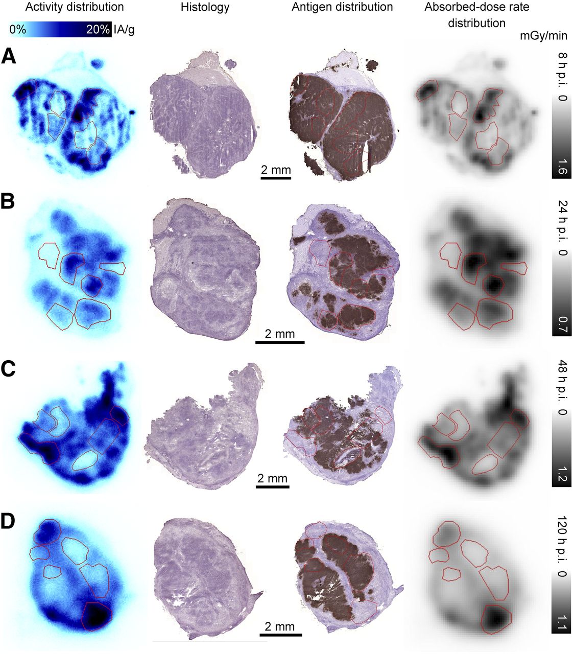
- FIGURE 1.
Representative tumor sections from group 2 at 4 time points after injection. From left to right: digital autoradiography images of 177Lu distribution scaled to %IA/g, adjacent sections stained with hematoxylin and eosin for histologic examination, adjacent sections stained with BR96 antibody to determine antigen distribution, and absorbed-dose rate distribution at time of sacrifice based on activity image (individually scaled). ROIs used in correlation analysis are outlined in red where applicable. Sections from group 2 are displayed due to visually better-quality staining for antigen distribution. Peripheral distribution of activity in each nodule is shown at 8 h after injection (A), and correlation between uptake and antigen is shown at 24 h after injection (B). At 48 h after injection (C), this correlation has given way to correlation between activity and antigen-negative granulation tissue, which is even more pronounced at 120 h after injection (D). p.i. = after injection.
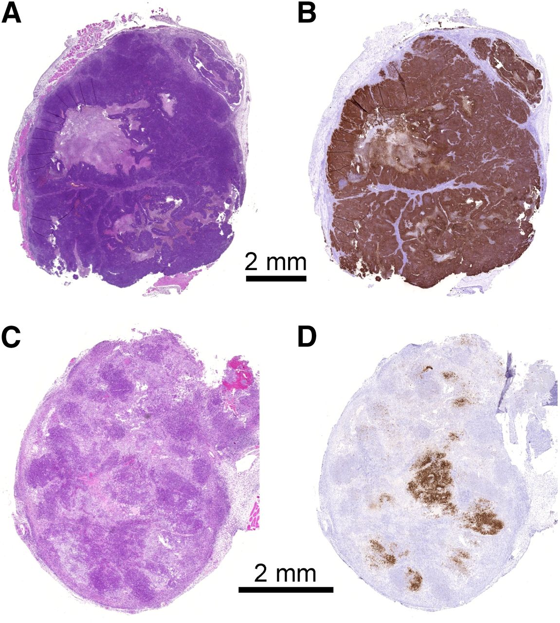
- FIGURE 2.
Representative tumor sections from untreated animals (top), and animals treated with 1.0 mg/kg dose of unlabeled mAb 48 h after injection (bottom). Sections are stained with hematoxylin and eosin (A and C) and BR96 antibody (B and D). Reduction in viable, antigen-expressing tumor cells is seen in tumor treated with unlabeled mAb.
- FIGURE 3.
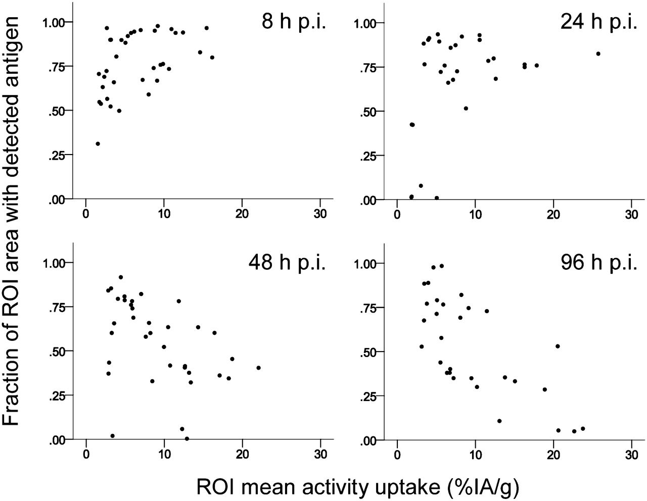
Scatter plots demonstrating changing correlation between antigen expression and activity over time. p.i. = after injection.


Tables
No. of animals sacrificed at each time point after injection Group Injected activity (MBq/kg) 2 h 8 h 24 h 48 h 72 h 96 h 120 h 168 h 1 50 3 3 2 3 — 3 3 — 2 25 — 3 3 3 3 3 3 3 177Lu-DOTA-BR96 activity (%IA/g) Time after injection (h) Tumor Blood 0 — 6.72 (5.82–8.70) 2 1.97 (1.54–2.13) — 8 4.37 (3.15–6.38) 4.29 (4.13–4.62) 24 6.95 (5.09–10.2) 2.81 (2.52–2.94) 48 8.43 (6.34–9.81) 2.12 (1.95–2.43) 72 14.9 (7.48–19.0) 1.81 (1.37–1.99) 96 10.6 (4.24–20.7) 1.45 (1.29–1.89) 120 7.80 (2.65–13.0) 1.56 (1.45–1.64) 168 28.3 (11.6–52.1) 1.17 (0.98–1.69) Data are median, followed by range in parentheses.
- TABLE 3
Estimated Absorbed-Dose Rates for Whole Tumor Sections and High- and Low-Uptake Regions (Relative to Each Section)
Mean absorbed-dose (mGy/min) Whole tumor sections High-uptake ROIs Low-uptake ROIs Time after injection (h) Group 1 Group 2 Group 1 Group 2 Group 1 Group 2 2 0.10 (0.10–0.20) — 0.40 (0.30–0.50) — 0.10 (0.00–0.10) — 8 0.37 (0.27–0.43) 0.49 (0.40–0.52) 0.67 (0.61–1.13) 1.01 (0.70–1.31) 0.18 (0.15–0.30) 0.27 (0.15–0.37) 24 (0.54–0.79) 0.48 (0.33–0.58) 1.12 (1.09–1.81) 0.81 (0.59–1.08) 0.47 (0.14–0.51) 0.25 (0.17–0.52) 48 0.60 (0.49–0.65) 0.53 (0.46–0.60) 1.06 (0.78–1.35) 0.90 (0.56–1.15) 0.33 (0.21–0.67) 0.28 (0.22–0.51) 72 — 0.71 (0.29–0.84) — 1.43 (0.51–1.65) — 0.27 (0.17–0.77) 96 0.43 (0.25–1.04) 0.39 (0.38–0.63) 0.78 (0.48–1.03) 0.93 (0.70–1.17) 0.23 (0.18–0.35) 0.35 (0.19–0.53) 120 0.38 (0.30–0.66) 0.37 (0.34–1.88) 0.78 (0.56–1.43) 0.79 (0.45–3.62) 0.23 (0.17–0.34) 0.29 (0.17–2.92) 168 — 1.10 (0.76–2.40) — 1.73 (1.23–5.31) — 0.52 (0.17–3.01) Data are median, followed by range in parentheses.
- TABLE 4
Correlation Between Activity Uptake and Absorbed-Dose Rate at Time of Sacrifice vs. Antigen-Positive Area Fraction in ROIs of Tumor Sections
Pearson correlation coefficient Time after injection(h) Antigen vs. activity uptake Antigen vs. absorbed-dose rate at time of sacrifice 8 0.450* 0.568† 24 0.384* 0.405* 48 −0.432* −0.490* 96 −0.711† −0.643† 120 −0.621† −0.572†
Supplemental Data
Files in this Data Supplement:


