
Main menu

  • Home
  • Content
    • Current
    • Ahead of print
    • Past Issues
    • JNM Supplement
    • SNMMI Annual Meeting Abstracts
    • Continuing Education
    • JNM Podcasts
  • Subscriptions
    • Subscribers
    • Institutional and Non-member
    • Rates
    • Journal Claims
    • Corporate & Special Sales
  • Authors
    • Submit to JNM
    • Information for Authors
    • Assignment of Copyright
    • AQARA requirements
  • Info
    • Reviewers
    • Permissions
    • Advertisers
  • About
    • About Us
    • Editorial Board
    • Contact Information
  • More
    • Alerts
    • Feedback
    • Help
    • SNMMI Journals
  • SNMMI
    • JNM
    • JNMT
    • SNMMI Journals
    • SNMMI

User menu

  • Subscribe
  • My alerts
  • Log in
  • My Cart

Search

  • Advanced search
Journal of Nuclear Medicine
  • SNMMI
    • JNM
    • JNMT
    • SNMMI Journals
    • SNMMI
  • Subscribe
  • My alerts
  • Log in
  • My Cart
Journal of Nuclear Medicine

Advanced Search

  • Home
  • Content
    • Current
    • Ahead of print
    • Past Issues
    • JNM Supplement
    • SNMMI Annual Meeting Abstracts
    • Continuing Education
    • JNM Podcasts
  • Subscriptions
    • Subscribers
    • Institutional and Non-member
    • Rates
    • Journal Claims
    • Corporate & Special Sales
  • Authors
    • Submit to JNM
    • Information for Authors
    • Assignment of Copyright
    • AQARA requirements
  • Info
    • Reviewers
    • Permissions
    • Advertisers
  • About
    • About Us
    • Editorial Board
    • Contact Information
  • More
    • Alerts
    • Feedback
    • Help
    • SNMMI Journals
  • View or Listen to JNM Podcast
  • Visit JNM on Facebook
  • Join JNM on LinkedIn
  • Follow JNM on Twitter
  • Subscribe to our RSS feeds
Research ArticleClinical Investigations

In Newly Diagnosed Diffuse Large B-Cell Lymphoma, Determination of Bone Marrow Involvement with 18F-FDG PET/CT Provides Better Diagnostic Performance and Prognostic Stratification Than Does Biopsy

Louis Berthet, Alexandre Cochet, Salim Kanoun, Alina Berriolo-Riedinger, Olivier Humbert, Michel Toubeau, Inna Dygai-Cochet, Caroline Legouge, Olivier Casasnovas and François Brunotte
Journal of Nuclear Medicine August 2013, 54 (8) 1244-1250; DOI: https://doi.org/10.2967/jnumed.112.114710
Louis Berthet
1Department of Nuclear Medicine, Centre Georges-François Leclerc, Dijon, France
  • Find this author on Google Scholar
  • Find this author on PubMed
  • Search for this author on this site
Alexandre Cochet
1Department of Nuclear Medicine, Centre Georges-François Leclerc, Dijon, France
2Le2i UMR CNRS 5158, Dijon, France; and
  • Find this author on Google Scholar
  • Find this author on PubMed
  • Search for this author on this site
Salim Kanoun
1Department of Nuclear Medicine, Centre Georges-François Leclerc, Dijon, France
  • Find this author on Google Scholar
  • Find this author on PubMed
  • Search for this author on this site
Alina Berriolo-Riedinger
1Department of Nuclear Medicine, Centre Georges-François Leclerc, Dijon, France
  • Find this author on Google Scholar
  • Find this author on PubMed
  • Search for this author on this site
Olivier Humbert
1Department of Nuclear Medicine, Centre Georges-François Leclerc, Dijon, France
2Le2i UMR CNRS 5158, Dijon, France; and
  • Find this author on Google Scholar
  • Find this author on PubMed
  • Search for this author on this site
Michel Toubeau
1Department of Nuclear Medicine, Centre Georges-François Leclerc, Dijon, France
  • Find this author on Google Scholar
  • Find this author on PubMed
  • Search for this author on this site
Inna Dygai-Cochet
1Department of Nuclear Medicine, Centre Georges-François Leclerc, Dijon, France
  • Find this author on Google Scholar
  • Find this author on PubMed
  • Search for this author on this site
Caroline Legouge
3Department of Clinical Hematology, Centre Hospitalier Régional Universitaire, Hôpital Le Bocage, Dijon, France
  • Find this author on Google Scholar
  • Find this author on PubMed
  • Search for this author on this site
Olivier Casasnovas
3Department of Clinical Hematology, Centre Hospitalier Régional Universitaire, Hôpital Le Bocage, Dijon, France
  • Find this author on Google Scholar
  • Find this author on PubMed
  • Search for this author on this site
François Brunotte
1Department of Nuclear Medicine, Centre Georges-François Leclerc, Dijon, France
2Le2i UMR CNRS 5158, Dijon, France; and
  • Find this author on Google Scholar
  • Find this author on PubMed
  • Search for this author on this site
  • Article
  • Figures & Data
  • Info & Metrics
  • PDF
Loading

Article Figures & Data

Figures

  • Tables
  • FIGURE 1.
    • Download figure
    • Open in new tab
    • Download powerpoint
    FIGURE 1.

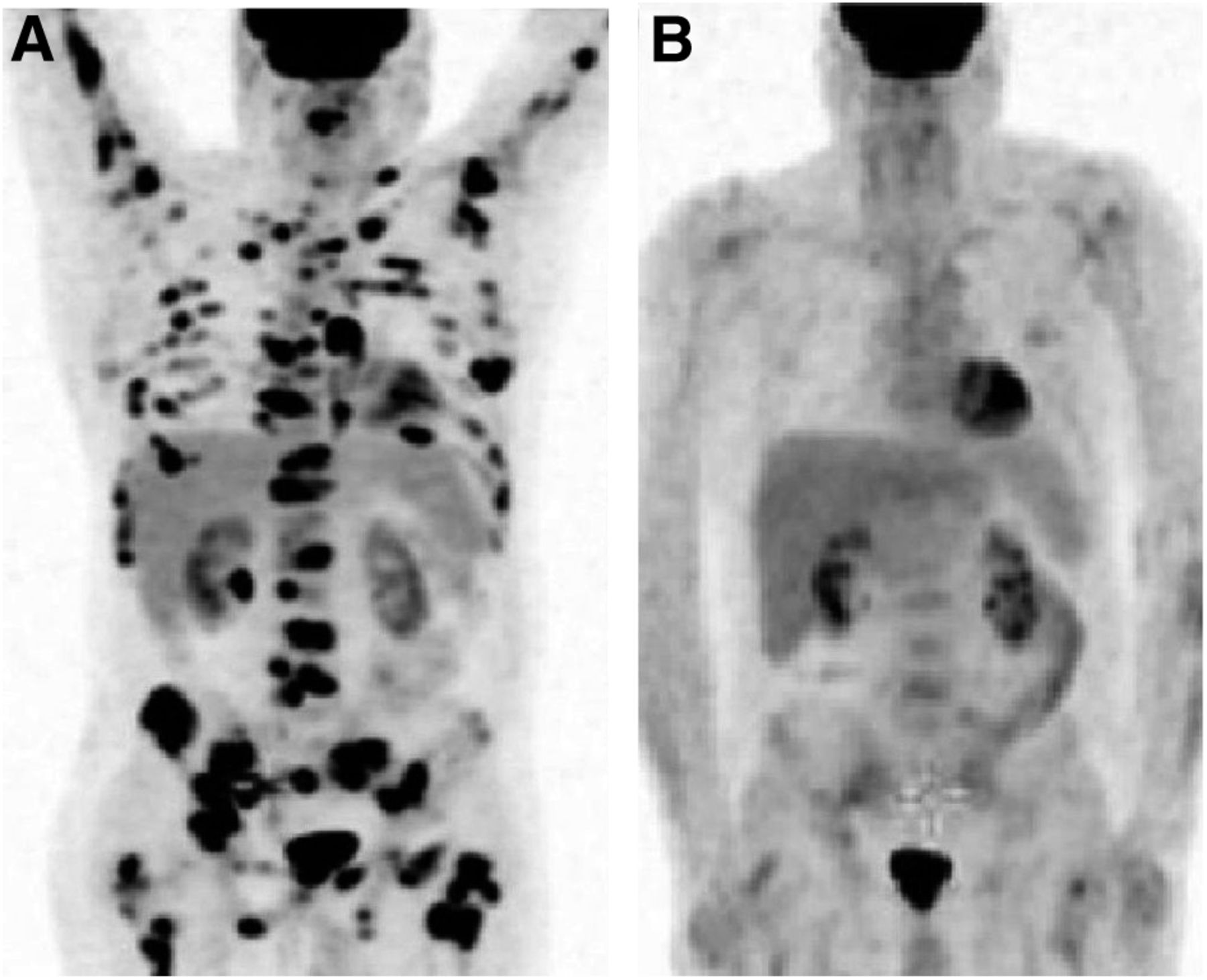
    Diffuse bone marrow uptake pattern in 18F-FDG PET/CT. (A and B) Uptake lower than (A) or similar to (B) that in liver was considered negative for BMI. (C) Uptake higher than that in liver was always linked to anemia or inflammatory processes and also considered negative for BMI.

  • FIGURE 2.
    • Download figure
    • Open in new tab
    • Download powerpoint
    FIGURE 2.

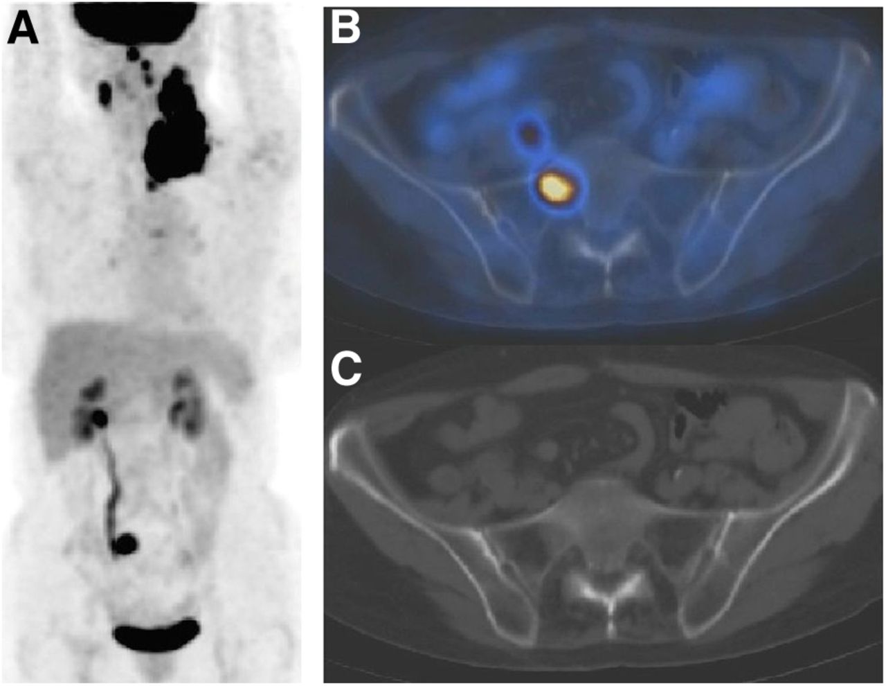
    Unifocal bone marrow uptake pattern in 18F-FDG PET/CT. Focal lesion on right pelvic bone (A) was shown to be located on right part of sacrum (B), with no underlying anomaly on CT (C). Usual BMB in left posterior iliac crest was negative. Targeted MR imaging confirmed BMI. According to our criteria, patient was considered to have BMI.

  • FIGURE 3.
    • Download figure
    • Open in new tab
    • Download powerpoint
    FIGURE 3.

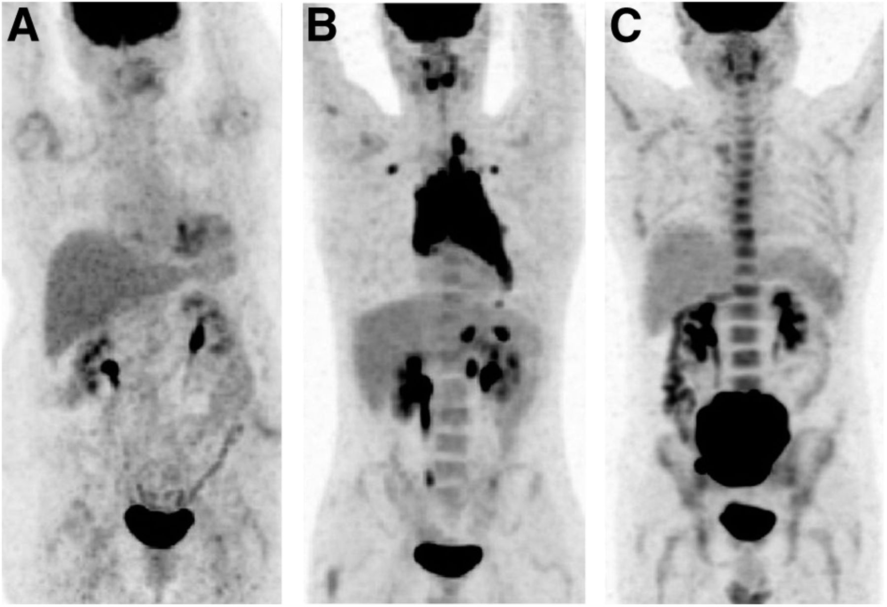
    Patient with negative initial BMB of left posterior iliac crest. (A) Initial 18F-FDG PET/CT highlighted multifocal uptake in bone marrow. Guided biopsy of right iliac crest came back positive. (B) 18F-FDG PET/CT monitoring revealed excellent metabolic response. According to our criteria, this patient was considered to have BMI.

  • FIGURE 4.
    • Download figure
    • Open in new tab
    • Download powerpoint
    FIGURE 4.

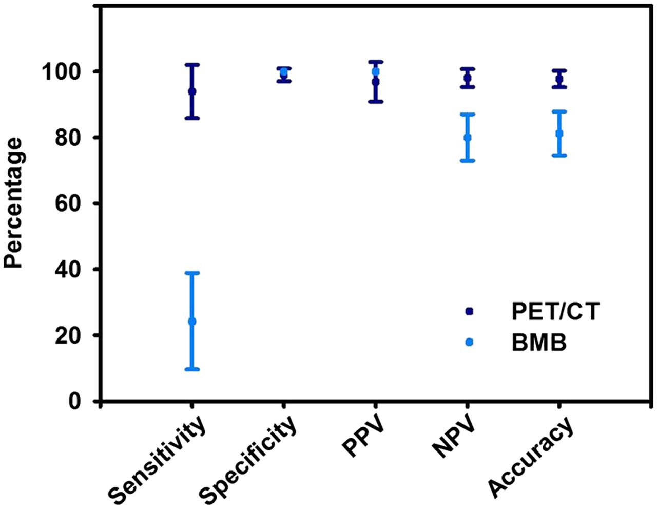
    Diagnostic performance of BMB and 18F-FDG PET/CT regarding BMI (95% CIs).
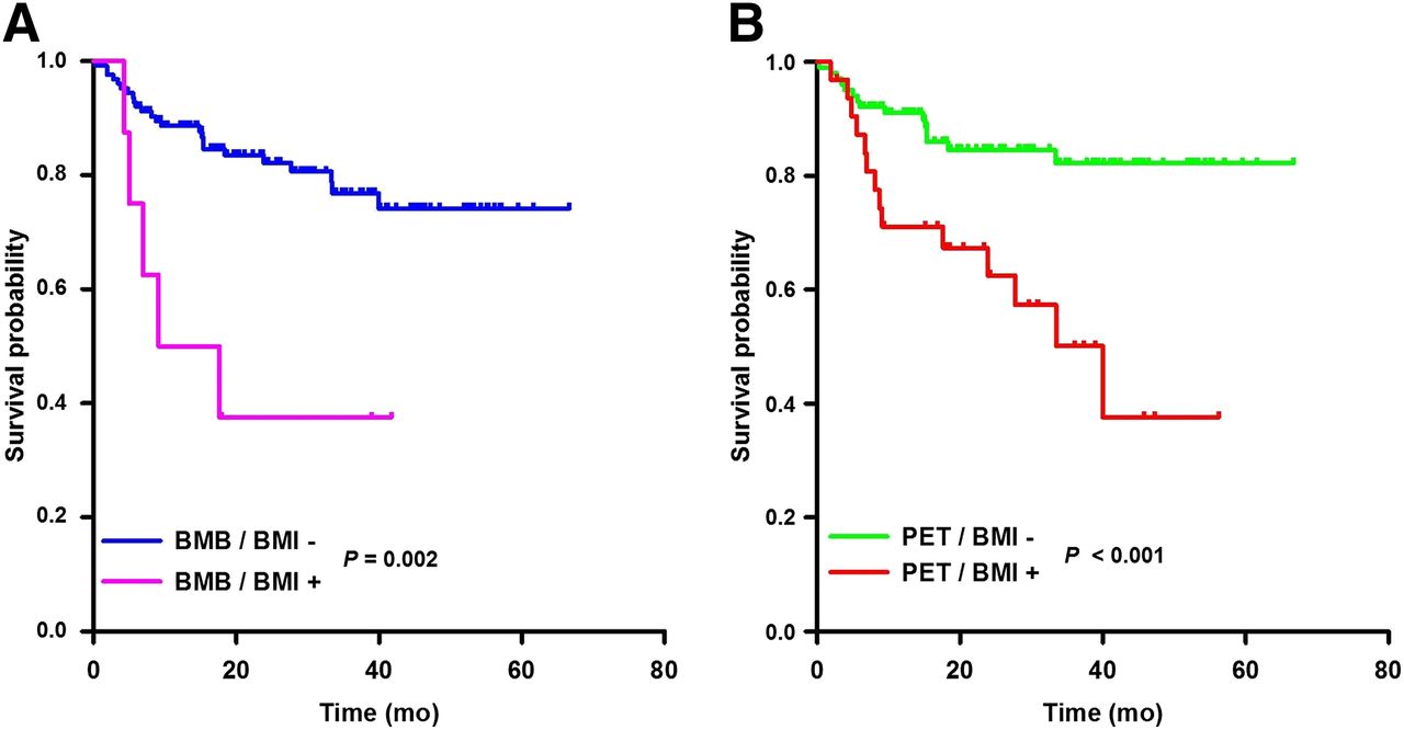
  • FIGURE 5.
    • Download figure
    • Open in new tab
    • Download powerpoint
    FIGURE 5.

    PFS according to BMB (A) or 18F-FDG PET/CT status (B).
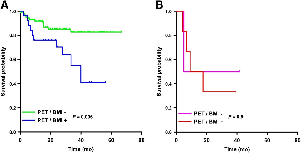
  • FIGURE 6.
    • Download figure
    • Open in new tab
    • Download powerpoint
    FIGURE 6.

    PFS of patients with negative BMB (A) or positive BMB (B) considering their 18F-FDG PET/CT status.

Tables

  • Figures
    • View popup
    TABLE 1

    Baseline Clinical Characteristics According to Bone Marrow Status by BMB or 18F-FDG PET/CT

    BMB18F-FDG PET/CT
    CharacteristicBMI+ (n = 8)BMI− (n = 125)PBMI+* (n = 32)BMI−† (n = 101)P
    Male sex4 (50)63 (50)121 (66)46 (46)0.07
    IPI > 25 (63)48 (38)0.517 (53)36 (36)0.1
    Age (y)59 ± 1157 ± 150.659 ± 1057 ± 160.3
    Age ≥ 60 y4 (50)64 (51)115 (47)53 (52)0.7
    LDH+5 (63)79 (63)123 (72)61 (60)0.3
    Stage III/IV8 (100)91 (73)0.128 (88)70 (70)0.06
    ECOG ≥ 22 (25)21 (17)0.67 (22)16 (16)0.4
    Extranodal involvement > 15 (63)23 (18)0.0111 (34)17 (17)0.05
    BMB/BMI+———6 (19)2 (2)0.003
    PET/BMI+6 (75)26 (21)0.003———
    • ↵* Presence of clear-cut 18F-FDG–avid bone marrow foci (unifocal of multifocal) consistent with BMI.

    • ↵† Absence of clear-cut 18F-FDG–avid bone marrow foci (intense but homogeneous uptake is considered negative).

    • Qualitative data are expressed as numbers, followed by percentages in parentheses; continuous data are expressed as mean ± SD.

    • View popup
    TABLE 2

    Cox Regression Analysis for PFS

    Univariate analysisMultivariate analysis
    CharacteristicHR95% CIPHR95% CIP
    BMB/BMI+4.861.62–14.580.052.200.80–6.040.1
    PET/BMI+2.891.20–6.980.022.481.15–5.330.02
    IPI > 24.541.73–11.930.0023.111.42–6.800.005
    Age > 60 y1.940.77–4.880.2———
    Stage III or IV2.380.69–8.130.2———
    ECOG ≥ 25.852.39–14.39<0.001———
    LDH+3.651.07–12.480.04———
    Extranodal site > 12.961.21–7.260.02———
    • View popup
    TABLE 3

    Cox regression Analysis for OS

    Univariate analysisMultivariate analysis
    CharacteristicHR95% CIPHR95% CIP
    BMB/BMI+4.101.36–12.360.012.670.87–8.210.09
    PET/BMI+2.811.16–6.780.022.170.88–5.330.09
    IPI > 24.591.75–12.030.0024.151.57–10.950.004
    Age > 60 y2.080.83–5.290.1———
    Stage III or IV2.530.74–8.690.1———
    ECOG ≥ 26.802.72–17.12<0.001———
    LDH+3.521.03–12.030.04———
    Extranodal site > 12.881.17–7.060.02———
    • View popup
    TABLE 4

    Correlation Between 18F-FDG PET/CT and BMB Results and Final Bone Marrow Status in Our Study

    18F-FDG PET/CTBMB
    Final statusBMI+BMI−BMI+BMI−
    BMI+312825
    BMI−1990100
    • n = 133.

    • View popup
    TABLE 5

    Correlation Between 18F-FDG PET/CT and BMB Results and Final Bone Marrow Status in 3 Studies

    18F-FDG PET/CTBMB
    StudynFinal statusBMI+BMI−BMI+BMI−
    Mittal et al. (23)60BMI+250196
    BMI−233035
    Muslimani et al. (16)57BMI+203——
    BMI−232——
    Pelosi et al. (15)120BMI+2141015
    BMI−095095
    Overall237 and 180BMI+6672921
    BMI−41600130
PreviousNext
Back to top

In this issue

Journal of Nuclear Medicine: 54 (8)
Journal of Nuclear Medicine
Vol. 54, Issue 8
August 1, 2013
  • Table of Contents
  • Table of Contents (PDF)
  • About the Cover
  • Index by author
Print
Download PDF
Article Alerts
Sign In to Email Alerts with your Email Address
Email Article

Thank you for your interest in spreading the word on Journal of Nuclear Medicine.

NOTE: We only request your email address so that the person you are recommending the page to knows that you wanted them to see it, and that it is not junk mail. We do not capture any email address.

Enter multiple addresses on separate lines or separate them with commas.
In Newly Diagnosed Diffuse Large B-Cell Lymphoma, Determination of Bone Marrow Involvement with 18F-FDG PET/CT Provides Better Diagnostic Performance and Prognostic Stratification Than Does Biopsy
(Your Name) has sent you a message from Journal of Nuclear Medicine
(Your Name) thought you would like to see the Journal of Nuclear Medicine web site.
Citation Tools
In Newly Diagnosed Diffuse Large B-Cell Lymphoma, Determination of Bone Marrow Involvement with 18F-FDG PET/CT Provides Better Diagnostic Performance and Prognostic Stratification Than Does Biopsy
Louis Berthet, Alexandre Cochet, Salim Kanoun, Alina Berriolo-Riedinger, Olivier Humbert, Michel Toubeau, Inna Dygai-Cochet, Caroline Legouge, Olivier Casasnovas, François Brunotte
Journal of Nuclear Medicine Aug 2013, 54 (8) 1244-1250; DOI: 10.2967/jnumed.112.114710

Citation Manager Formats

  • BibTeX
  • Bookends
  • EasyBib
  • EndNote (tagged)
  • EndNote 8 (xml)
  • Medlars
  • Mendeley
  • Papers
  • RefWorks Tagged
  • Ref Manager
  • RIS
  • Zotero
Share
In Newly Diagnosed Diffuse Large B-Cell Lymphoma, Determination of Bone Marrow Involvement with 18F-FDG PET/CT Provides Better Diagnostic Performance and Prognostic Stratification Than Does Biopsy
Louis Berthet, Alexandre Cochet, Salim Kanoun, Alina Berriolo-Riedinger, Olivier Humbert, Michel Toubeau, Inna Dygai-Cochet, Caroline Legouge, Olivier Casasnovas, François Brunotte
Journal of Nuclear Medicine Aug 2013, 54 (8) 1244-1250; DOI: 10.2967/jnumed.112.114710
Twitter logo Facebook logo LinkedIn logo Mendeley logo
  • Tweet Widget
  • Facebook Like
  • Google Plus One
Bookmark this article

Jump to section

  • Article
    • Abstract
    • MATERIALS AND METHODS
    • RESULTS
    • DISCUSSION
    • CONCLUSION
    • DISCLOSURE
    • Acknowledgments
    • Footnotes
    • REFERENCES
  • Figures & Data
  • Info & Metrics
  • PDF

Related Articles

  • This Month in JNM
  • PubMed
  • Google Scholar

Cited By...

  • Discordant bone marrow involvement in non-Hodgkin lymphoma
  • Reply to B. Bennani-Baiti et al, H.J.A. Adams et al, E. Laffon et al, and E.A. Hawkes et al
  • Do Not Abandon the Bone Marrow Biopsy Yet in Diffuse Large B-Cell Lymphoma
  • Combined PET and Biopsy Evidence of Marrow Involvement Improves Prognostic Prediction in Diffuse Large B-Cell Lymphoma
  • Recommendations for Initial Evaluation, Staging, and Response Assessment of Hodgkin and Non-Hodgkin Lymphoma: The Lugano Classification
  • Role of Imaging in the Staging and Response Assessment of Lymphoma: Consensus of the International Conference on Malignant Lymphomas Imaging Working Group
  • Reply: Prognostic Implications of Imaging-Based Bone Marrow Assessment in Lymphoma: 18F-FDG PET, MR Imaging, or 18F-FDG PET/MR Imaging?
  • Prognostic Implications of Imaging-Based Bone Marrow Assessment in Lymphoma: 18F-FDG PET, MR Imaging, or 18F-FDG PET/MR Imaging?
  • Google Scholar

More in this TOC Section

  • Cardiac Presynaptic Sympathetic Nervous Function Evaluated by Cardiac PET in Patients with Chronotropic Incompetence Without Heart Failure
  • Validation and Evaluation of a Vendor-Provided Head Motion Correction Algorithm on the uMI Panorama PET/CT System
  • Left Ventricular Strain from Myocardial Perfusion PET Imaging: Method Development and Comparison to 2-Dimensional Echocardiography
Show more Clinical Investigations

Similar Articles

Keywords

  • PET/CT
  • diffuse large B-cell lymphoma
  • bone marrow
  • biopsy
SNMMI

© 2025 SNMMI

Powered by HighWire