


Article Figures & Data
Figures
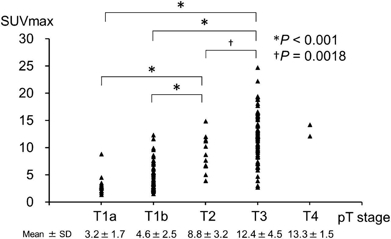
- FIGURE 1.
SUVmax of primary lesions among different pT stages.
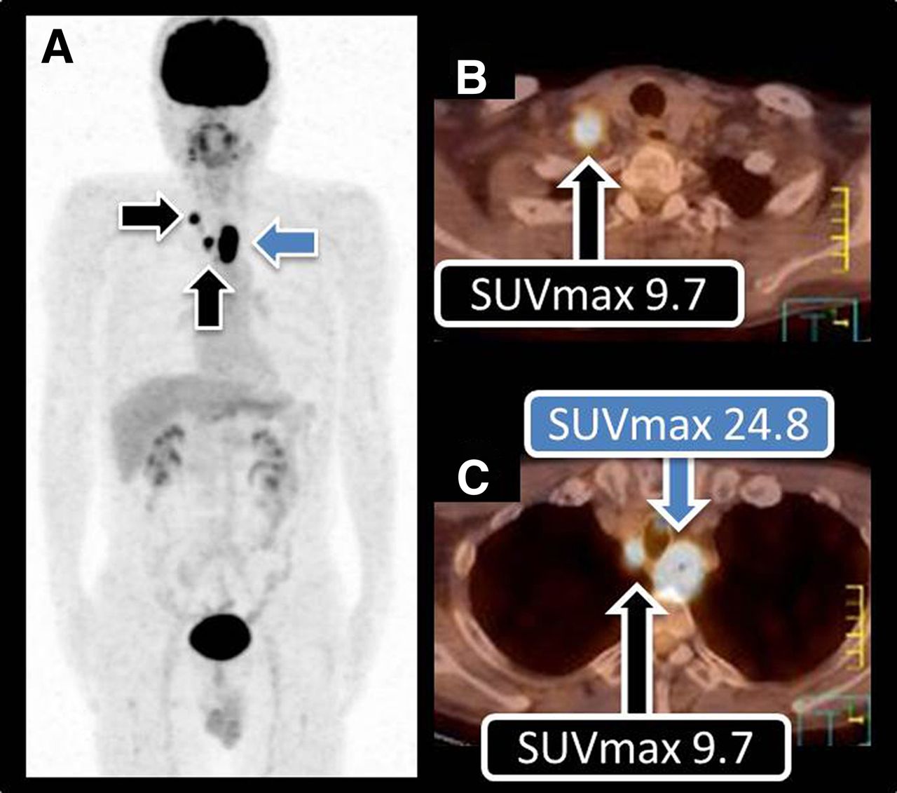
- FIGURE 2.
Representative 18F-FDG PET images of 52-y-old man with SCC of upper thoracic esophagus showing high 18F-FDG avidity in primary lesion. (A) Whole-body maximum-intensity-projection image demonstrating high 18F-FDG avidity in primary lesion (blue arrow; SUVmax, 24.8) and LN uptake in the cervical and thoracic regions (black arrows). (B) Axial image of cervical region showing abnormal 18F-FDG uptake in metastatic LN (SUVmax, 9.7). (C) Axial image of upper thoracic region showing abnormal 18F-FDG uptake in metastatic LN (SUVmax, 9.7). Pathologic analyses disclosed T3, LN metastasis of cervical and thoracic regions, which were concordant with 18F-FDG PET/CT findings.
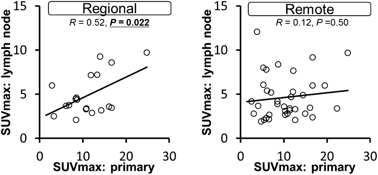
- FIGURE 3.
Representative SUVmax of regional and remote LN metastases was plotted against SUVmax of primary lesions. Representative value of LN SUVmax was defined as highest SUVmax of metastatic LNs in each region (cervical, thoracic, and abdominal).
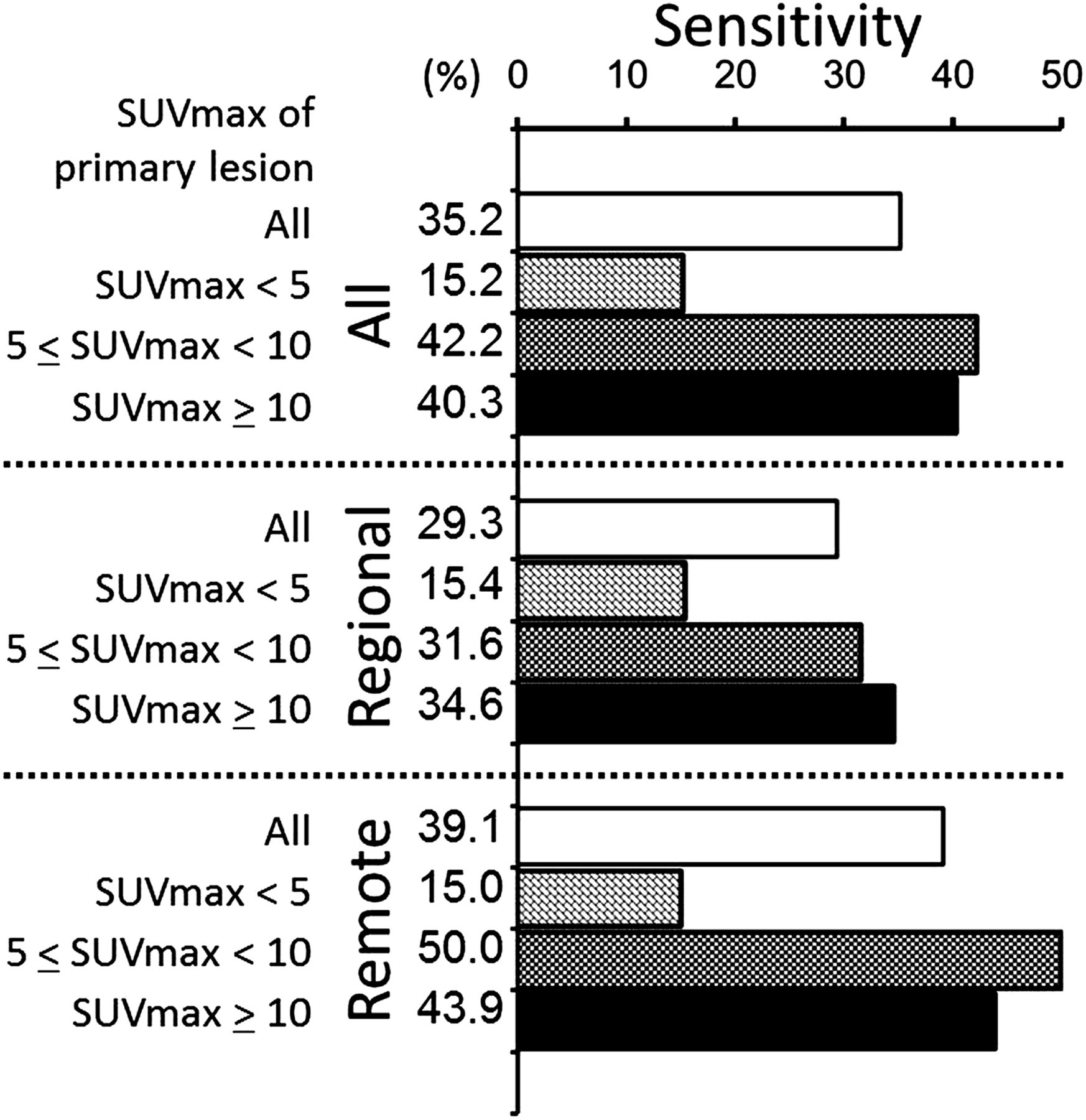
- FIGURE 4.
Comparison of sensitivities with visual analysis among different 18F-FDG avidity in primary lesions.
- FIGURE 5.
Results of ROC analyses: all patients (A), patients with low 18F-FDG avidity in primary lesions (SUVmax < 5) (B), and patients with mid to high 18F-FDG avidity in primary lesions (SUVmax ≥ 5) (C).


Tables
pT stage 18F-FDG positive Total Sensitivity (%) T1a 10 16 62.5 T1b 42 57 73.7 T2 12 12 100 T3 69 69 100 T4 2 2 100 Total 135 156 86.5 N stage PET Pathology N0 100 67 N1 50 46 N2 6 23 N3 0 20 Avidity N stage Low (SUVmax < 5) Mid (5 ≤ SUVmax < 10) High (10 < SUVmax) N0 35 (60.3) 15 (36.6) 17 (29.8) N1 15 (25.9) 10 (24.4) 21 (36.8) N2 6 (10.3) 9 (22.0) 8 (14.0) N3 2 (3.4) 7 (17.1) 11 (19.3) Total 58 41 57 Data in parentheses are percentages.
All nodes Regional nodes Remote nodes Parameter Low Mid to high Low Mid to high Low Mid to high Sensitivity (%) 15.2 41.5 15.4 33.7 15.0 46.9 Specificity (%) 95.7 92.3 91.1 89.6 97.9 93.5 Positive predictive value (%) 45.5 76.3 33.3 74.0 60.0 78.3 Negative predictive value (%) 82.8 71.3 78.8 61.6 84.7 76.4 Low = SUVmax < 5; mid to high = SUVmax ≥ 5.
- TABLE 5
Diagnostic Value of LN Metastasis Using Best-Cutoff Criteria in Mid to High Avidity in Primary Lesions
Parameter All (n = 73) Regional (n = 26) Remote (n = 47) Sensitivity (%) 66.1 70.0 64.1 Specificity (%) 85.7 66.7 100.0 Positive predictive value (%) 95.1 87.5 100.0 Negative predictive value (%) 37.5 40.0 36.4 Best-cutoff criteria (SUVmax, 3.3) was determined from all nodes and then applied to both regional and remote groups.


Jump to section
Related Articles
Cited By...
- No citing articles found.