


Article Figures & Data
Figures
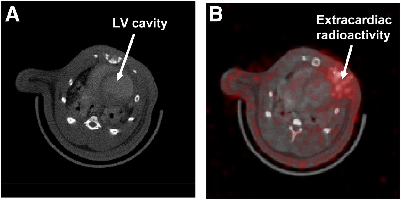
- FIGURE 1.
Illustrations of in vivo CT short-axis slice before LV cavity segmentation (A) and SPECT/CT fusion showing severe extracardiac activity in SPECT (B).
- FIGURE 2.
Illustrations of CT epicardial edge (A), correction map for extracardiac activity correction (B), LV myocardial volume in CT with unit activities assigned (C), and correction map for partial-volume effect generated by convolving binary CT image shown in C with PSF (D).
- FIGURE 3.
Illustrations of in vivo CT short-axis slice with segmented LV cavity colored in red (A), in vivo CT short-axis slice with LV myocardium (in green) superimposed (B), and in vivo SPECT short slice of αvβ3-targeted image with LV myocardium superimposed (C).
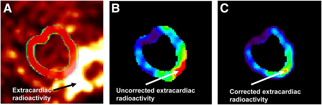
- FIGURE 4.
Demonstrations of in vivo SPECT short-axis slice of αvβ3-targeted image with region of interest (A), segmented LV myocardium volume in same SPECT slice before extracardiac activity correction (B), and SPECT slice after extracardiac activity correction (C).
- FIGURE 5.
Comparisons between SPECT-quantified and well-counted radioactivities for representative SPECT slice shown in Figure 4. Green curve shows true well-counted radioactivity. Red and black curves demonstrate SPECT-quantified radioactivity with and without corrections, respectively.
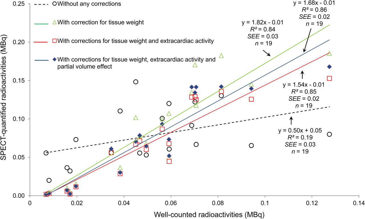
- FIGURE 6.
Correlations between SPECT-quantified and well-counted radioactivities with step-by-step corrections.


Additional Files
Supplemental Data
Files in this Data Supplement:


Jump to section
Related Articles
Cited By...
- No citing articles found.