


Article Figures & Data
Figures
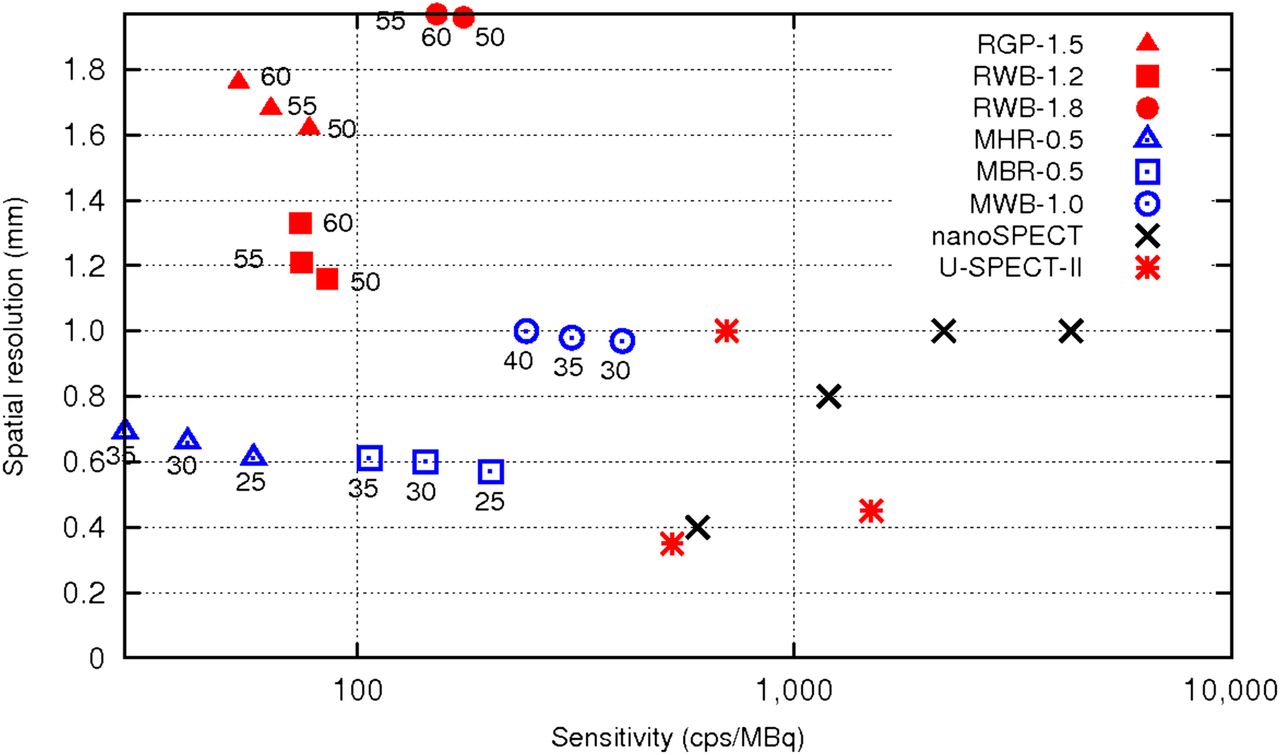
- FIGURE 1.
Tomographic spatial resolution as function of sensitivity measured with tested rat and mouse collimator sets: 1-RGP-1.5, 3-RWB-1.2, 3-RWB-1.8, 1-MHR-0.5, 5-MBR-0.5, and 5-MWB-1.0. RORs at which sensitivity and resolution measurements were performed are indicated in figure. Performance for 2 other micro-SPECT scanners, U-SPECT-II and nanoSPECT, are also reported for comparison. For U-SPECT-II, ultrahigh-resolution whole-body/focused mouse collimator (UHR-M), with aperture of 0.35 mm; general-purpose collimator for mouse (GP-M), with aperture of 0.6 mm; and ultrahigh sensitivity collimator for mouse (UHS-M), with aperture of 1 mm, were used. For nanoSPECT, UHR-M collimator, with aperture of 0.6 mm, and HR-M, with aperture of 1 mm, were used.
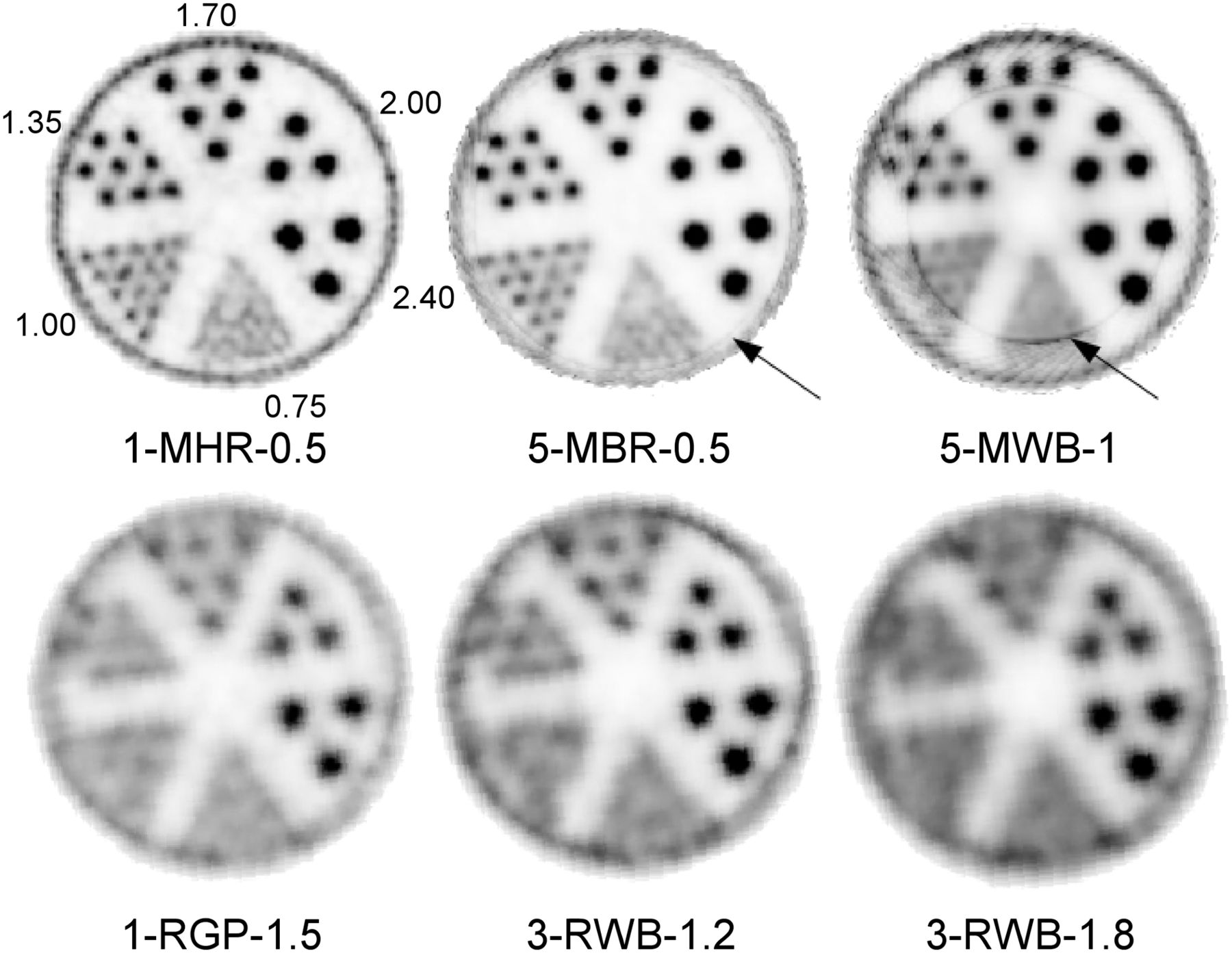
- FIGURE 2.
Derenzo phantom images obtained with 6 studied collimators. Observe ring artifacts obtained with 2 mouse 5-pinhole collimator plates due to limited FOV.
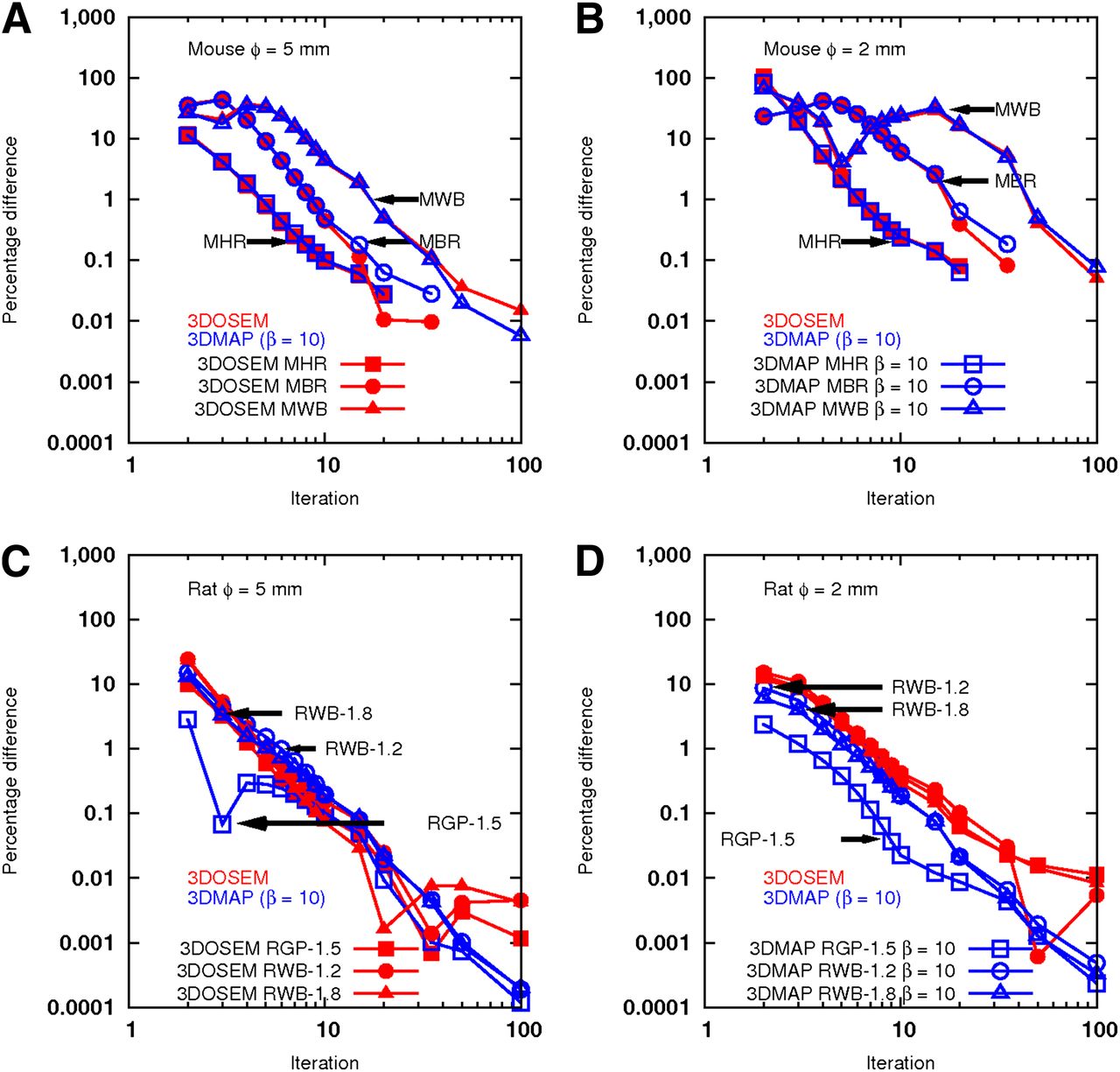
- FIGURE 3.
Absolute percentage differences of activity measured between 2 consecutive 3DOSEM/3DMAP (β = 10) iterations in 2- and 5-mm rods of IQ phantom obtained with tested mouse and rat collimator sets.
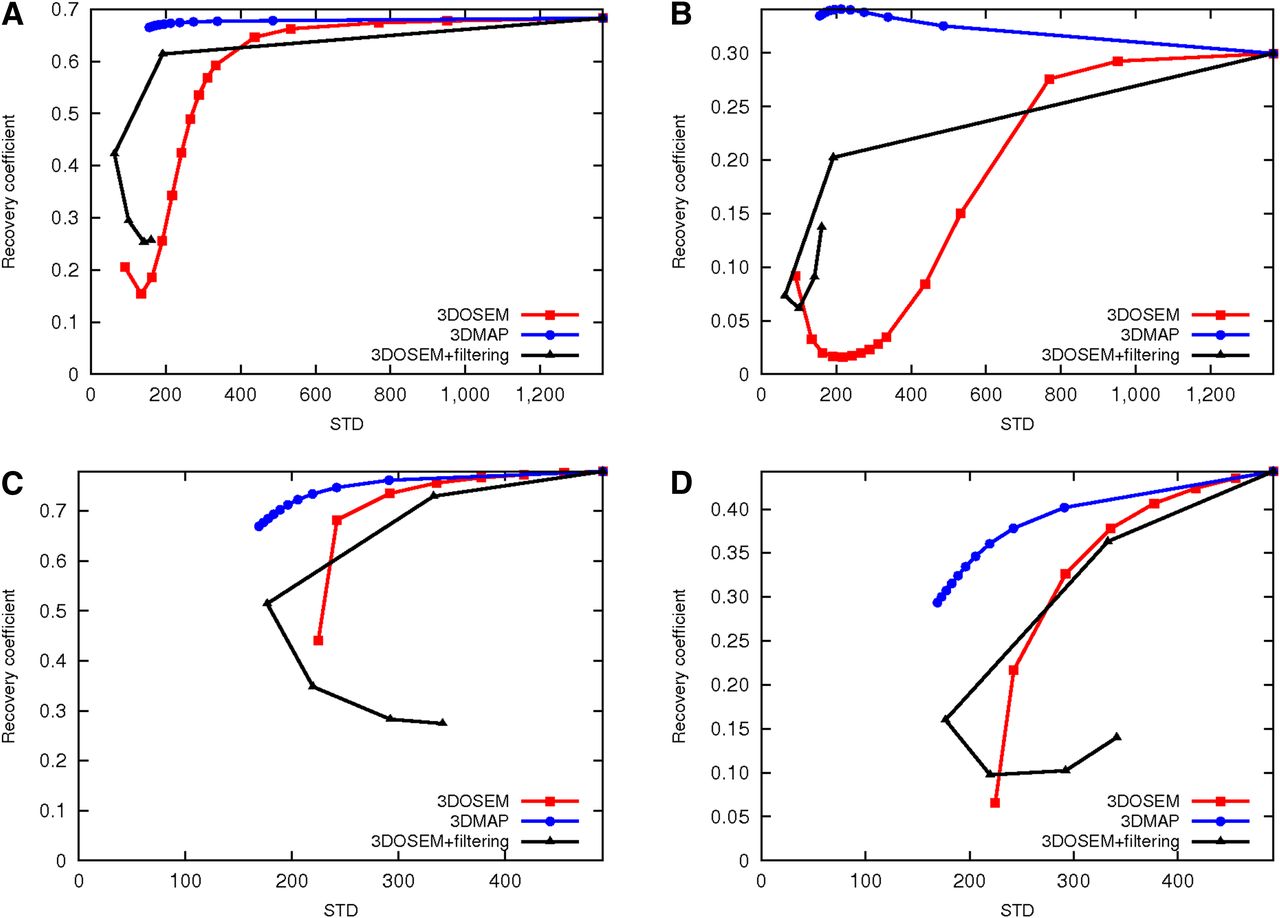
- FIGURE 4.
RCs measured at convergence from 3DOSEM images and for mouse (A) and rat (B) collimator sets.
- FIGURE 5.
(A and B) RCs obtained with 5-MWB-1 mouse collimator for 5- and 2-mm rods vs. SD measured from 3DOSEM, 3DMAP, and 3DOSEM followed by gaussian smoothing images, respectively. (C and D) RCs obtained with 3-RWB-1.2 rat collimators for 5- and 2-mm rods vs. SD measured from 3DOSEM, 3DMAP, and 3DOSEM followed by gaussian smoothing images, respectively.
- FIGURE 6.
99mTc-sestamibi images in mice obtained with collimator plates dedicated for mouse (A) and rat (B) imaging.


Tables
Collimation type Diameter of aperture (mm) Name ROR (mm) Focal length (mm) Mouse Single pinholes 0.5 1-MHR-0.5* 25, 30, and 35 97.2 1 1-MGP-1 25, 30, and 35 NM 2 1-MHS-2 25, 30, and 35 NM 3 1-MME-3 30, 35, and 40 NM Multipinholes 5 × 0.5 5-MBR-0.5* 25, 30, and 35 94.3 5 × 1 5-MWB-1* 30, 35, and 40 89.9 Rat Single pinhole 1.5 1-RGP-1.5* 50, 55, and 60 70 Multipinholes 3 × 1.2 3-RWB-1.2* 50, 55, and 60 70.4 3 × 1.8 3-RWB-1.8* 50, 55, and 60 70.5 ↵* Collimator sets used in present study.
NM = not measured.
Supplemental Data
Files in this Data Supplement:


