


Article Figures & Data
Figures
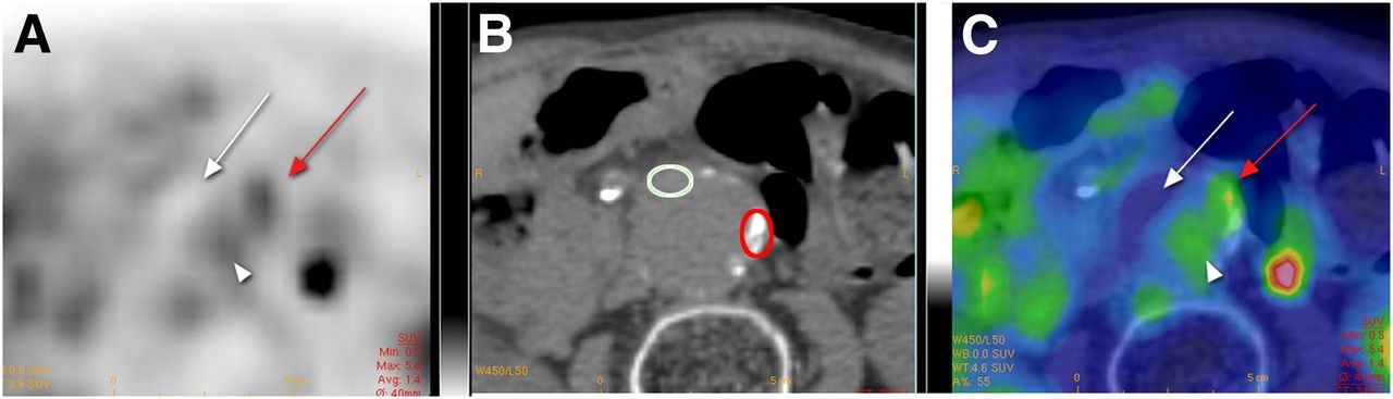
- FIGURE 1.
Example of positive 18F-FDG PET/CT: transaxial PET (A), CT (B), and fused PET/CT images (C). Focus of increased activity is visible in left lateral aspect of aortic wall (red arrow). SUVmax is 5.4 at this level and 2.8 in liver (not shown). There is absence of uptake by thrombus (white arrow) and mild uptake by intraaortic blood pool (arrowhead). Red circle indicates tissue sampling during surgery in positive site and white circle in negative site after procedure of anatomic localization described in detail in supplemental data (Supplemental Fig. 1).
- FIGURE 2.
Preoperative circulating CRP: median concentration (mg/L) in PET0 patients (● = PET0 without comorbidity and ○ = PET0 with comorbidity as listed in Table 2) and in PET+ patients (■ = PET+ without comorbidity and □ = PET+ with comorbidity). *P < 0.01, Mann–Whitney U test.
- FIGURE 3.
H/E staining (A), α-SMA immunostaining (B), and Ki67 immunostaining (C) of full-thickness sections from representative patients with negative PET (PET0) or with positive PET (PET+) at negative and positive site. Bar for H/E and α-SMA sections = 200 μm; bar for Ki67 sections = 50 μm. Arrow in A, bottom, points to tertiary lymphoid organ. a = adventitia; m = media.
- FIGURE 4.
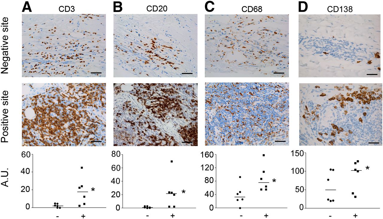
Immunolabeling of T lymphocytes (CD3, A), B lymphocytes (CD20, B), macrophages (CD68, C), and plasmocytes (CD138, D) in negative and positive sites of 18F-FDG uptake from same patient. Semiquantifications were performed in 6 pairs of samples. *P < 0.05, Wilcoxon signed-rank test. Bar = 50 μm.
- FIGURE 5.
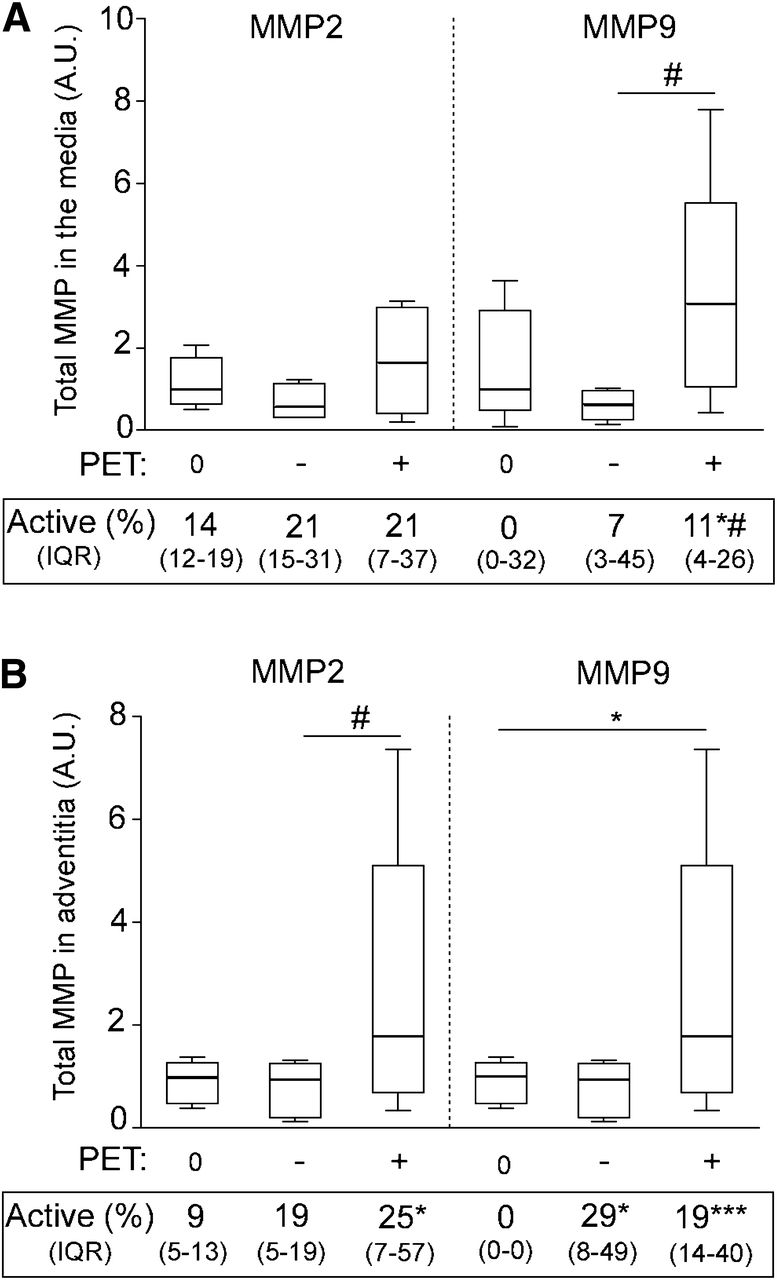
Quantification of MMP2 and MMP9 zymography analyses of samples collected from media and adventitia of PET0 patients (0) and PET+ patients at negative (−) or at positive site (+). Representative zymography gels are illustrated in Supplemental Figure 3. Box plot is of total MMP2 and MMP9 in media (A) and in adventitia (B). Median percentage of activated form is indicated below each group of samples (IQR). *P < 0.05. ***P < 0.001 vs. PET0 samples, Mann–Whitney U test. #P < 0.05, positive site vs. negative corresponding site, Wilcoxon signed-rank test. IQR = interquartile range.


Tables
- TABLE 1
Characteristics of Patients with No 18F-FDG Uptake (PET0) and with Positive 18F-FDG Uptake (PET+)
Patient characteristic PET0 (n = 10) PET+ (n = 8) Age (y) Median 75 78 IQR 71–80 69–81 Body mass index (kg/m2) Median 26.1 25.2 IQR 24.0–28.7 23.0–27.7 Deciding factor for surgery Size ≥ 55 mm 8 2 Growth/pain 1 4 Anxiety 1 2 Sex distribution Male 10 6 Female 0 2 Cardiovascular events 6 1 Hypertension 8 4 Smoker Current 3 3 Former 5 4 COPD 5 4 Diabetes 1 0 Hyperlipidemia 6 4 Statins 8 4 β-blocker 5 2 Calcium channel blocker 3 2 ACEI 2 0 NSAID 0 2 IQR = interquartile range; COPD = chronic obstructive pulmonary disease; ACEI = angiotensin-converting enzyme inhibitor; NSAID = nonsteroidal antiinflammatory drug.
Patient no. Deciding factor AAA size (mm) rSUV Time between PET and surgery (d) No 18F-FDG uptake 1 ≥55 mm 67 0.58 20 2 ≥55 mm 70 0.33 4 3 ≥55 mm 60 0.33 5 4 ≥55 mm 55* 0.41 36 5 ≥55 mm 57* 0.18 18 6 Growth 54† 0.54 6 7 ≥55 mm 80* 0.76 28 8 ≥55 mm 56 0.43 8 9 Anxiety 53 0.58 26 10 ≥55 mm 71 0.70 18 Median 58.5 0.49 18 IQR 55.3–69.3 0.35–0.58 7–25 Positive for 18F-FDG uptake 11 Anxiety 52 1.04 44 12 Growth 54 0.89 7 13 Pain 40* 1.93 19 14 Growth 55 1.22 28 15 ≥55 mm 69 1.06 20 16 ≥55 mm 67 0.89 5 17 Anxiety 57 0.97 21 18‡ Growth 53 0.84 11 Median 54.5 1.00§ 19 IQR 52.8–59.5 0.89–1.10 10–23
Supplemental Data
Files in this Data Supplement:


Jump to section
Related Articles
Cited By...
- Validation and reproducibility of mapping positron emission tomography uptake in the aortic wall and thrombus
- Radiomic and clinical predictors of cachexia in non-small cell lung cancer patients treated with immunotherapy
- Greater aortic inflammation and calcification in abdominal aortic aneurysmal disease than atherosclerosis: a prospective matched cohort study
- Systematic Review of Circulating, Biomechanical, and Genetic Markers for the Prediction of Abdominal Aortic Aneurysm Growth and Rupture
- 18F-Sodium Fluoride Uptake in Abdominal Aortic Aneurysms: The SoFIA3 Study
- 18F-FDG Uptake in Less Affected Lung Field Provides Prognostic Stratification in Patients with Interstitial Lung Disease
- High Structural Stress and Presence of Intraluminal Thrombus Predict Abdominal Aortic Aneurysm 18F-FDG Uptake: Insights From Biomechanics
- Monitoring the biological activity of abdominal aortic aneurysms Beyond Ultrasound
- Differential Gene Expression in Coiled versus Flow-Diverter-Treated Aneurysms: RNA Sequencing Analysis in a Rabbit Aneurysm Model
- Novel Molecular Imaging Approaches to Abdominal Aortic Aneurysm Risk Stratification
- Severity of Psoriasis Associates With Aortic Vascular Inflammation Detected by FDG PET/CT and Neutrophil Activation in a Prospective Observational Study
- Evidence of Cyclic Changes in the Metabolism of Abdominal Aortic Aneurysms During Growth Phases: 18F-FDG PET Sequential Observational Study
- Predicting Aortic Aneurysm Expansion by PET
- PET Imaging of Abdominal Aortic Aneurysm with 64Cu-Labeled Anti-CD105 Antibody Fab Fragment
- Imaging Vessel Wall Biology to Predict Outcome in Abdominal Aortic Aneurysm