


Article Figures & Data
Figures
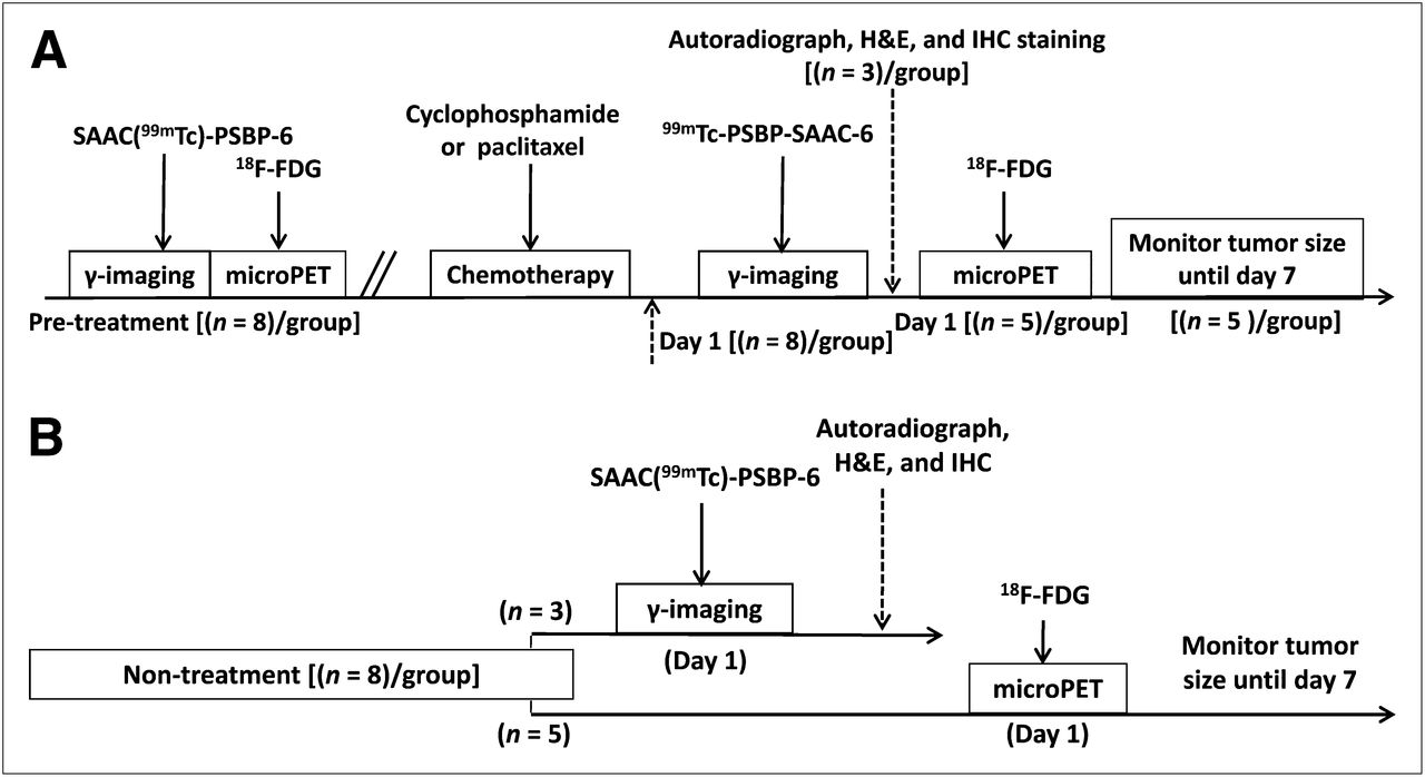
- FIGURE 1.
Design of study comparing γ-imaging using SAAC(99mTc)-PSBP-6 with small-animal PET using 18F-FDG in tumor-bearing mice after chemotherapy: TG (A) and N-TG (B). IHC = immunohistochemical.
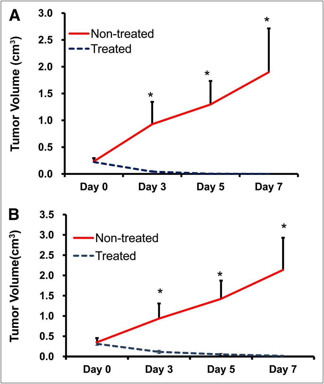
- FIGURE 2.
Tumor growth delays after chemotherapy. (A) C3H/HeJ mice bearing murine 38C13 B-cell lymphoma were treated with intraperitoneal injection of cyclophosphamide at dose of 100 mg/kg. (B) Nude mice bearing B16/F10 melanoma were treated with intravenous injection of PG-TXL at equivalent paclitaxel dose of 80 mg/kg. *P < 0.01. IHC = immunohistochemical.
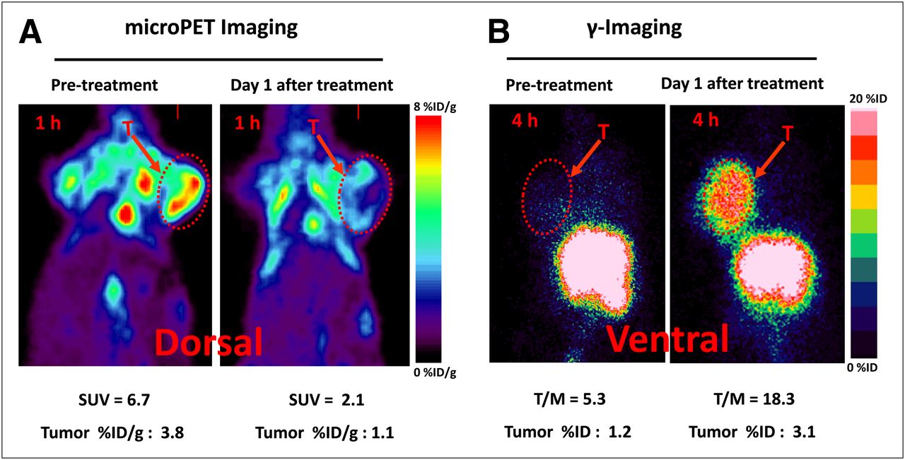
- FIGURE 3.
Representative images of 38C13 tumor–bearing mice before and 1 d after treatment with cyclophosphamide (intraperitoneal, 100 mg/kg). (A) Small-animal PET images acquired 1 h after 18F-FDG injection. (B) γ-images obtained 4 h after SAAC(99mTc)-PSBP-6 injection. SUV = standardized uptake value; T = tumors; T/M = tumor-to-muscle ratio.
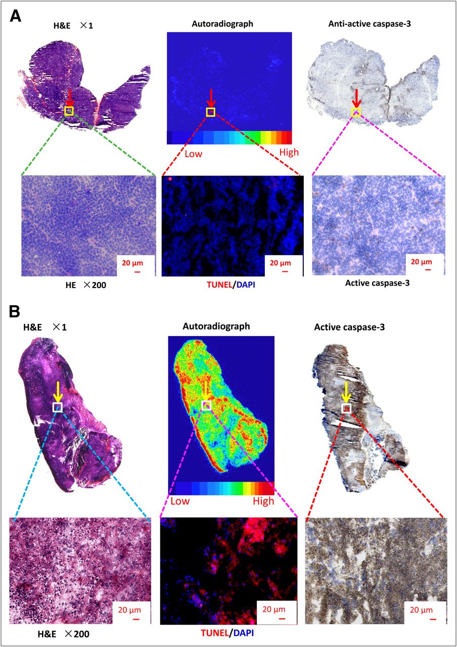
- FIGURE 4.
Representative autoradiographs and photographs of H&E-, TUNEL-, and active caspase 3–stained slides of 38C13 tumors from nontreated mouse (A) and cyclophosphamide-treated mouse (B). Tumors were removed at 4 h after intravenous injection of SAAC(99mTc)-PSBP-6. Red in fluorescent microphotographs shows TUNEL-positive apoptotic cells; blue represents 6-diamidino-2-phenylindole (DAPI)–stained cells.
- FIGURE 5.
Representative images of B16/F10 tumor–bearing mice before and 1 d after treatment with PG-TXL (intravenous, equivalent TXL dose = 80 mg/kg). (A) Small-animal PET images acquired 1 h after 18F-FDG injection. (B) γ-images obtained 4 h after SAAC(99mTc)-PSBP-6 injection. SUV = standardized uptake value; T = tumors; T/M = tumor-to-muscle ratio.

Additional Files
Supplemental Data
Files in this Data Supplement:


