


Abstract
513
Objectives To demonstrate the feasibility of retrospectively fused 18F FDG-PET and MRI (PET/MRI) in diagnosing pancreas tumor, in particular differentiating malignant tumor from benign lesions. We also evaluated the additional value of fused FDG-PET/MRI for detecting complicated lesions such as inflammations or cysts.
Methods We analyzed 119 patients retrospectively. Evaluated lesions were composed of 97 cancers and 22 benign lesions. Fused FDG-PET/MRI images (PET/T1- weighted image or PET/T2-weighted image) were made by dedicated software using 3T MRI image and FDG-PET images. They were interpreted and compared with FDG-PET/CT images respectively by two well-trained radiologists without the knowledge of clinical information. We compared diagnostic ability between PET/CT and fused FDG-PET/MRI image. In addition, we evaluated the complication which we were able to detect only by PET/MRI.
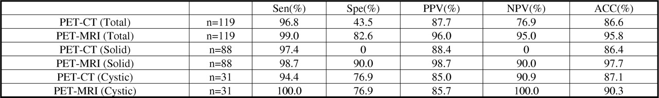
Results Using PET/MRI fused image, accuracy significantly improved from PET/CT (86.6% vs. 95.8%). In 65.9 % of solid lesions, invasion to pancreatic duct was additionally diagnosed by PET/MRI. In addition, invasion to surrounding tissues was additionally diagnosed in 43.1% of solid lesions. As for cystic lesions, intra-tumor structures such as mural nodule (38.7%) or intra-cystic septum (74.2%) were additionally diagnosed. Besides, PET/MRI detected benign lesions that were not diagnosed by PET/CT, such as complicated inflammation (13.6% in solid lesion) or additional cysts (9.1% in solid lesion and 9.7 % in cystic lesion).
Conclusions In diagnosing pancreatic lesions, fused PET/MRI image was useful in differentiating cancer from benign lesion. It was also useful in diagnosing cancer invasion to surrounding tissues. As a further utility, detecting additional complicated benign lesions were also confirmed

Comparison of diagnostic capability between PET/CT and PET/MRI fused image (Differential diagnosis)
In this issue


Jump to section
Related Articles
Cited By...
- No citing articles found.