
Main menu

  • Home
  • Content
    • Current
    • Ahead of print
    • Past Issues
    • JNM Supplement
    • SNMMI Annual Meeting Abstracts
    • Continuing Education
    • JNM Podcasts
  • Subscriptions
    • Subscribers
    • Institutional and Non-member
    • Rates
    • Journal Claims
    • Corporate & Special Sales
  • Authors
    • Submit to JNM
    • Information for Authors
    • Assignment of Copyright
    • AQARA requirements
  • Info
    • Reviewers
    • Permissions
    • Advertisers
  • About
    • About Us
    • Editorial Board
    • Contact Information
  • More
    • Alerts
    • Feedback
    • Help
    • SNMMI Journals
  • SNMMI
    • JNM
    • JNMT
    • SNMMI Journals
    • SNMMI

User menu

  • Subscribe
  • My alerts
  • Log in
  • My Cart

Search

  • Advanced search
Journal of Nuclear Medicine
  • SNMMI
    • JNM
    • JNMT
    • SNMMI Journals
    • SNMMI
  • Subscribe
  • My alerts
  • Log in
  • My Cart
Journal of Nuclear Medicine

Advanced Search

  • Home
  • Content
    • Current
    • Ahead of print
    • Past Issues
    • JNM Supplement
    • SNMMI Annual Meeting Abstracts
    • Continuing Education
    • JNM Podcasts
  • Subscriptions
    • Subscribers
    • Institutional and Non-member
    • Rates
    • Journal Claims
    • Corporate & Special Sales
  • Authors
    • Submit to JNM
    • Information for Authors
    • Assignment of Copyright
    • AQARA requirements
  • Info
    • Reviewers
    • Permissions
    • Advertisers
  • About
    • About Us
    • Editorial Board
    • Contact Information
  • More
    • Alerts
    • Feedback
    • Help
    • SNMMI Journals
  • View or Listen to JNM Podcast
  • Visit JNM on Facebook
  • Join JNM on LinkedIn
  • Follow JNM on Twitter
  • Subscribe to our RSS feeds
Meeting ReportTechnologist

Variability and accuracy of two common CT vessel quantification techniques and comparison with pathology

Dawn Holley, William Stoffregen, Doug Follett and Erik Mittra
Journal of Nuclear Medicine May 2012, 53 (supplement 1) 2527;
Dawn Holley
1LyChron, Mountain View, CA
  • Find this author on Google Scholar
  • Find this author on PubMed
  • Search for this author on this site
William Stoffregen
2Pathology, Boston Scientific Corp., San Jose, CA
  • Find this author on Google Scholar
  • Find this author on PubMed
  • Search for this author on this site
Doug Follett
1LyChron, Mountain View, CA
  • Find this author on Google Scholar
  • Find this author on PubMed
  • Search for this author on this site
Erik Mittra
3Radiology, Stanford University, Stanford, CA
  • Find this author on Google Scholar
  • Find this author on PubMed
  • Search for this author on this site
  • Article
  • Figures & Data
  • Info & Metrics
Loading

Abstract

2527

Objectives Accurate quantification of pulmonary vein measurements on CT is needed for a variety of preclinical and clinical applications. Several different processing techniques are available, but their comparability and accuracy is not well known. The objective was to evaluate 2 commercially available processing techniques and compare with pathology results in a large-animal model.

Methods Gated cardiac CT scans were done on 5 canines, both pre- and post-pulmonary vein ablation, using a 64-slice GE Discovery VCT. Multiple pulmonary veins were measured twice at the ostia on these animals using (1) a fully automated vessel analysis (VA) software and (2) a partially automated multiplanar reformat (MPR) software, both on the GE Advanced Windows Workstation. The animals were then euthanized and the software results were compared to pathology measurements. Unpaired T-tests were done to compare differences between these techniques.

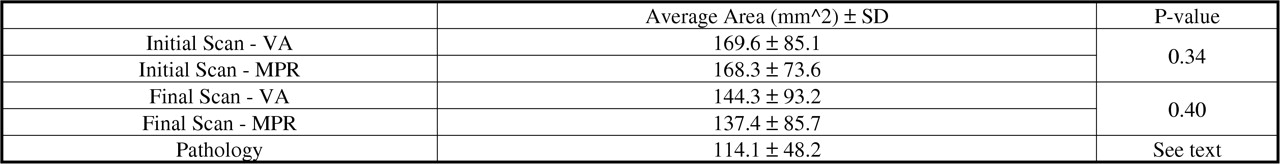
Results Both the VA and MPR techniques performed well and were not significantly different from each other (Table). The MPR technique showed slightly lower numbers than VA, both pre- and post-pulmonary ablation. The pathology measurements were smaller than either automated technique. However, neither the VA (p=0.69) nor the MPR (p=0.62) was significantly different from the pathology measurements. Fairly high variances in the data could account for the lack of significance.

Conclusions The results of this study show that if done rigorously, both the fully automated VA software and the partially automated MPR software provide accurate and comparable results for the noninvasive measurement of pulmonary veins in canines. However, the MPR software, more than VA, trended toward the true pathology measurements. Ultimately, other factors (processing time, ease of use, data ouput, cost, etc.) may influence the choice of software

Figure
  • Download figure
  • Open in new tab
  • Download powerpoint

Table

Key: VA: Vessel Analysis, MPR: Multiplanar Reformat; SD: Standard Deviation

Back to top

In this issue

Journal of Nuclear Medicine
Vol. 53, Issue supplement 1
May 2012
  • Table of Contents
  • Index by author
Article Alerts
Sign In to Email Alerts with your Email Address
Email Article

Thank you for your interest in spreading the word on Journal of Nuclear Medicine.

NOTE: We only request your email address so that the person you are recommending the page to knows that you wanted them to see it, and that it is not junk mail. We do not capture any email address.

Enter multiple addresses on separate lines or separate them with commas.
Variability and accuracy of two common CT vessel quantification techniques and comparison with pathology
(Your Name) has sent you a message from Journal of Nuclear Medicine
(Your Name) thought you would like to see the Journal of Nuclear Medicine web site.
Citation Tools
Variability and accuracy of two common CT vessel quantification techniques and comparison with pathology
Dawn Holley, William Stoffregen, Doug Follett, Erik Mittra
Journal of Nuclear Medicine May 2012, 53 (supplement 1) 2527;

Citation Manager Formats

  • BibTeX
  • Bookends
  • EasyBib
  • EndNote (tagged)
  • EndNote 8 (xml)
  • Medlars
  • Mendeley
  • Papers
  • RefWorks Tagged
  • Ref Manager
  • RIS
  • Zotero
Share
Variability and accuracy of two common CT vessel quantification techniques and comparison with pathology
Dawn Holley, William Stoffregen, Doug Follett, Erik Mittra
Journal of Nuclear Medicine May 2012, 53 (supplement 1) 2527;
Twitter logo Facebook logo LinkedIn logo Mendeley logo
  • Tweet Widget
  • Facebook Like
  • Google Plus One
Bookmark this article

Jump to section

  • Article
  • Figures & Data
  • Info & Metrics

Related Articles

  • No related articles found.
  • Google Scholar

Cited By...

  • No citing articles found.
  • Google Scholar

More in this TOC Section

Technologist

  • 68Ga DOTA TATE PET/CT in patients with neuroendocrine tumors: a technologist’s perspective
  • Quantitative accuracy of F-18 FDG injection compared among three automated infusion devices
  • A novel gallium-68 radioisotope generator
Show more Technologist

Technologist Scientific Papers IV

  • Variability and accuracy of two FDG quantification techniques: A combined phantom and pre-clinical study
  • Bio-distribution based on SPECT/CT of 188Re-liposome in SD rat
Show more Technologist Scientific Papers IV

Similar Articles

SNMMI

© 2025 SNMMI

Powered by HighWire