


Article Figures & Data
Figures
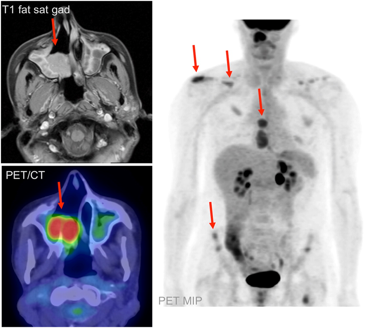
- FIGURE 1.
Patient 4 was a 46-y-old man with recurrent esthesioneuroblastoma. MRI and PET/CT demonstrate postoperative inflammation in the left maxillary sinus, whereas PET/CT demonstrates unsuspected bilateral cervical nodal metastasis (arrows). This finding prompted bilateral radical neck dissection, with pathologic confirmation of PET/CT findings.
- FIGURE 2.
Patient 8 was a 70-y-old man with recurrent esthesioneuroblastoma. MRI was interpreted as showing only postoperative change, whereas PET/CT demonstrated extensive local recurrence, associated bony remodeling, and right orbital involvement (arrows). Patient opted for conservative management.
- FIGURE 3.
Patient 6 was a 44-y-old woman with recurrent esthesioneuroblastoma. MRI and PET/CT demonstrated local recurrence in right nasal cavity and maxillary sinus (arrow). PET/CT demonstrated unsuspected, extensive osseous metastases (arrows), altering management from possible surgical resection of local recurrence to salvage chemotherapy.
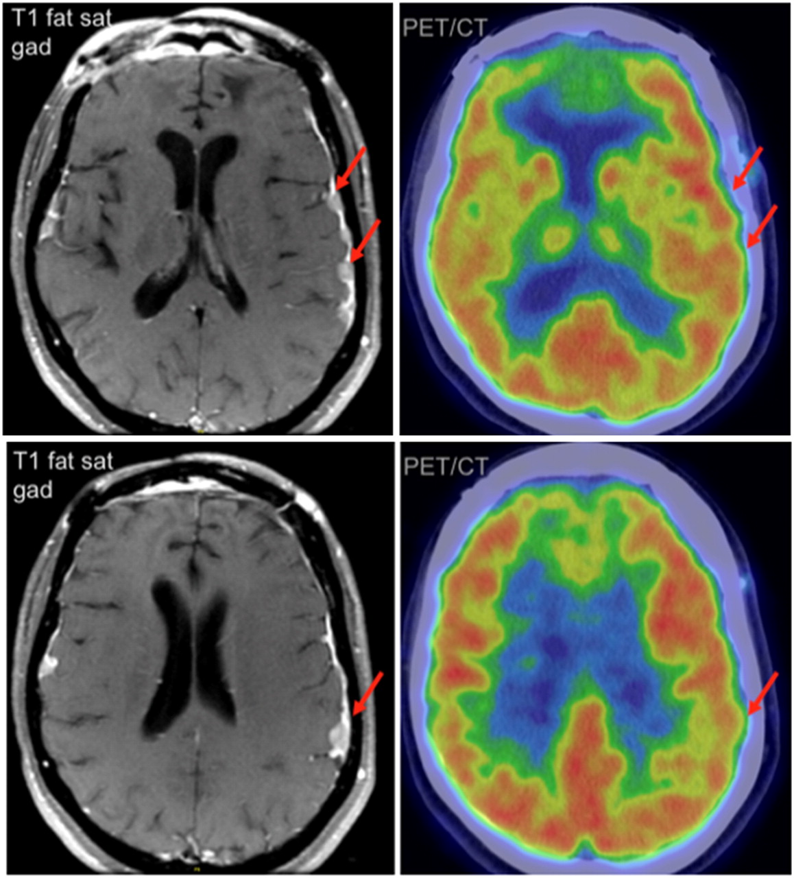
- FIGURE 4.
Patient 12 was a 47-y-old man with intracranial esthesioneuroblastoma metastases. MRI demonstrated nodular, enhancing metastases to left frontal and parietal dura, whereas PET/CT failed to demonstrate focal increase in 18F-FDG activity in these regions. Metastatic esthesioneuroblastoma was confirmed by surgical biopsy.
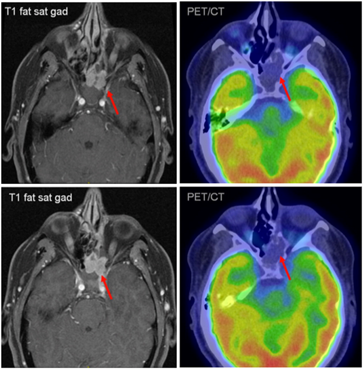
- FIGURE 5.
Patient 14 was a 40-y-old woman with locally recurrent esthesioneuroblastoma. MRI demonstrated local recurrence in left ethmoid and sphenoid sinuses, whereas PET/CT showed soft-tissue mass in this region without increased 18F-FDG activity (arrows). Pathologic analysis of subsequently resected specimen demonstrated low-grade esthesioneuroblastoma.


Tables
Stage Description A Tumor confined to nasal cavity B Tumor confined to nasal cavity and paranasal sinuses C Tumor extent beyond nasal cavity and paranasal sinuses, including involvement of cribriform plate, base of skull, orbits, and intracranial cavity D Tumor with metastasis to cervical lymph nodes or distant site Adapted from Kadish et al. (3) and Morita et al. (6).
Characteristic Value Age (y) Mean 52.3 ± 10 Range 23–81 Sex (n) Male 11 (39%) Female 17 (61%) PET/CT indication (n) Staging 11 (39%) Restaging 17 (61%) Hyams pathologic grade (n) No grade 9 (32%) Low grade (I–II) 10 (36%) High grade (III–IV) 9 (32%) Modified Kadish stage (n) Stage A 0 (0%) Stage B 4 (14%) Stage C 10 (36%) Stage D 14 (50%) Patient no. Age (y) Sex Kadish stage at time of examination PET/CT Number/type conventional imaging Anatomic site of discordance Clinical impact 1 50 M D Restaging 2 MRI Levels I and II TP, bilateral radical neck dissection 2 5 F D Restaging 1 MRI Level I TP, selective neck dissection 3 5 F D Restaging 1 MRI/1CT Level I TP, selective neck dissection 4 46 M D Restaging 1 MRI Levels I, II, III, and IV, RPLN TP, bilateral modified neck dissection 4 47 M D Restaging 1 MRI R PPF TP, γ-knife therapy 5 66 F D Restaging 2 MRI Level II/lumbar epidural TP, prompted LN Bx; palliative spinal XRT given carcinomatosis 6 44 F D Restaging 1 MRI Distant osseous TP, palliative radiation 6 45 F D Restaging 1 MRI Distant osseous TP, palliative radiation 6 44 F D Restaging 1 MRI Distant osseous TP, prompted bone Bx; treatment changed to salvage chemotherapy 7 44 M C Staging 1 MRI/1 CT Level II TP, systemic chemotherapy given stage C primary 7 44 M D Restaging 2 MRI Level II TP, prompted Bx; neoadjuvant chemotherapy 8 70 M D Restaging 3 MRI Maxillary sinus TP, prompted Bx; conservative management 9 42 M D Restaging 1 CT Level I/distant osseous TP, conservative management 4 48 M D Restaging 2 MRI L PPF/infraclavicular LN TP, palliative care 10 80 F D Restaging 1 MRI ACF FN, therapy based on MRI 10 81 F D Restaging 2 MRI ACF FN, therapy based on MRI 11 37 F C Restaging 1 CT/1 MRI ACF FN, therapy based on MRI 12 48 M C Restaging 2 MRI Frontal/parietal dura FN, therapy based on MRI 4 47 M D Restaging 1 MRI Ethmoid sinus FN, therapy based on MRI 13 40 F B Restaging 1 MRI L sphenoid/ethmoid FN, therapy based on MRI 14 50 M C Staging 1 MRI Level III FP, pathology showed reactive node 14 50 M C Restaging 1 MRI Cribriform plate/ACF TN, MRI false-positive; no recurrence to date TP = true-positive; RPLN = retropharyngeal lymph node; PPF = pterygopalatine fossa; LN = lymph node; Bx = biopsy; XRT = radiation therapy; ACF= anterior cranial fossa; FN = false-negative; FP = false-positive; TN = true-negative.


