


Article Figures & Data
Figures
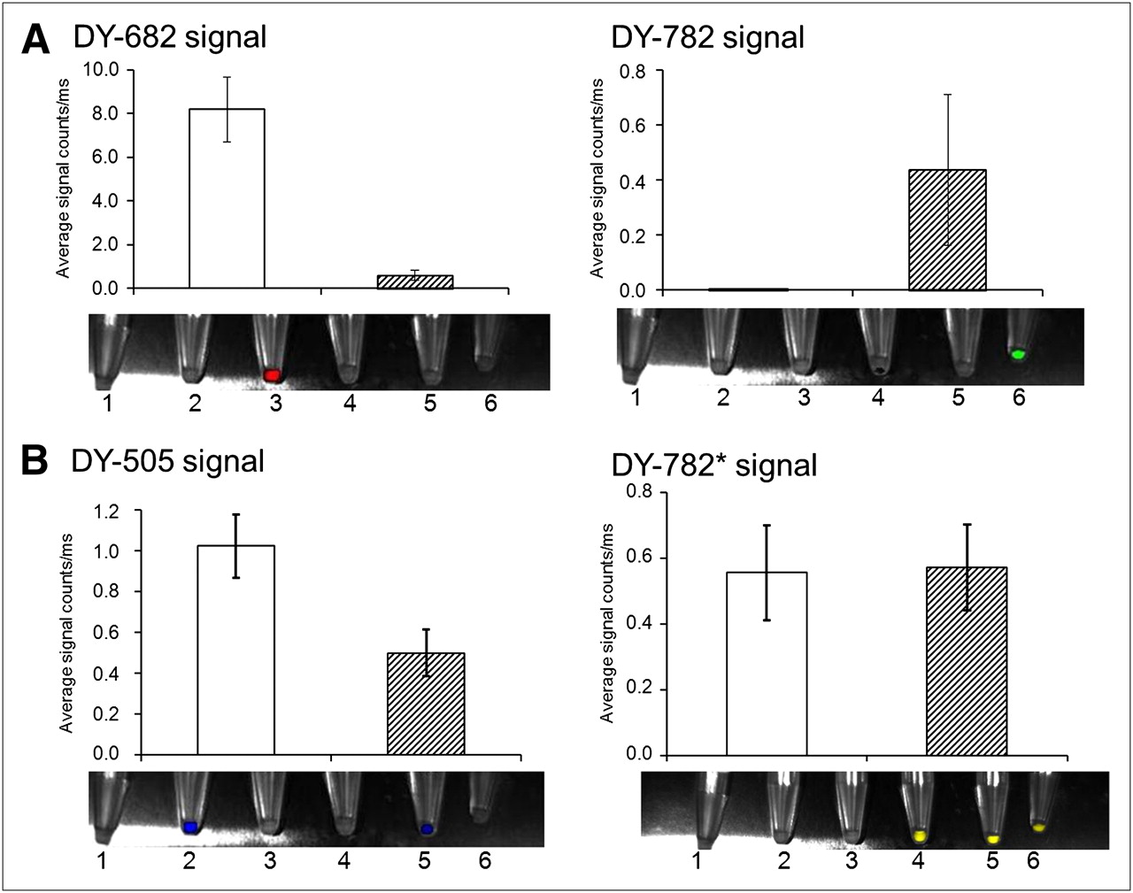
- FIGURE 1.
Acceptor fluorescence shows FRET behavior at defined concentrations of donors vs. acceptors. Defined concentrations (0.002–0.2 corresponding to 1.3–130 pmol/cm2) of DY-505, DY-682, DY-782 or combinations of DY-505 with DY-782 and DY-682 with DY-782 were spotted onto adsorbing membrane (1-to-1 stoichiometry), and fluorescence was imaged using whole-body NIRF imaging system. (A) Spectrally discriminated images of spots displaying signals derived from donor (DY-682: red, DY-505: blue) and acceptor probe (DY-782: green). (B and C) Semiquantitative analysis of acceptor fluorescence intensity alone (white bars) or in combination with donor (hatched bars). (D) Acceptor fluorescence of IgG-DY couples. d = estimated donor-to-acceptor distance; conc = concentration of each dye per spot. Data are from 3 independent experiments.
- FIGURE 2.
FRET effects can also be observed for DY-682 with DY-782 after internalization of dyes by murine macrophages J774. After incubation of J774 macrophages for 24 h with IgG-coupled dyes (1.2 nmol/mL), cells were pelleted and imaged using whole-body NIRF imaging system. Semiquantitative analysis of fluorescence intensity was performed for each fluorophore alone (white bars) or in combination (hatched bars) (A: IgG-DY-682 with DY-782; B: IgG-DY-505 with DY-782). Values are mean average of 3 different cell pellets and SEM. Bottom of each graph shows superimposed light and color-coded NIRF images of cell pellets after dye incubation.
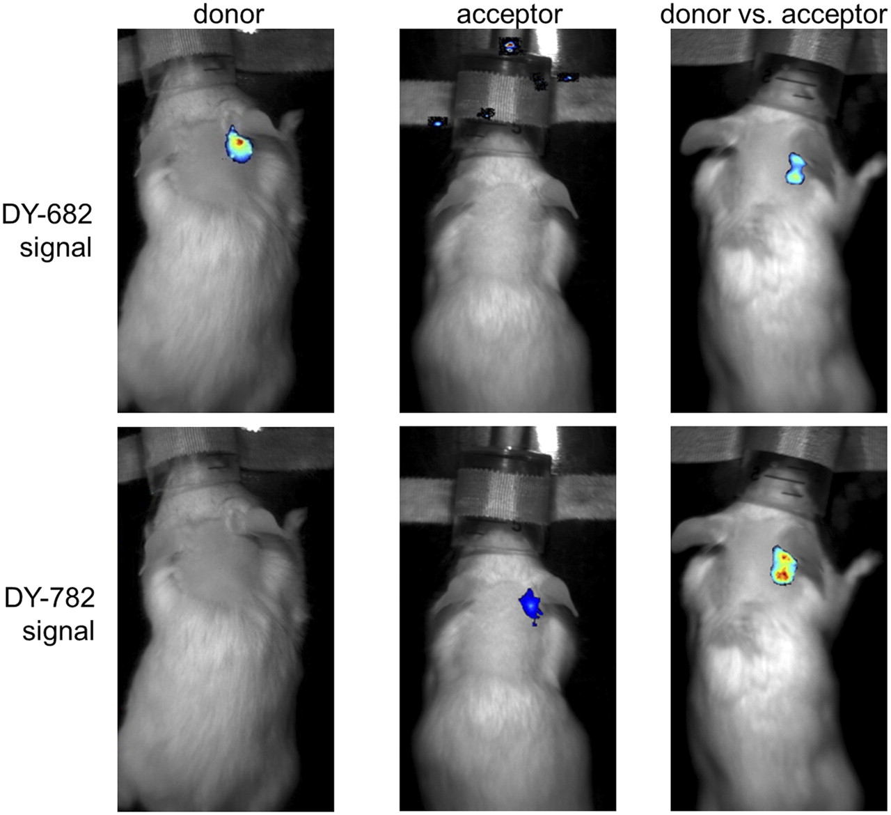
- FIGURE 3.
FRET effects can also be demonstrated after inoculation of labeled macrophages into mice. J774 macrophages were incubated with dye (1.2 nmol/mL)-coupled IgG probes (each probe alone or in combination) for 24 h and injected subcutaneously on backs of mice. Representative visible and superimposed NIRF (false colors: blue [low fluorescence intensity] and red [high fluorescence intensity]) images of mice were obtained at 30 min after inoculation using whole-body NIRF imaging system. (Top) DY-682 and DY-782 signals of mouse injected with macrophages labeled with IgG-DY-682 and IgG-DY-782. (Bottom) DY-505 and DY-782* signals corresponding to macrophages labeled with IgG-DY-505 and IgG-DY-782.
- FIGURE 4.
After systemic probe application of IgG-DY-682 vs. IgG-DY-782, FRET effects are visible in mouse model of inflammatory ear edema. Representative images are shown of DY-682 and DY-782 signals of edematous mouse injected with IgG-DY-682 and IgG-DY-782 (55 nmol of dye per kilogram of body weight) 6 h after application using whole-body NIRF imaging system. Overlay is of fluorescence (false colors: blue [low fluorescence intensity] and red [high fluorescence intensity]) and white-light images. From left to right are shown localization of donor, acceptor, and donor in presence of acceptor.


Tables
- TABLE 1.
Spectroscopic Properties of Free Dyes and Their IgG Conjugates in Phosphate-Buffered Saline
Wavelength of dye absorption maximum (nm) Wavelength of dye emission maximum (nm) Fluorescence quantum yield* Dye–IgG Dye Free dye Dye–IgG Free dye Dye–IgG Free dye Dye–IgG Free-dye molar decadic absorption coefficient at main absorption maximum (M−1 cm−1) Dye-to-protein ratio† Spectral overlap integral‡ (nm4 mol L−1) Förster radius‡ (nm) DY-682 674 682 712 713 0.20 0.18§ 1.11 × 10⁵ 2.9 19.3 × 10¹⁵ 6.4 DY-505 498 504 529 532 0.78 0.40§ 0.70 × 10⁵ 2.3 1.09 × 10¹⁵ 4.5 DY-782 752 763 796 802 0.06 0.04§ 1.12 × 10⁵ 3.1 — — ↵* Determined against standard dye 1,1′,3,3,3′,3′-hexamethylindotricarbocyanine iodide (yield = 0.33, EtOH) for DY-682 and DY-782 and against rhodamine 6G (yield = 0.89, EtOH) for DY-505.
↵† Determined photometrically.
↵‡ For FRET from respective donor dye to acceptor dye DY-782.
↵§ Fluorescence quantum yields of IgG conjugates were corrected for dimer absorption.
Supplemental Data
Files in this Data Supplement:


Jump to section
Related Articles
Cited By...
- No citing articles found.