Abstract
The opioid and serotonergic systems are closely involved in pain processing and mood disorders. The aim of this study was to assess the influence of systemic morphine on cerebral serotonin 2A receptor (5-HT2A) binding in dogs using SPECT with the 5-HT2A radioligand 123I-5I-R91150. Methods: 5-HT2A binding was estimated with and without morphine pretreatment in 8 dogs. The 5-HT2A binding indices in the frontal, parietal, temporal, and occipital cortex and in the subcortical region were obtained by semiquantification. Results: A significantly decreased 5-HT2A binding index was found in the morphine group for the right (morphine, 1.41 ± 0.06; control, 1.52 ± 0.10) and left (morphine, 1.44 ± 0.08; control, 1.55 ± 0.11) frontal cortices, with P = 0.012 and P = 0.040, respectively. No significant differences were noted for the other regions. Conclusion: Morphine decreased the frontocortical 5-HT2A availability, confirming an interaction between the 5-HTergic and the opioid systems. Whether this interaction is caused by decreased receptor density due to direct internalization or is the result of indirect actions, such as increased endogenous serotonin release, remains to be elucidated.
It is well documented that there is a close interaction between the brain opioid and serotonin (5-hydroxytryptamine [5-HT]) neurotransmitter systems (1–3). Activation of the μ-opioid receptor system leads to 5-HTergic changes, including increased release of synaptic 5-HT, as was shown in several brain regions and in the spinal cord in various in vivo microdialysis studies (1,2). Furthermore, pharmacologic and behavioral studies pointed out that the 5-HTergic system is implicated in spinal pain transmission, modulation, and control and particularly in the mediation of opioid analgesia (3,4). Morphine-induced analgesia is thought to rely in part on the indirect activation of descending inhibitory 5-HTergic pathways (3). Consequently, antidepressants that inhibit 5-HT reuptake, such as fluvoxamine and fluoxetine, are also known to have analgesic properties, especially in chronic pain states (5,6).
Beside its role in pain modulation (7–9), 5-HT2A receptor (5-HT2A) gained interest over the last few decades due to its involvement in several neurologic disorders in humans, including anxiety (10), depression (11), schizophrenia (12), and obsessive–compulsive disorder (13). More specifically, atypical antipsychotic drugs acting as 5-HT2A antagonists and inverse agonists were seen to be effective in reducing the symptoms of these diseases (10,14). Because the opioid system has been shown to be involved in several of these mood disorders as well (15), the interaction between μ-opioid receptors and 5-HT2A at an emotional or cognitive level gained interest, partly with regard to common opioid side effects such as tolerance and addiction that are thought to have a 5-HTergic component as well. Consequently, several in vitro and agonist–antagonist behavioral studies were conducted on rodents, giving further evidence for this opioid–serotonin interaction (13,16–18).
Functional brain imaging is a valuable tool for the investigation of neurotransmitter systems in vivo and is thus commonly used in research involving both neurologic disorders and pain. The visualization of the brain 5-HT2A system in dogs with SPECT and the radioligand 123I-5I-R91150 was proven to be efficacious in several studies. Studies regarding the role of the 5-HT2A system in canine anxiety, aggression, and aging supported the application of a dog model as a suitable model for human disease (19–21).
The aim of the present study was to assess the influence of morphine on central 5-HT2A in dogs. More specifically, the possible change in 5-HT2A availability after an acute dose of morphine was investigated by means of functional brain imaging.
MATERIALS AND METHODS
Animals
This study was approved by the Ghent University Ethical Committee (EC 2008-143). All guidelines for animal welfare, imposed by the Ethical Committee, were respected. To minimize sex, breed, and age influence, 8 female neutered beagle dogs approximately 5 y old were used for this study. The dogs weighed 13.04 ± 1.06 kg (mean ± SD). None of the dogs had a history of previous major disease or neurologic disorder.
Tracer
123I-5I-R91150 (123I-labeled 4-amino-N-[1-[3-(4-fluorophenoxy)propyl]-4-methyl-4-piperidinyl]-5-iodo-2-methoxybenzamide) was synthesized by electrophilic substitution on the 5-position of the methoxybenzamide group of R91150, followed by purification with high-performance liquid chromatography, and was obtained from the Nuclear Medicine and PET Research Department of the VU University Medical Centre. The product had a radiochemical purity of more than 99% and was sterile and pyrogen-free. A specific activity of 370 GBq/μmol was obtained. The radioligand is a 5-HT2A antagonist with high affinity (dissociation constant, 0.11 nM) and selectivity for 5-HT2A. The selectivity of the ligand for 5-HT2A with regard to other neurotransmitter receptors such as other 5-HT receptors, including 5-HT2C and 5-HT1A, dopamine receptors, adrenergic receptors, and histamine receptors, is at least a factor of 50. The tracer is displaceable with ketanserin, a 5-HT2 antagonist (22), and has been shown to be 5-HT2A–specific in dogs (23,24).
Procedure
To assess the possible influence of morphine on the binding index of the 5-HT2A radioligand, binding indices obtained by SPECT after morphine pretreatment were compared with binding indices obtained without morphine pretreatment. Each dog was scanned twice (nonrandomized), with a washout period of at least 2 mo between the scans. Images were acquired 90 min after radioligand injection, based on previous results for optimal scanning time (24). The injected tracer activity was 14.73 ± 3.71 MBq/kg of body weight for the control group and 15.42 ± 1.21 MBq/kg of body weight for the morphine group (mean ± SD). In the morphine group, pretreatment (0.5 mg/kg of body weight, morphine HCl; Sterop) was administered through manual intravenous injection over 1 min, 30 min before radioligand injection. The dose was based on a clinically relevant dose in dogs (25).
Anesthetic Protocol
All dogs were sedated before undergoing general anesthesia with intramuscular dexmedetomidine (Dexdomitor; Orion Corp.), 375 μg/m2 of body surface area. Anesthesia was induced with intravenous propofol (Propovet; Abbott Laboratories), 2.22 ± 0.40 mg/kg of body weight for the control group and 1.55 ± 0.31 mg/kg of body weight for the morphine group (mean ± SD). Anesthesia was maintained, after endotracheal intubation, with isoflurane (Isoflo; Abbott Laboratories) (1.4% end-tidal concentration) in oxygen using a rebreathing system. The acquisition started 10 min after induction of anesthesia. The dogs were mechanically ventilated by intermittent positive pressure ventilation to maintain eucapnia, that is, end-tidal CO2 within 35–45 mm Hg, to limit the influence of partial pressure of CO2 on cerebral perfusion.
Acquisition
The dogs were positioned in ventral recumbence with the head fixed in a preformed cushion, to prevent individual positioning artifacts. SPECT was performed with a triple-head γ-camera (Triad Trionix) equipped with ultra-high-resolution parallel-hole collimators (8 mm in full width at half maximum). Images were acquired 90 min after injection of the radioligand. The total acquisition time was 30 min. For each acquisition, 90 projection images were obtained on a 128 × 128 matrix using a step-and-shoot mode (20 s/step, 4° steps). Camera and table positioning were recorded to ensure optimal intraindividual comparison.
Processing Protocol
The radioligand data were reconstructed using the HybridRecon program (Hermes Medical Solutions). We applied an ordered-subset expectation maximization-based reconstruction algorithm with attenuation (using a CT transmission scan), collimator response, and Monte Carlo–based scatter correction. The details of the reconstruction algorithm can be found in a publication by Sohlberg et al. (26).
The 123I-5I-R91150 emission data were fitted to a template based on previously obtained cerebral perfusion data using multimodality software (version 5.0; Nuclear Diagnostics AB). This software displays images in a dual-window setting and allows for manual coregistration by providing tools for scaling, rotating, and translating images in all 3 dimensions. The perfusion data provide the necessary anatomic references using the region-of-interest (ROI) approach. The following ROIs were included: right and left frontal, temporal, parietal, and occipital cortices, as well as the subcortical and cerebellar region. The cerebellum, a region with low densities of 5-HT2A, was used as a reference region for nonspecifically bound and free radioligand. Consequently, the measured radioactivity in the cortical regions represents the total activity, that is, specific and nonspecific bound and free radioligand. The binding index was operationally calculated as ([counts per pixel in ROI] − [counts per pixel in cerebellum])/(counts per pixel in cerebellum). This binding index is proportional to the in vivo density of available receptors under pseudoequilibrium conditions (27).
Statistical Analysis
The injected radioligand dose per kilogram of body weight and the injected propofol dose were compared between the 2 treatment groups using the Student t test. Changes in 5-HT2A binding with and without morphine pretreatment were compared using the mixed model, with “treatment” and “ROI” as categoric fixed effects and “dog” as a random effect. Left–right differences within groups were compared using a paired t test. Data are presented as mean ± SD. The level of significance was set at a P value of less than 0.05.
RESULTS
No severe adverse reactions were seen after the morphine administration. All dogs were variably sedated. Some dogs showed mild nausea (n = 4) and panting (n = 4), and 1 dog had a short period of mild excitation. None of the dogs vomited.
No significant differences were found between the 2 treatment groups for the injected radioligand dose per kilogram of body weight. A significant difference was found for the propofol dose per kilogram of body weight between groups; namely dogs in the morphine group required a lower propofol dose for induction (P = 0.0013).
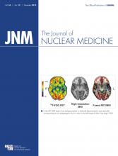
Table 1 represents the 5-HT2A binding indices in the different ROIs for both groups. A significantly lower binding index in the left (P = 0.040) and right (P = 0.012) frontal cortices was found in the dogs pretreated with morphine, compared with the control group (Table 1). The decreased 5-HT2A availability in the frontal cortex after morphine treatment can be visually appreciated in Figure 1.
5-HT2A Binding Indices With and Without Morphine Pretreatment (0.5 mg/kg Intravenously) in 8 Dogs
Sagittal (left) and transversal (right) 123I-5I-R91150 SPECT images of 1 dog. Compared with control images (A and B), 5-HT2A availability was decreased after morphine pretreatment (C and D) in frontal cortex (delineated region).
The left–right comparison within groups showed no significant differences.
DISCUSSION
A 123I-5I-R91150 SPECT study was conducted on dogs to examine the influence of a single intravenous bolus of morphine on 5-HT2A binding, and a decline in 5-HT2A availability was observed in the frontocortical region in dogs treated with morphine, compared with the untreated group.
The binding indices were decreased significantly only in the frontal cortex, even though there was an overall decrease in 5-HT2A binding after morphine treatment (Fig. 1). Both μ-opioid receptor and 5-HT2A are widely distributed throughout the cortex (28,29). The frontal cortex, however, is known to be a region with both high μ-opioid receptor expression and high 5-HT2A expression (28,29). The frontocortical region is involved in the control of the cognitive and emotional aspects of pain. Not only do μ-opioid receptor analgesics exert their action through peripheral and spinal μ-opioid receptor activation, which alters the transmission of the noxious stimulus, but also through central μ-opioid receptor activation which influences the perception of pain. Therefore, the frontal cortex is, not surprisingly, rich in μ-opioid receptors. In the frontal cortex, 5-HT2A is well represented as well (28,29). Frontocortical 5-HT2A has been shown to play a role in several mood disorders, such as anxiety, aggression, and depression, not only in humans (10,11) but also in dogs (19,21). These are mood disorders that often arise during opioid addiction and dependence, and a 5-HTergic dysfunction is suggested to represent a main mechanism contributing to mood disorders in opiate abstinence (30). Additionally, 5-HT2A blockade is thought to restore some of the neurochemical modifications induced by long-term use of drugs of abuse, such as morphine (31).
The decreased 5-HT2A binding indices found in the frontal cortex could be due to morphine-induced 5-HT release in the cortex, hence competing or interfering with 5-HT2A radioligand binding. μ-opioid receptor agonists affect, next to their direct inhibitory action on neurons, several other neurotransmitter systems that contribute to opioid effects. One of these neurotransmitter systems is the 5-HTergic system, greatly involved in mood regulation. It has been well established that the analgesic action of morphine relies in part on the activation of descending bulbospinal 5-HTergic fibers. Supraspinal μ-opioid receptor activation by morphine leads to 5-HT release in the spinal cord (1). Supraspinal μ-opioid receptor activation also causes increased 5-HT levels in several brain regions (2). In rodents, for instance, systemic morphine increased extracellular 5-HT in the frontal cortex (32). Another sequel of the increased synaptic 5-HT release could be downregulation or internalization of 5-HT2A. However, the in vitro study from Van Oekelen et al. demonstrated that cell pretreatment with 5-HT for 15 min and 48 h did not provoke reduced 5-HT2A numbers (33). This finding pleads against downregulation or internalization of 5-HT2A by morphine-invoked release of 5-HT.
Furthermore, it has been demonstrated that both receptors share a common intracellular pathway involving protein kinase C activation (18). The latter study additionally reported that 5-HT2A activation resulted in a faster μ-opioid receptor desensitization, internalization, and downregulation (18). By analogy, we might be tempted to interpret our results in a similar although opposite way, namely that μ-opioid receptor activation might increase 5-HT2A downregulation or internalization. An interaction between both receptors on glutamatergic neurons in the neocortex has been suggested by the findings of Marek et al. (16).
Both receptors appear to regulate glutamate release presynaptically: 5-HT2A activation promotes glutamate release, whereas μ-opioid receptor activation inhibits this enhanced glutamate release (16). The glutamate release induced by 5-HT2A activation is thought to mediate a typical behavioral pattern of head shaking and twitching, which is seen after frontocortical 5-HT2A activation (34). Accordingly, this behavior was attenuated after μ-opioid receptor agonist (buprenorphine, fentanyl) administration (17).
This might not be the only cause for the lower binding indices found. SPECT is an elegant technique to quantify neurotransmitter systems in vivo, albeit a degree of caution is warranted when interpreting the images since the present findings may reflect other types of interactions as well. Direct interactions involving direct binding of morphine to 5-HT2A may be another possibility, since some studies have found direct interactions between opioids and 5-HTergic receptors. For instance, morphine was shown to interact with human 5-HT3A (35), and fentanyl shows an affinity, albeit low, for 5-HT1A (36). To our knowledge, however, no such interaction has been described for morphine and 5-HT2A.
In a recent study comparing the effects of morphine on 5-HTergic neurons in midbrain areas in the presence or absence of acute noxious stimulation, it was found that those 5-HTergic neurons responded differently to morphine when pain was present; that is, they showed a higher neuronal activity (37). The influence of morphine on 5-HT2A availability might be different in the presence of acute noxious stimulation.
The interference of anesthetics with radioligand binding also needs consideration (38). However, both groups underwent the same anesthetic protocol, with the exception of the propofol dose, thus eliminating interference of anesthetics with radioligand binding as a possible confounding factor. Several studies on the influence of different anesthetic agents on the central 5-HTergic system have shown that isoflurane reduces 5-HT release (39), whereas propofol increases synaptic 5-HT levels by blocking the 5-HT transporter (40). In the present study, the control group was administered more propofol than the morphine group. However, recent unpublished findings from our research group suggested no difference in 5-HT2A binding between low and high propofol doses, implying that the difference in propofol dose between the 2 treatment groups in this study would be of minor importance. Additionally, we found that morphine did not influence regional cerebral blood flow in dogs (unpublished data); therefore, this factor cannot account for the found changes in 5-HT2A binding indices.
CONCLUSION
Our findings support the existence of an interaction between morphine and 5-HT2A binding in the canine frontal cortex. In the absence of pain and mood disorders, morphine decreased 5-HT2A availability. Future studies investigating the exact mechanism of this interaction would be of great interest, including the influence of morphine on the serotonin transporter. Overall, the present study supports the possible beneficial effect of 5-HT2A drugs in the prevention of common opiate side effects such as dependence and addiction, as well as the use of opioids in mood disorders.
DISCLOSURE STATEMENT
The costs of publication of this article were defrayed in part by the payment of page charges. Therefore, and solely to indicate this fact, this article is hereby marked “advertisement” in accordance with 18 USC section 1734.
Acknowledgments
This study was supported by Special Research Fund Grant 01J06109 from Ghent University. No other potential conflict of interest relevant to this article was reported.
Footnotes
Published online Oct. 22, 2012.
- © 2012 by the Society of Nuclear Medicine and Molecular Imaging, Inc.
REFERENCES
- Received for publication October 4, 2010.
- Accepted for publication June 28, 2012.


