
Main menu

  • Home
  • Content
    • Current
    • Ahead of print
    • Past Issues
    • JNM Supplement
    • SNMMI Annual Meeting Abstracts
    • Continuing Education
    • JNM Podcasts
  • Subscriptions
    • Subscribers
    • Institutional and Non-member
    • Rates
    • Journal Claims
    • Corporate & Special Sales
  • Authors
    • Submit to JNM
    • Information for Authors
    • Assignment of Copyright
    • AQARA requirements
  • Info
    • Reviewers
    • Permissions
    • Advertisers
  • About
    • About Us
    • Editorial Board
    • Contact Information
  • More
    • Alerts
    • Feedback
    • Help
    • SNMMI Journals
  • SNMMI
    • JNM
    • JNMT
    • SNMMI Journals
    • SNMMI

User menu

  • Subscribe
  • My alerts
  • Log in
  • Log out
  • My Cart

Search

  • Advanced search
Journal of Nuclear Medicine
  • SNMMI
    • JNM
    • JNMT
    • SNMMI Journals
    • SNMMI
  • Subscribe
  • My alerts
  • Log in
  • Log out
  • My Cart
Journal of Nuclear Medicine

Advanced Search

  • Home
  • Content
    • Current
    • Ahead of print
    • Past Issues
    • JNM Supplement
    • SNMMI Annual Meeting Abstracts
    • Continuing Education
    • JNM Podcasts
  • Subscriptions
    • Subscribers
    • Institutional and Non-member
    • Rates
    • Journal Claims
    • Corporate & Special Sales
  • Authors
    • Submit to JNM
    • Information for Authors
    • Assignment of Copyright
    • AQARA requirements
  • Info
    • Reviewers
    • Permissions
    • Advertisers
  • About
    • About Us
    • Editorial Board
    • Contact Information
  • More
    • Alerts
    • Feedback
    • Help
    • SNMMI Journals
  • View or Listen to JNM Podcast
  • Visit JNM on Facebook
  • Join JNM on LinkedIn
  • Follow JNM on Twitter
  • Subscribe to our RSS feeds
Meeting ReportInstrumentation & Data Analysis: Data Analysis & Management

Computer assisted cancer diagnosis from PET-CT using dynamic threshold adjustment method

Takako Sato, Haruomi Kimura, Hiroshi Arisawa, Momoko Okasaki, Ryogo Minamimoto and Tomio Inoue
Journal of Nuclear Medicine May 2011, 52 (supplement 1) 2096;
Takako Sato
1Graduate School of Environment and Information Sciences, Yokohama National University, Yokohama, Japan
  • Find this author on Google Scholar
  • Find this author on PubMed
  • Search for this author on this site
Haruomi Kimura
1Graduate School of Environment and Information Sciences, Yokohama National University, Yokohama, Japan
  • Find this author on Google Scholar
  • Find this author on PubMed
  • Search for this author on this site
Hiroshi Arisawa
1Graduate School of Environment and Information Sciences, Yokohama National University, Yokohama, Japan
  • Find this author on Google Scholar
  • Find this author on PubMed
  • Search for this author on this site
Momoko Okasaki
2Department of Radiology, Yokohama City University School of Medicine, Yokohama, Japan
  • Find this author on Google Scholar
  • Find this author on PubMed
  • Search for this author on this site
Ryogo Minamimoto
3Department of Radiology, National Center for Global Health and Medicine, Tokyo, Japan
  • Find this author on Google Scholar
  • Find this author on PubMed
  • Search for this author on this site
Tomio Inoue
2Department of Radiology, Yokohama City University School of Medicine, Yokohama, Japan
  • Find this author on Google Scholar
  • Find this author on PubMed
  • Search for this author on this site
  • Article
  • Figures & Data
  • Info & Metrics
Loading

Abstract

2096

Objectives In Cancer Diagnosis from PET-CT images, the radiologists seek abnormal areas as isolated accumulations of FDG. However the color and contrast of regions sometimes depend on the color-mappings on the viewer set by an engineer. To avoid that, we have developed a method for detecting abnormal area and presenting them in a standardized color and density.

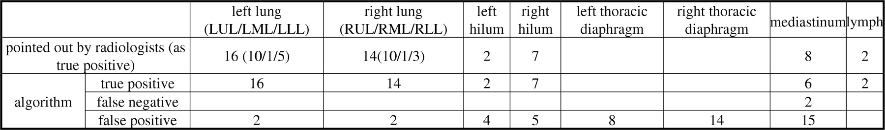
Methods The method consists of 2 phases. First the 3-dimetional outline of each organ is extracted on PET images automatically. Second, in each organ, “cliff” areas (CA) are detected. CA means an area which has a larger SUV than a organ-specific value and also shows a sudden rise of SUV. Then the CA is extended to neighboring regions which have larger SUV than the minimum SUV in the original CA. To verify the accuracy of algorithm, we compare the algorithm result in lung and mediastinum with the report written by 2 experienced radiologist (over 10 years). The study included 26 patients (F:M 4:22; age range 36-89, 72 ± 12 years) who have cancer(s) in lung or mediastinum. Radiologist pointed out 49 areas.

Results Algorithm can detect 47 areas as abnormal in 49 ‘true’ positives. But 2 area is detected as in another organ. False negative: 2 (all in mediastinum), False positive: 50 areas (18 in thoracic diaphragm, 14 in mediastinum, 9 in hilum). Sensitivity is 95.9%, PPV is 48.5%. Specificity of organs: 52.2%(left lung), 44.0%(right lung), 48.6%(mediastinum), so there is no difference between organs.

Conclusions The above algorithm could detect all abnormal areas involving comparatively-low SUV areas. More precise detection of outline of organs is expected

Figure
  • Download figure
  • Open in new tab
  • Download powerpoint
Back to top

In this issue

Journal of Nuclear Medicine
Vol. 52, Issue supplement 1
May 2011
  • Table of Contents
  • Index by author
Article Alerts
Sign In to Email Alerts with your Email Address
Email Article

Thank you for your interest in spreading the word on Journal of Nuclear Medicine.

NOTE: We only request your email address so that the person you are recommending the page to knows that you wanted them to see it, and that it is not junk mail. We do not capture any email address.

Enter multiple addresses on separate lines or separate them with commas.
Computer assisted cancer diagnosis from PET-CT using dynamic threshold adjustment method
(Your Name) has sent you a message from Journal of Nuclear Medicine
(Your Name) thought you would like to see the Journal of Nuclear Medicine web site.
Citation Tools
Computer assisted cancer diagnosis from PET-CT using dynamic threshold adjustment method
Takako Sato, Haruomi Kimura, Hiroshi Arisawa, Momoko Okasaki, Ryogo Minamimoto, Tomio Inoue
Journal of Nuclear Medicine May 2011, 52 (supplement 1) 2096;

Citation Manager Formats

  • BibTeX
  • Bookends
  • EasyBib
  • EndNote (tagged)
  • EndNote 8 (xml)
  • Medlars
  • Mendeley
  • Papers
  • RefWorks Tagged
  • Ref Manager
  • RIS
  • Zotero
Share
Computer assisted cancer diagnosis from PET-CT using dynamic threshold adjustment method
Takako Sato, Haruomi Kimura, Hiroshi Arisawa, Momoko Okasaki, Ryogo Minamimoto, Tomio Inoue
Journal of Nuclear Medicine May 2011, 52 (supplement 1) 2096;
Twitter logo Facebook logo LinkedIn logo Mendeley logo
  • Tweet Widget
  • Facebook Like
  • Google Plus One
Bookmark this article

Jump to section

  • Article
  • Figures & Data
  • Info & Metrics

Related Articles

  • No related articles found.
  • Google Scholar

Cited By...

  • No citing articles found.
  • Google Scholar

More in this TOC Section

Instrumentation & Data Analysis: Data Analysis & Management

  • Variability in SUV biodistribution in delayed PET-MR studies
  • Automated temporal subtraction scheme of torso FDG-PET scans by using a statistical shape model for normal cases
  • Impact of partial volume correction on FDG treatment response measures in head and neck and advanced ovarian cancer
Show more Instrumentation & Data Analysis: Data Analysis & Management

Data Analysis and Management Posters

  • Quantitative differences of pancreatic lesions in cilinical simultaneous [18F]FDG PET/MR imaging:TOF versus non-TOF measurements
  • Extension of the MIAKAT analysis software package to non-brain and pre-clinical PET analysis.
  • Robustness of radiomic features in 18F-FDG PET/CT imaging of Nasopharyngeal Carcinoma: impact of parameter settings on different feature matrices
Show more Data Analysis and Management Posters

Similar Articles

SNMMI

© 2025 SNMMI

Powered by HighWire