
Main menu

  • Home
  • Content
    • Current
    • Ahead of print
    • Past Issues
    • JNM Supplement
    • SNMMI Annual Meeting Abstracts
    • Continuing Education
    • JNM Podcasts
  • Subscriptions
    • Subscribers
    • Institutional and Non-member
    • Rates
    • Journal Claims
    • Corporate & Special Sales
  • Authors
    • Submit to JNM
    • Information for Authors
    • Assignment of Copyright
    • AQARA requirements
  • Info
    • Reviewers
    • Permissions
    • Advertisers
  • About
    • About Us
    • Editorial Board
    • Contact Information
  • More
    • Alerts
    • Feedback
    • Help
    • SNMMI Journals
  • SNMMI
    • JNM
    • JNMT
    • SNMMI Journals
    • SNMMI

User menu

  • Subscribe
  • My alerts
  • Log in
  • My Cart

Search

  • Advanced search
Journal of Nuclear Medicine
  • SNMMI
    • JNM
    • JNMT
    • SNMMI Journals
    • SNMMI
  • Subscribe
  • My alerts
  • Log in
  • My Cart
Journal of Nuclear Medicine

Advanced Search

  • Home
  • Content
    • Current
    • Ahead of print
    • Past Issues
    • JNM Supplement
    • SNMMI Annual Meeting Abstracts
    • Continuing Education
    • JNM Podcasts
  • Subscriptions
    • Subscribers
    • Institutional and Non-member
    • Rates
    • Journal Claims
    • Corporate & Special Sales
  • Authors
    • Submit to JNM
    • Information for Authors
    • Assignment of Copyright
    • AQARA requirements
  • Info
    • Reviewers
    • Permissions
    • Advertisers
  • About
    • About Us
    • Editorial Board
    • Contact Information
  • More
    • Alerts
    • Feedback
    • Help
    • SNMMI Journals
  • View or Listen to JNM Podcast
  • Visit JNM on Facebook
  • Join JNM on LinkedIn
  • Follow JNM on Twitter
  • Subscribe to our RSS feeds
Research ArticleBasic Science Investigations

Multimodal Imaging with 18F-FDG PET and Cerenkov Luminescence Imaging After MLN4924 Treatment in a Human Lymphoma Xenograft Model

Robbie Robertson, Melissa Saylor Germanos, Mark G. Manfredi, Peter G. Smith and Matthew D. Silva
Journal of Nuclear Medicine November 2011, 52 (11) 1764-1769; DOI: https://doi.org/10.2967/jnumed.111.091710
Robbie Robertson
  • Find this author on Google Scholar
  • Find this author on PubMed
  • Search for this author on this site
Melissa Saylor Germanos
  • Find this author on Google Scholar
  • Find this author on PubMed
  • Search for this author on this site
Mark G. Manfredi
  • Find this author on Google Scholar
  • Find this author on PubMed
  • Search for this author on this site
Peter G. Smith
  • Find this author on Google Scholar
  • Find this author on PubMed
  • Search for this author on this site
Matthew D. Silva
  • Find this author on Google Scholar
  • Find this author on PubMed
  • Search for this author on this site
  • Article
  • Figures & Data
  • Supplemental
  • Info & Metrics
  • PDF
Loading

Article Figures & Data

Figures

  • Additional Files
  • FIGURE 1.
    • Download figure
    • Open in new tab
    • Download powerpoint
    FIGURE 1.

    PET images from representative pairs of animals. Reduction in signal intensity can be observed after treatment, indicating reduction in uptake of 18F-FDG in tumor. Arrows refer to tumor location.
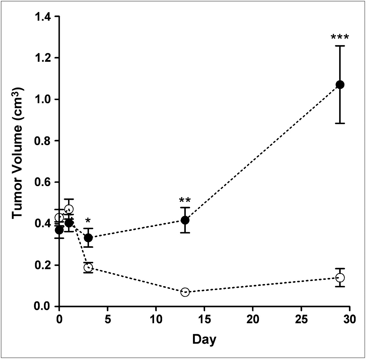
  • FIGURE 2.
    • Download figure
    • Open in new tab
    • Download powerpoint
    FIGURE 2.

    MLN4924 treatment results in reduction in tumor volume from baseline of 56% on day 3 and 84% on day 13 in OCI-Ly10 xenograft model. Treatment was halted on day 15 of study, and volume reduction is durable when compared with both baseline (−64%) and vehicle group (−87%). *P < 0.05. **P < 0.01. ***P < 0.001.
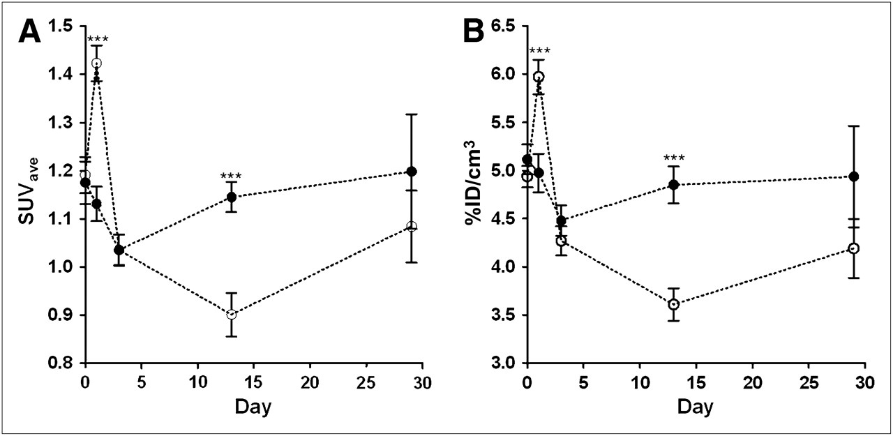
  • FIGURE 3.
    • Download figure
    • Open in new tab
    • Download powerpoint
    FIGURE 3.

    SUVave (A) and decay-corrected %ID/cm3 (B). On day 13, all PET parameters are significantly reduced, as compared with vehicle treatment, by 21.3% and 25.6% for SUVave and %ID/cm3, respectively (P < 0.0001). ***P < 0.001.
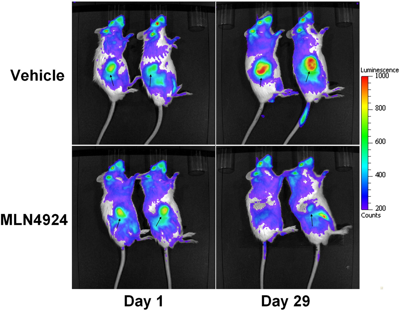
  • FIGURE 4.
    • Download figure
    • Open in new tab
    • Download powerpoint
    FIGURE 4.

    Cerenkov luminescence images from representative pairs of animals. As observed on PET data, there is visual reduction in image signal intensity in MLN4924 treatment group that is consistent with reduction in 18F-FDG uptake (Fig. 1).

  • FIGURE 5.
    • Download figure
    • Open in new tab
    • Download powerpoint
    FIGURE 5.

    Cerenkov luminescence image analysis: radiance (A) and radiance/ID (B). On day 13, CLI parameters for MLN4924-treated group were all significantly reduced from baseline (30.9% and 30.4%) and from vehicle-treated values (37.5% and 37.2%) for radiance and radiance/ID, respectively (all P < 0.0001). **P < 0.01. ***P < 0.001.
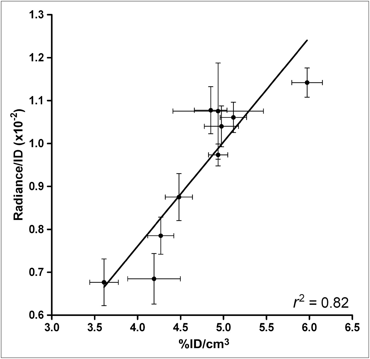
  • FIGURE 6.
    • Download figure
    • Open in new tab
    • Download powerpoint
    FIGURE 6.

    There was strong correlation between PET %ID/cm3 and CLI radiance/ID (r2 = 0.82). Correlations between PET %ID/cm3 and CLI radiance (r2 = 0.83) and other parameters are shown in Supplemental Figure 5.

Additional Files

  • Figures
  • Supplemental Data

    Files in this Data Supplement:

    • Supplemental Data
PreviousNext
Back to top

In this issue

Journal of Nuclear Medicine: 52 (11)
Journal of Nuclear Medicine
Vol. 52, Issue 11
November 1, 2011
  • Table of Contents
  • Table of Contents (PDF)
  • About the Cover
  • Index by author
Print
Download PDF
Article Alerts
Sign In to Email Alerts with your Email Address
Email Article

Thank you for your interest in spreading the word on Journal of Nuclear Medicine.

NOTE: We only request your email address so that the person you are recommending the page to knows that you wanted them to see it, and that it is not junk mail. We do not capture any email address.

Enter multiple addresses on separate lines or separate them with commas.
Multimodal Imaging with 18F-FDG PET and Cerenkov Luminescence Imaging After MLN4924 Treatment in a Human Lymphoma Xenograft Model
(Your Name) has sent you a message from Journal of Nuclear Medicine
(Your Name) thought you would like to see the Journal of Nuclear Medicine web site.
Citation Tools
Multimodal Imaging with 18F-FDG PET and Cerenkov Luminescence Imaging After MLN4924 Treatment in a Human Lymphoma Xenograft Model
Robbie Robertson, Melissa Saylor Germanos, Mark G. Manfredi, Peter G. Smith, Matthew D. Silva
Journal of Nuclear Medicine Nov 2011, 52 (11) 1764-1769; DOI: 10.2967/jnumed.111.091710

Citation Manager Formats

  • BibTeX
  • Bookends
  • EasyBib
  • EndNote (tagged)
  • EndNote 8 (xml)
  • Medlars
  • Mendeley
  • Papers
  • RefWorks Tagged
  • Ref Manager
  • RIS
  • Zotero
Share
Multimodal Imaging with 18F-FDG PET and Cerenkov Luminescence Imaging After MLN4924 Treatment in a Human Lymphoma Xenograft Model
Robbie Robertson, Melissa Saylor Germanos, Mark G. Manfredi, Peter G. Smith, Matthew D. Silva
Journal of Nuclear Medicine Nov 2011, 52 (11) 1764-1769; DOI: 10.2967/jnumed.111.091710
Twitter logo Facebook logo LinkedIn logo Mendeley logo
  • Tweet Widget
  • Facebook Like
  • Google Plus One
Bookmark this article

Jump to section

  • Article
    • Abstract
    • MATERIALS AND METHODS
    • RESULTS
    • DISCUSSION
    • CONCLUSION
    • DISCLOSURE STATEMENT
    • Acknowledgments
    • Footnotes
    • REFERENCES
  • Figures & Data
  • Supplemental
  • Info & Metrics
  • PDF

Related Articles

  • This Month in JNM
  • PubMed
  • Google Scholar

Cited By...

  • Optical Imaging Modalities: Principles and Applications in Preclinical Research and Clinical Settings
  • Targeted PET imaging strategy to differentiate malignant from inflamed lymph nodes in diffuse large B-cell lymphoma
  • {beta}-Radioluminescence Imaging: A Comparative Evaluation with Cerenkov Luminescence Imaging
  • Cerenkov-Specific Contrast Agents for Detection of pH In Vivo
  • In Vivo Localization of 90Y and 177Lu Radioimmunoconjugates Using Cerenkov Luminescence Imaging in a Disseminated Murine Leukemia Model
  • Google Scholar

More in this TOC Section

  • PET/MRI of Hypoxic Atherosclerosis Using 64Cu-ATSM in a Rabbit Model
  • Tumor Uptake of Anti-CD20 Fabs Depends on Tumor Perfusion
  • How Sensitive Is the Upper Gastrointestinal Tract to 90Y Radioembolization? A Histologic and Dosimetric Analysis in a Porcine Model
Show more Basic Science Investigations

Similar Articles

SNMMI

© 2025 SNMMI

Powered by HighWire