


Article Figures & Data
Figures
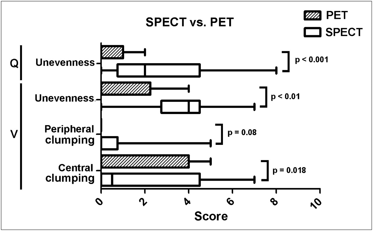
- FIGURE 1.
Box plot comparing qualitative image scores. Score ranges from 0 to 10, with 0 representing none or normal.
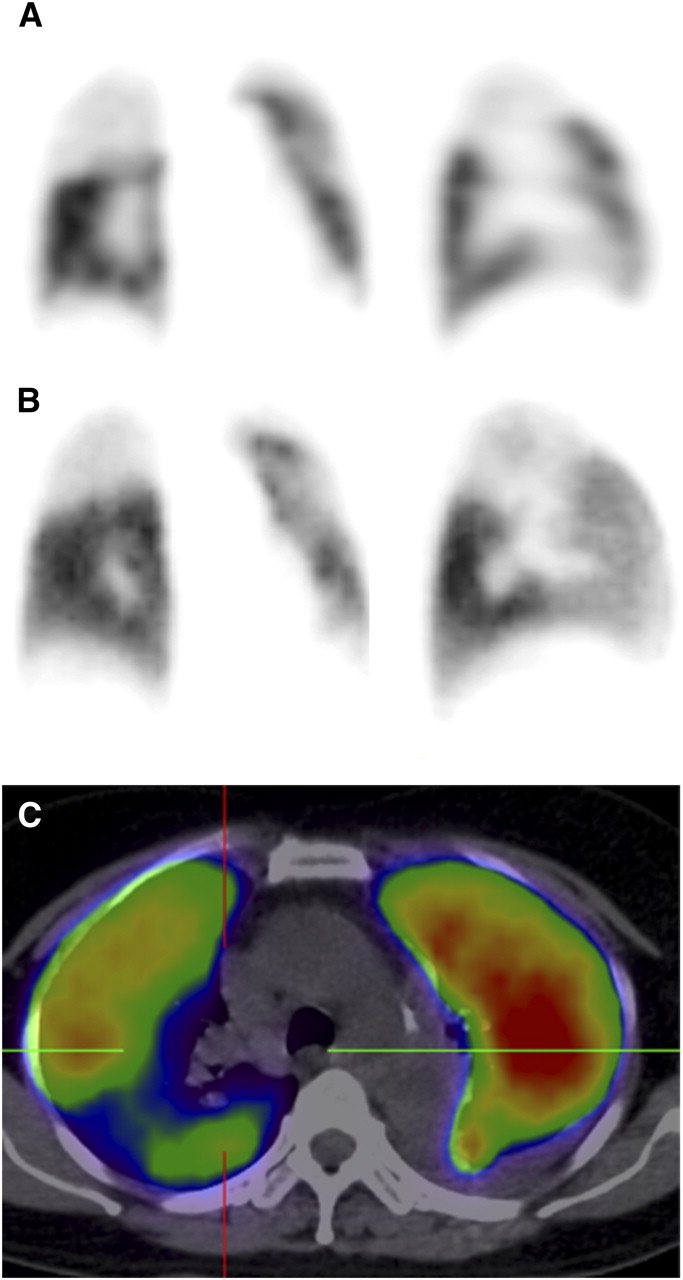
- FIGURE 2.
Representative coronal slice of patient 9, with severe chronic obstructive airway disease. (A) SPECT ventilation image was deemed nondiagnostic because of marked clumping and poor peripheral distribution of radiotracer. (B) PET image also demonstrates clumping, but peripheral distribution is more homogeneous, with corresponding matched changes on perfusion imaging enabling exclusion of PE with higher confidence. (C and D) Fused PET/CT (C) and low-dose CT (D) demonstrate no evidence of consolidation.
- FIGURE 3.
Coronal (left) and sagittal (right) SPECT (A), PET (B), and PET/CT (C) perfusion images of patient 5, demonstrating multiple segmental unmatched perfusion defects with near-normal ventilation (not shown). Diagnosis of PE was confirmed on follow-up. PET perfusion study was performed 8 d after SPECT study, accounting for resolution of some perfusion abnormalities.
- FIGURE 4.
Coronal and sagittal SPECT perfusion (A), PET perfusion (B), and axial PET/CT (C) images in patient with non–small cell lung carcinoma. Right upper lobe shows large unmatched perfusion defect (normal ventilation not shown). On its own, this may be interpreted as consistent with PE, but correlative CT demonstrates extensive mediastinal lymphadenopathy with extrinsic compression of right upper lobe pulmonary artery, a more likely explanation for findings than PE. Contrast-enhanced CT confirmed findings.


Tables
Patient no. Age (y) Symptoms of DVT PE most or equally likely diagnosis HR > 100 Immobilization (≥3 d) or surgery in previous 4 wk Prior PE or DVT Hemoptysis Malignancy Prior radiotherapy to chest 1 61 N N Y N N N Y Y 2 24 N Y Y N N N Y Y 3 55 N N Y N N N Y N 4 41 N N N N N N Y N 5 62 Y Y Y Y Y N N N 6 68 N N Y Y N N Y N 7 57 N N N N N N Y N 8 26 N N Y N N N Y Y 9 70 N Y N N N N Y Y 10 54 Y Y Y Y N N Y N DVT = deep venous thrombosis; HR = heart rate.
Patient no. Days between PET and SPECT Conventional V/Q result PET/CT result CTPA result Final diagnosis on follow-up Length of follow-up (d) Technique PE Ancillary findings 1 0 SPECT/CT Absent Airway disease Concordant — No PE 151 2 0 SPECT/CT Absent Atelectasis, mediastinal LN, pericardial effusion Concordant Equivocal No PE 178 3 0 SPECT/CT Absent Extrinsic RUL artery compression, postradiotherapy change Concordant — No PE 140 4 2 SPECT/CT Absent Concordant No PE 44 5 8 SPECT Bilateral Concordant Bilateral PE PE 172 6 0 SPECT/CT Absent Concordant — No PE 147 7 6 SPECT/CT Absent Concordant — No PE 137 8 0 SPECT/CT Absent Postradiotherapy changes Concordant — No PE 107 9 1 SPECT/CT Nondiagnostic Airway disease PE absent — No PE 102 10 1 SPECT/CT Bilateral Postradiotherapy change, bronchiectasis PE absent — PE 81 RUL = right upper lobe.
Supplemental Data
Files in this Data Supplement:


Jump to section
Related Articles
Cited By...
- A Technical Overview of Technegas as a Lung Ventilation Agent
- Single-arm prospective interventional study assessing feasibility of using gallium-68 ventilation and perfusion PET/CT to avoid functional lung in patients with stage III non-small cell lung cancer
- Correlation of 68Ga Ventilation-Perfusion PET/CT with Pulmonary Function Test Indices for Assessing Lung Function