
Main menu

  • Home
  • Content
    • Current
    • Ahead of print
    • Past Issues
    • JNM Supplement
    • SNMMI Annual Meeting Abstracts
    • Continuing Education
    • JNM Podcasts
  • Subscriptions
    • Subscribers
    • Institutional and Non-member
    • Rates
    • Journal Claims
    • Corporate & Special Sales
  • Authors
    • Submit to JNM
    • Information for Authors
    • Assignment of Copyright
    • AQARA requirements
  • Info
    • Reviewers
    • Permissions
    • Advertisers
  • About
    • About Us
    • Editorial Board
    • Contact Information
  • More
    • Alerts
    • Feedback
    • Help
    • SNMMI Journals
  • SNMMI
    • JNM
    • JNMT
    • SNMMI Journals
    • SNMMI

User menu

  • Subscribe
  • My alerts
  • Log in
  • Log out
  • My Cart

Search

  • Advanced search
Journal of Nuclear Medicine
  • SNMMI
    • JNM
    • JNMT
    • SNMMI Journals
    • SNMMI
  • Subscribe
  • My alerts
  • Log in
  • Log out
  • My Cart
Journal of Nuclear Medicine

Advanced Search

  • Home
  • Content
    • Current
    • Ahead of print
    • Past Issues
    • JNM Supplement
    • SNMMI Annual Meeting Abstracts
    • Continuing Education
    • JNM Podcasts
  • Subscriptions
    • Subscribers
    • Institutional and Non-member
    • Rates
    • Journal Claims
    • Corporate & Special Sales
  • Authors
    • Submit to JNM
    • Information for Authors
    • Assignment of Copyright
    • AQARA requirements
  • Info
    • Reviewers
    • Permissions
    • Advertisers
  • About
    • About Us
    • Editorial Board
    • Contact Information
  • More
    • Alerts
    • Feedback
    • Help
    • SNMMI Journals
  • View or Listen to JNM Podcast
  • Visit JNM on Facebook
  • Join JNM on LinkedIn
  • Follow JNM on Twitter
  • Subscribe to our RSS feeds
Research ArticleClinical Investigation

Detection of Hepatocellular Carcinoma with PET/CT: A Prospective Comparison of 18F-Fluorocholine and 18F-FDG in Patients with Cirrhosis or Chronic Liver Disease

Jean-Noël Talbot, Laetitia Fartoux, Sona Balogova, Valérie Nataf, Khaldoun Kerrou, Fabrice Gutman, Virginie Huchet, David Ancel, Jean-Didier Grange and Olivier Rosmorduc
Journal of Nuclear Medicine November 2010, 51 (11) 1699-1706; DOI: https://doi.org/10.2967/jnumed.110.075507
Jean-Noël Talbot
  • Find this author on Google Scholar
  • Find this author on PubMed
  • Search for this author on this site
Laetitia Fartoux
  • Find this author on Google Scholar
  • Find this author on PubMed
  • Search for this author on this site
Sona Balogova
  • Find this author on Google Scholar
  • Find this author on PubMed
  • Search for this author on this site
Valérie Nataf
  • Find this author on Google Scholar
  • Find this author on PubMed
  • Search for this author on this site
Khaldoun Kerrou
  • Find this author on Google Scholar
  • Find this author on PubMed
  • Search for this author on this site
Fabrice Gutman
  • Find this author on Google Scholar
  • Find this author on PubMed
  • Search for this author on this site
Virginie Huchet
  • Find this author on Google Scholar
  • Find this author on PubMed
  • Search for this author on this site
David Ancel
  • Find this author on Google Scholar
  • Find this author on PubMed
  • Search for this author on this site
Jean-Didier Grange
  • Find this author on Google Scholar
  • Find this author on PubMed
  • Search for this author on this site
Olivier Rosmorduc
  • Find this author on Google Scholar
  • Find this author on PubMed
  • Search for this author on this site
  • Article
  • Figures & Data
  • Info & Metrics
  • PDF
Loading

Article Figures & Data

Figures

  • Tables
  • FIGURE 1.
    • Download figure
    • Open in new tab
    • Download powerpoint
    FIGURE 1.

    Maximum-intensity-projection images showing 2 liver foci; the smallest, in inferior part of liver, was 18F-fluorocholine– and 18F-FDG–positive. On transaxial slices of upper part of liver, center of largest lesion was photopenic on both 18F-fluorocholine (left) and 18F-FDG (right) PET/CT images and corresponded to hemorrhagic necrosis. At periphery of this lesion, there was definite 18F-FDG uptake, and 18F-fluorocholine was taken up but with lesser intensity than for noncancerous liver parenchyma (18F-fluorocholine tumor–to–non-tumor ratio T/NTR = 0.97). Postsurgical histology confirmed well-differentiated HCC in this part of lesion. Thus, as compared with nonmalignant liver, HCC tissue was hypermetabolic for 18F-FDG but hypometabolic for 18F-fluorocholine.
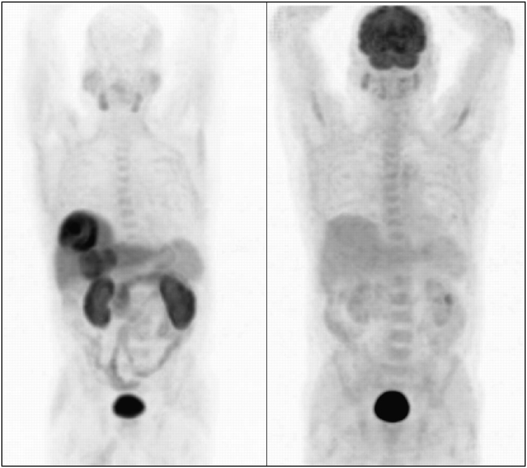
  • FIGURE 2.
    • Download figure
    • Open in new tab
    • Download powerpoint
    FIGURE 2.

    Maximum-intensity-projection images showing 2 liver foci, both hot on 18F-fluorocholine PET (left) but not visible on 18F-FDG PET (right) images. Both liver foci corresponded to untreated well-differentiated HCC.
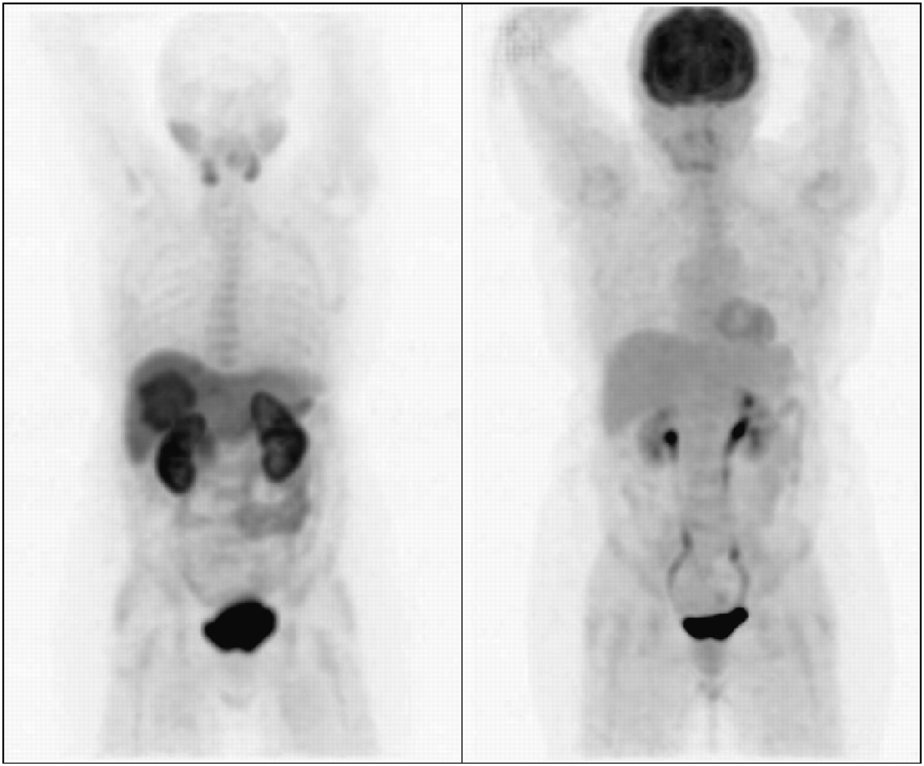
  • FIGURE 3.
    • Download figure
    • Open in new tab
    • Download powerpoint
    FIGURE 3.

    Resection of left liver for hepatocholangiocarcinoma; 20-mm nodule subsequently developed in remaining liver (arrow). Nodule appeared hypometabolic for 18F-fluorocholine (bottom left) and avid for 18F-FDG (bottom right); hepatocholangiocarcinoma was confirmed by biopsy. On PET images of thorax (top left, 18F-fluorocholine; top right, 18F-FDG), widespread lung foci were discovered, corresponding to 1 metastasis but also to lesions of anthracosilicosis, and 1 thyroid nodule, which was benign.

  • FIGURE 4.
    • Download figure
    • Open in new tab
    • Download powerpoint
    FIGURE 4.

    Transaxial liver slice of metastasis of colon cancer: photopenic on 18F-fluorocholine PET/CT (left) and hot on 18F-FDG PET/CT (right) images.

  • FIGURE 5.
    • Download figure
    • Open in new tab
    • Download powerpoint
    FIGURE 5.

    Maximum intensity projection in case of FNH: 1 liver lesion positive on 18F-fluorocholine PET (left) and not visible on 18F-FDG PET (right) images.

Tables

  • Figures
    • View popup
    TABLE 1.

    Diagnostic Performance of 18F-Fluorocholine and 18F-FDG PET/CT for Detection of HCC or Other Malignancies in Patients with Liver Nodules on Cirrhosis or Chronic Liver Disease

    18F-fluorocholine PET/CT18F-FDG PET/CT
    ParameterValue95% CIValue95% CIMcNemar test
    Patient-based sensitivity for HCC or hepatocholangiocarcinoma (n = 34)88%73%–97%68%50%–83%NS (P = 0.07)
    Detection rate in patients with other malignancies (n = 8)88%47%–100%88%47%–100%NS
    Patient-based specificity in case of benignity (n = 17)47%23%–72%94%71%–100%P < 0.01
    Overall site-based sensitivity for HCC or hepatocholangiocarcinoma (n = 70)84%74%–92% (hot or photopenic site evocative of malignancy)67%55%–78% (hot site evocative of malignancy)P = 0.01
    Site-based sensitivity for well-differentiated HCC (n = 32)94%79%–99%59%41%–76%P = 0.001
    Site-based sensitivity for poorly differentiated HCC or hepatocholangiocarcinoma (n = 38)76%60%–89%74%57%–87%NS
    Detection rate in other malignant sites (n = 18)78%52%–94%89%65%–99%NS
    Site-based specificity in case of benignity (n = 34)62%44%–78%91%76%–98%P < 0.01
    • NS = nonsignificant.

PreviousNext
Back to top

In this issue

Journal of Nuclear Medicine: 51 (11)
Journal of Nuclear Medicine
Vol. 51, Issue 11
November 1, 2010
  • Table of Contents
  • Table of Contents (PDF)
  • About the Cover
  • Index by author
Print
Download PDF
Article Alerts
Sign In to Email Alerts with your Email Address
Email Article

Thank you for your interest in spreading the word on Journal of Nuclear Medicine.

NOTE: We only request your email address so that the person you are recommending the page to knows that you wanted them to see it, and that it is not junk mail. We do not capture any email address.

Enter multiple addresses on separate lines or separate them with commas.
Detection of Hepatocellular Carcinoma with PET/CT: A Prospective Comparison of 18F-Fluorocholine and 18F-FDG in Patients with Cirrhosis or Chronic Liver Disease
(Your Name) has sent you a message from Journal of Nuclear Medicine
(Your Name) thought you would like to see the Journal of Nuclear Medicine web site.
Citation Tools
Detection of Hepatocellular Carcinoma with PET/CT: A Prospective Comparison of 18F-Fluorocholine and 18F-FDG in Patients with Cirrhosis or Chronic Liver Disease
Jean-Noël Talbot, Laetitia Fartoux, Sona Balogova, Valérie Nataf, Khaldoun Kerrou, Fabrice Gutman, Virginie Huchet, David Ancel, Jean-Didier Grange, Olivier Rosmorduc
Journal of Nuclear Medicine Nov 2010, 51 (11) 1699-1706; DOI: 10.2967/jnumed.110.075507

Citation Manager Formats

  • BibTeX
  • Bookends
  • EasyBib
  • EndNote (tagged)
  • EndNote 8 (xml)
  • Medlars
  • Mendeley
  • Papers
  • RefWorks Tagged
  • Ref Manager
  • RIS
  • Zotero
Share
Detection of Hepatocellular Carcinoma with PET/CT: A Prospective Comparison of 18F-Fluorocholine and 18F-FDG in Patients with Cirrhosis or Chronic Liver Disease
Jean-Noël Talbot, Laetitia Fartoux, Sona Balogova, Valérie Nataf, Khaldoun Kerrou, Fabrice Gutman, Virginie Huchet, David Ancel, Jean-Didier Grange, Olivier Rosmorduc
Journal of Nuclear Medicine Nov 2010, 51 (11) 1699-1706; DOI: 10.2967/jnumed.110.075507
Twitter logo Facebook logo LinkedIn logo Mendeley logo
  • Tweet Widget
  • Facebook Like
  • Google Plus One
Bookmark this article

Jump to section

  • Article
    • Abstract
    • MATERIALS AND METHODS
    • RESULTS
    • DISCUSSION
    • CONCLUSION
    • Acknowledgments
    • REFERENCES
  • Figures & Data
  • Info & Metrics
  • PDF

Related Articles

  • No related articles found.
  • PubMed
  • Google Scholar

Cited By...

  • 18F-Fluorocholine PET/CT as an Imaging Biomarker in Patients With Hepatocellular Carcinoma Receiving Atezolizumab Plus Bevacizumab
  • Transcriptomics Associates Molecular Features with 18F-Fluorocholine PET/CT Imaging Phenotype and Its Potential Relationship to Survival in Hepatocellular Carcinoma
  • Impact of Organic Cation Transporters (OCT-SLC22A) on Differential Diagnosis of Intrahepatic Lesions
  • Evaluating Treatment Response of Radioembolization in Intermediate-Stage Hepatocellular Carcinoma Patients Using 18F-Fluoroethylcholine PET/CT
  • PET/MR Imaging: A Critical Appraisal
  • Oncologic PET/MRI, Part 1: Tumors of the Brain, Head and Neck, Chest, Abdomen, and Pelvis
  • Google Scholar

More in this TOC Section

  • First-in-Human Study of 18F-Labeled PET Tracer for Glutamate AMPA Receptor [18F]K-40: A Derivative of [11C]K-2
  • Detection of HER2-Low Lesions Using HER2-Targeted PET Imaging in Patients with Metastatic Breast Cancer: A Paired HER2 PET and Tumor Biopsy Analysis
  • [11C]Carfentanil PET Whole-Body Imaging of μ-Opioid Receptors: A First in-Human Study
Show more Clinical Investigation

Similar Articles

SNMMI

© 2025 SNMMI

Powered by HighWire