
Main menu

  • Home
  • Content
    • Current
    • Ahead of print
    • Past Issues
    • JNM Supplement
    • SNMMI Annual Meeting Abstracts
    • Continuing Education
    • JNM Podcasts
  • Subscriptions
    • Subscribers
    • Institutional and Non-member
    • Rates
    • Journal Claims
    • Corporate & Special Sales
  • Authors
    • Submit to JNM
    • Information for Authors
    • Assignment of Copyright
    • AQARA requirements
  • Info
    • Reviewers
    • Permissions
    • Advertisers
  • About
    • About Us
    • Editorial Board
    • Contact Information
  • More
    • Alerts
    • Feedback
    • Help
    • SNMMI Journals
  • SNMMI
    • JNM
    • JNMT
    • SNMMI Journals
    • SNMMI

User menu

  • Subscribe
  • My alerts
  • Log in
  • My Cart

Search

  • Advanced search
Journal of Nuclear Medicine
  • SNMMI
    • JNM
    • JNMT
    • SNMMI Journals
    • SNMMI
  • Subscribe
  • My alerts
  • Log in
  • My Cart
Journal of Nuclear Medicine

Advanced Search

  • Home
  • Content
    • Current
    • Ahead of print
    • Past Issues
    • JNM Supplement
    • SNMMI Annual Meeting Abstracts
    • Continuing Education
    • JNM Podcasts
  • Subscriptions
    • Subscribers
    • Institutional and Non-member
    • Rates
    • Journal Claims
    • Corporate & Special Sales
  • Authors
    • Submit to JNM
    • Information for Authors
    • Assignment of Copyright
    • AQARA requirements
  • Info
    • Reviewers
    • Permissions
    • Advertisers
  • About
    • About Us
    • Editorial Board
    • Contact Information
  • More
    • Alerts
    • Feedback
    • Help
    • SNMMI Journals
  • View or Listen to JNM Podcast
  • Visit JNM on Facebook
  • Join JNM on LinkedIn
  • Follow JNM on Twitter
  • Subscribe to our RSS feeds
Research ArticleCLINICAL INVESTIGATIONS

Directions and Magnitudes of Misregistration of CT Attenuation-Corrected Myocardial Perfusion Studies: Incidence, Impact on Image Quality, and Guidance for Reregistration

John A. Kennedy, Ora Israel and Alex Frenkel
Journal of Nuclear Medicine September 2009, 50 (9) 1471-1478; DOI: https://doi.org/10.2967/jnumed.109.062141
John A. Kennedy
  • Find this author on Google Scholar
  • Find this author on PubMed
  • Search for this author on this site
Ora Israel
  • Find this author on Google Scholar
  • Find this author on PubMed
  • Search for this author on this site
Alex Frenkel
  • Find this author on Google Scholar
  • Find this author on PubMed
  • Search for this author on this site
  • Article
  • Figures & Data
  • Info & Metrics
  • PDF
Loading

Article Figures & Data

Figures

  • Tables
  • FIGURE 1. 
    • Download figure
    • Open in new tab
    • Download powerpoint
    FIGURE 1. 

    QC images for SPECT/CT registration in AC-MPI. (A) CT: Hounsfield units obtained from CT are converted to appropriate attenuation coefficient values. (B) Non–AC-MPI. (C) Fused SPECT/CT: MPI contours are shown in red. Software-enabled control and correction of emission–transmission registration before AC image reconstruction.
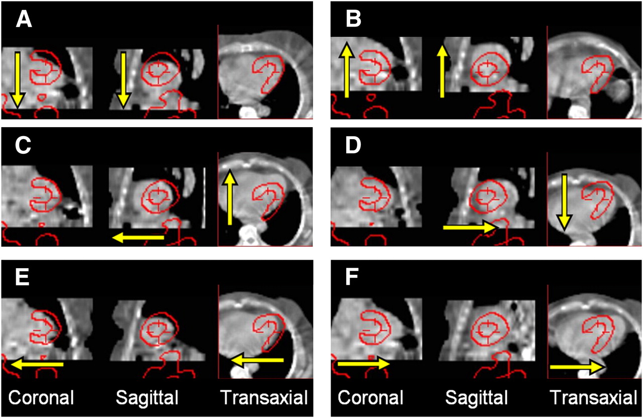
  • FIGURE 2. 
    • Download figure
    • Open in new tab
    • Download powerpoint
    FIGURE 2. 

    Artificial CT shift along 3 axes and 6 directions by 3 pixels with respect to SPECT (red contour). Directions were caudal, −z-axis (A), cephalad, +z-axis (B), ventral, −y-axis (C), dorsal, +y-axis (D), right, −x-axis (E), and left, +x-axis (F).
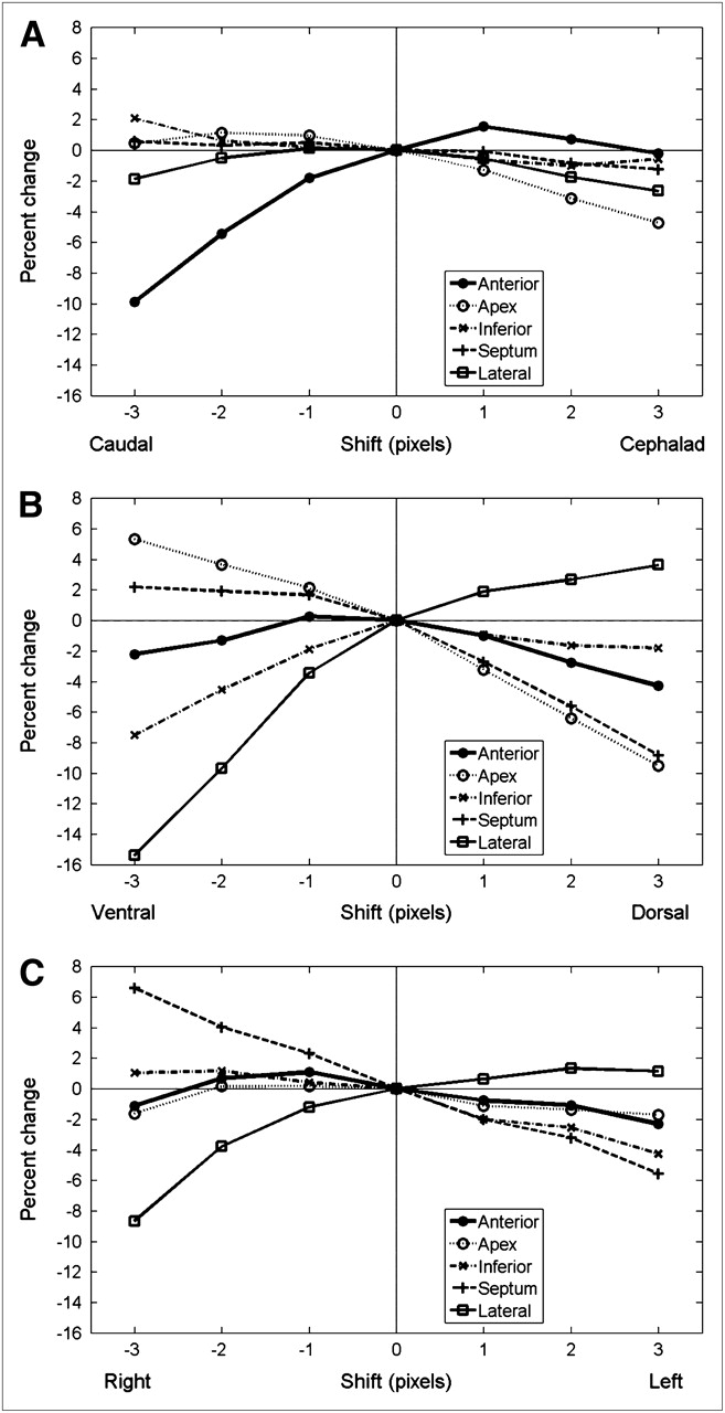
  • FIGURE 3. 
    • Download figure
    • Open in new tab
    • Download powerpoint
    FIGURE 3. 

    Percentage change in polar map segment score as function of misregistration of CT with respect to SPECT. For each axis, most severe changes are caused by 3-pixel shift as follows: anterior wall by a caudal shift (A), lateral wall by ventral shift (B), and lateral wall by right shift (C). All severe changes can be visually identified as misregistration, with myocardium on SPECT overlapping lung tissue on CT.

Tables

  • Figures
    • View popup
    TABLE 1

    Number of Studies with Given Direction and Magnitude of Misregistration

    Direction of misregistrationMagnitude of misregistration (pixels)
    >1>2>3
    On any axis181 (72.9%)57 (22.9%)10 (4.0%)
    Most severe direction on any axis*152 (61.3%)40 (16.1%)7 (2.8%)
    ≥2 axes65 (26.2%)5 (2.0%)0
    Most severe direction* ≥2 axes3 (1.2%)00
    • ↵* In different experiment, caudal, ventral, or right artificial shift of CT was found to induce most severe changes in polar map scoring.

    • Number in parentheses is percentage of 248 consecutive clinical studies.

    • View popup
    TABLE 2

    Relative Effect of CT Misregistration on Change in AC-MPI Polar Map Score

    Magnitude of misregistration (pixels)Region
    Direction of misregistrationAnteriorApexInferiorSeptumLateral
    Cephalad (+z-axis)3−−−−−
    2+−−−−
    1+−−−−
    Caudal (−z-axis)1−++++
    2− −+++−
    3− −+++−
    Dorsal (+y-axis)3−− −−− −+
    2−− −−− −+
    1−−−−+
    Ventral (−y-axis)1−+−+−
    2−+−+− −
    3−++− −+− − −
    Left (+x-axis)3−−−− −+
    2−−−−+
    1−−−−+
    Right (−x-axis)1++++−
    2++++−
    3−−++ +− −
    • Difference with respect to registered SPECT/CT: + + + = positive change of ≥15%; − − − = negative change of ≤−15%; + + = positive change of <15% and ≥5%; − − = negative change of >−15% and ≤−5%; + = positive change of <5% and >0%; − = negative change of >−5% and <0%.

    • View popup
    TABLE 3

    κ-Statistics and Significance of McNemar Differences of SSS for Each Artificial Misregistration

    No. of patients changing category*
    Misregistration directionMisregistration magnitude (pixels)Abnormal to normalNormal to abnormalκ-valueMcNemar test
    Cephalad (+z-axis)3230.440NS
    2410.510NS
    1430.300NS
    Caudal (–z-axis)1300.706NS
    2330.394NS
    3270.043P < 0.05
    Dorsal (+y-axis)328−0.075P < 0.05
    2170.400P < 0.05
    1340.286NS
    Ventral (–y-axis)1410.510NS
    2420.406NS
    3230.490NS
    Left (+x-axis)3340.286NS
    2220.596NS
    1310.604NS
    Right (– x-axis)1320.500NS
    2240.381NS
    3170.140P < 0.05
    • ↵* Registered case: 9 normal (SSS < 4), 11 abnormal (SSS ≥ 4).

    • NS = no significant difference (P > 0.05).

    • View popup
    TABLE 4

    Directionality: Misregistrations with Significant Difference Between Positive and Negative 3-Pixel Shifts (by Student t test)

    Misregistration directionRegion
    AnteriorApexInferiorSeptumLateral
    Cephalad/caudal (z-axis)P < 0. 001P < 0.001P < 0.025NSNS
    Dorsal/ventral (y-axis)NSP < 0.001P < 0.002P < 0.001P < 0.001
    Left/right (x-axis)NSNSP < 0.001P < 0.001P < 0.004
    • NS = no significant difference between positive and negative shifts (P > 0.05).

PreviousNext
Back to top

In this issue

Journal of Nuclear Medicine: 50 (9)
Journal of Nuclear Medicine
Vol. 50, Issue 9
September 2009
  • Table of Contents
  • Table of Contents (PDF)
  • About the Cover
  • Index by author
Print
Download PDF
Article Alerts
Sign In to Email Alerts with your Email Address
Email Article

Thank you for your interest in spreading the word on Journal of Nuclear Medicine.

NOTE: We only request your email address so that the person you are recommending the page to knows that you wanted them to see it, and that it is not junk mail. We do not capture any email address.

Enter multiple addresses on separate lines or separate them with commas.
Directions and Magnitudes of Misregistration of CT Attenuation-Corrected Myocardial Perfusion Studies: Incidence, Impact on Image Quality, and Guidance for Reregistration
(Your Name) has sent you a message from Journal of Nuclear Medicine
(Your Name) thought you would like to see the Journal of Nuclear Medicine web site.
Citation Tools
Directions and Magnitudes of Misregistration of CT Attenuation-Corrected Myocardial Perfusion Studies: Incidence, Impact on Image Quality, and Guidance for Reregistration
John A. Kennedy, Ora Israel, Alex Frenkel
Journal of Nuclear Medicine Sep 2009, 50 (9) 1471-1478; DOI: 10.2967/jnumed.109.062141

Citation Manager Formats

  • BibTeX
  • Bookends
  • EasyBib
  • EndNote (tagged)
  • EndNote 8 (xml)
  • Medlars
  • Mendeley
  • Papers
  • RefWorks Tagged
  • Ref Manager
  • RIS
  • Zotero
Share
Directions and Magnitudes of Misregistration of CT Attenuation-Corrected Myocardial Perfusion Studies: Incidence, Impact on Image Quality, and Guidance for Reregistration
John A. Kennedy, Ora Israel, Alex Frenkel
Journal of Nuclear Medicine Sep 2009, 50 (9) 1471-1478; DOI: 10.2967/jnumed.109.062141
Twitter logo Facebook logo LinkedIn logo Mendeley logo
  • Tweet Widget
  • Facebook Like
  • Google Plus One
Bookmark this article

Jump to section

  • Article
    • Abstract
    • MATERIALS AND METHODS
    • RESULTS
    • DISCUSSION
    • CONCLUSION
    • Acknowledgments
    • Footnotes
    • References
  • Figures & Data
  • Info & Metrics
  • PDF

Related Articles

  • This Month in JNM
  • PubMed
  • Google Scholar

Cited By...

  • Deep Learning-Based Attenuation Correction Improves Diagnostic Accuracy of Cardiac SPECT
  • Assessment of Myocardial CZT SPECT Recording in a Forward-Leaning Bikerlike Position
  • Google Scholar

More in this TOC Section

  • Cardiac Presynaptic Sympathetic Nervous Function Evaluated by Cardiac PET in Patients with Chronotropic Incompetence Without Heart Failure
  • Validation and Evaluation of a Vendor-Provided Head Motion Correction Algorithm on the uMI Panorama PET/CT System
  • Prognostic Role of 68Ga-PSMA11 PET–Based Response in Patients with Prostate Cancer Undergoing Taxane-Based Chemotherapy
Show more Clinical Investigations

Similar Articles

SNMMI

© 2025 SNMMI

Powered by HighWire