


Article Figures & Data
Figures
- FIGURE 1.
Binding and affinity analysis of ZEGFR:1907 monomer and dimer using surface plasmon resonance (Biacore) and flow cytometry. (A) Biacore sensorgrams obtained after injection of purified ZEGFR:1907 monomer (▵) and ZEGFR:1907 dimer (□) at 62.5 nM over sensor chip flow-cell surface containing amine-coupled EGFR-ECD. (B and C) Flow cytometric analysis and affinity measurements of EGFR-binding Affibody molecules to EGFR on A431 cells. Affibody molecules were fluorescently labeled site specifically to C-terminally introduced cysteine with Alexa Fluor 488. Equilibrium-binding curve for ZEGFR:1907 monomer (B) and dimer (C). Data are average from 3 experiments and SD.
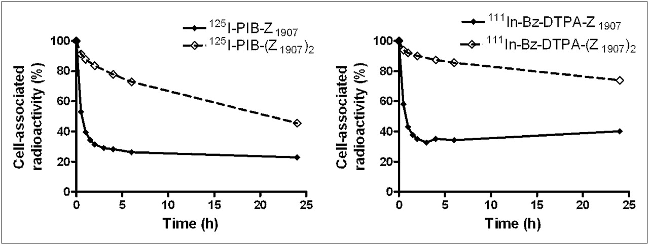
- FIGURE 2.
Cellular retention of radioactivity after interrupted incubation of EGFR-expressing A431 cells with EGFR-targeting Affibody molecules. Data are average value from 3 Petri dishes and SD. Error bars might not be seen because they are smaller than point symbols.
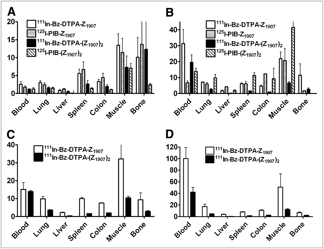
- FIGURE 3.
Tumor-to-organ ratios after injection of radiolabeled anti-EGFR Affibody molecules in BALB/c nu/nu mice bearing EGFR-expressing xenografts at 4 h after injection and injected dose of 3 μg (A), 24 h after injection and injected dose of 3 μg (B), 4 h after injection and injected dose of 50 μg (C), and 24 h after injection and injected dose of 50 μg (D). Data are average of 4 animals and SD.
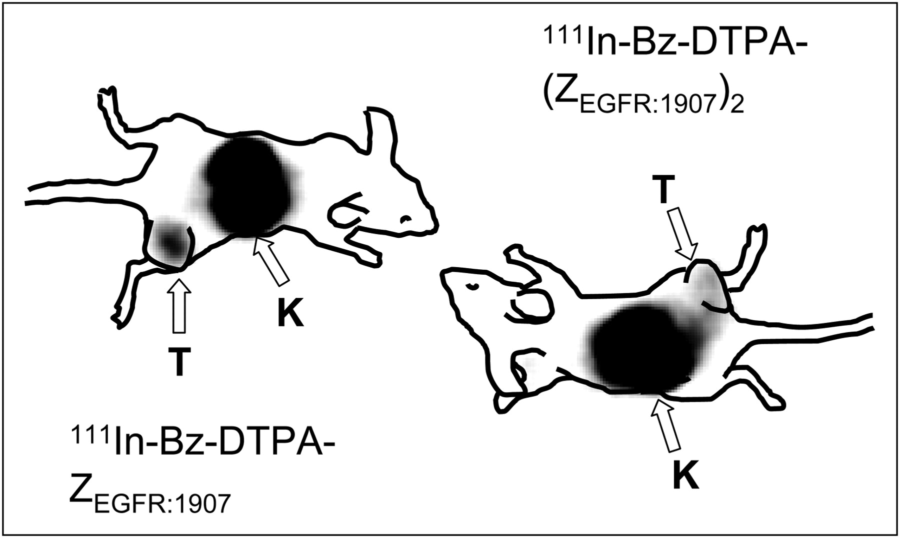
- FIGURE 4.
Imaging of EGFR expression in A431 xenografts in BALB/c nude mice using 111In-Bz-DTPA-Z1907 and 111In-Bz-DTPA-(Z1907)2. Planar γ-camera images were collected 24 h after administration of tracers. Arrows point to tumors (T) and kidneys (K).


Tables
- TABLE 1
Biodistribution of 111In-Labeled Tracers (Injected Dose, 3 μg) 4 and 24 Hours After Injection in BALB/c nu/nu Mice Bearing EGFR-Expressing A431 Xenografts
4 h after injection 24 h after injection Organ or tissue 111In-Bz-DTPA-ZEGFR:1907 111In-Bz-DTPA-ZTaq 111In-Bz-DTPA-(ZEGFR:1907)2 111In-Bz-DTPA-(ZAβ)2 111In-Bz-DTPA-ZEGFR:1907 111In-Bz-DTPA-(ZEGFR:1907)2 Blood 1.5 ± 0.5* 0.03 ± 0.01 1.5 ± 0.7 0.037 ± 0.002 0.07 ± 0.02* 0.07 ± 0.02† Lung 1.3 ± 0.2*‡ 0.25 ± 0.04 1.1 ± 0.2† 0.15 ± 0.01 0.28 ± 0.04*‡ 0.5 ± 0.2† Liver 5 ± 1*‡ 1.4 ± 0.2 18 ± 5† 0.6 ± 0.2 1.2 ± 0.3*‡ 6 ± 1†‡ Spleen 0.7 ± 0.2* 0.41 ± 0.06 1.1 ± 0.2† 0.18 ± 0.03 0.33 ± 0.04* 0.76 ± 0.05† Colon 1.2 ± 0.2* 0.30 ± 0.05 1.6 ± 0.5 0.23 ± 0.05 0.44 ± 0.08* 1.35 ± 0.09† Kidney 136 ± 11*‡ 189 ± 19 73 ± 7† 207 ± 35 102 ± 2* 59 ± 7† Tumor 3.73 ± 0.08*‡ 0.4 ± 0.2 1.57 ± 0.07 0.19 ± 0.07 2.0 ± 0.3*‡ 1.23 ± 0.07† Muscle 0.29 ± 0.06* 0.06 ± 0.01 0.24 ± 0.07 0.06 ± 0.04 0.09 ± 0.02* 0.20 ± 0.05† Bone 0.5 ± 0.3 0.3 ± 0.2§ 0.7 ± 0.2 0.16 ± 0.10 0.3 ± 0.2‡ 0.6 ± 0.2† Brain 0.06 ± 0.02* 0.004 ± 0.002 0.06 ± 0.03 NM 0.01 ± 0.01‡ 0.04 ± 0.03 GI tract‖ 3.6 ± 0.8*‡ 1.0 ± 0.4 6.0 ± 0.7† 1.0 ± 0.3 1.2 ± 0.2*‡ 6 ± 1† Carcass‖ 13 ± 1* 3 ± 1 12 ± 2† 1.8 ± 0.3 4.9 ± 0.5‡ 9.6 ± 0.5† ↵* Significant difference (P < 0.05, paired t test) between 111In-Bz-DTPA-ZEGFR:1907 and 125I-PIB-ZEGFR:1907.
↵† Significant difference (P < 0.05, paired t test) between 111In-Bz-DTPA-(ZEGFR:1907)2 and 125I-PIB-(ZEGFR:1907)2.
↵‡ Significant difference (P < 0.05, unpaired t test) between 111In-Bz-DTPA-ZEGFR:1907 and 111In-Bz-DTPA- (ZEGFR:1907)2.
↵§ No significant difference (P > 0.05, unpaired t test) between 111In-Bz-DTPA-ZEGFR:1907 and 111In-Bz-DTPA-ZTaq.
↵‖ Data for gastrointestinal (GI) tract and carcass are presented as %IA per sample.
NM = nonmeasureable.
Data are presented as average %IA/g and SD (n = 4). Nonspecific Affibody molecules ZTaq and (ZAβ)2 have been included as negative control for 111In-Bz-DTPA-ZEGFR:1907 and 111In-Bz-DTPA-(ZEGFR:1907)2, respectively.
- TABLE 2
Biodistribution of 125I-Labeled Tracers (Injected Dose, 3 μg) 4 and 24 Hours After Injection in BALB/c nu/nu Mice Bearing EGFR-Expressing A431 Xenografts
4 h after injection 24 h after injection Blood or tissue 125I-PIB-ZEGFR:1907 125I-PIB-ZTaq 125I-PIB-(ZEGFR:1907)2 125I-PIB-(ZAβ)2 125I-PIB-ZEGFR:1907 125I-PIB-(ZEGFR:1907)2 Blood 1.2 ± 0.3* 0.25 ± 0.02 1.1 ± 0.5 0.052 ± 0.003 0.04 ± 0.01* 0.04 ± 0.01† Lung 0.9 ± 0.2* 0.12 ± 0.03 1.0 ± 0.2† 0.04 ± 0.01 0.05 ± 0.02* 0.05 ± 0.02† Liver 2.1 ± 0.7*‡ 0.21 ± 0.03 6 ± 2† 0.05 ± 0.01 0.06 ± 0.01*‡ 0.3 ± 0.2† Spleen 0.31 ± 0.08* 0.11 ± 0.02 0.4 ± 0.2† 0.065 ± 0.002 0.05 ± 0.02* 0.04 ± 0.02† Colon 0.47 ± 0.10*‡ 0.10 ± 0.07 0.73 ± 0.10 0.05 ± 0.02 0.02 ± 0.01* 0.09 ± 0.10† Kidney 6.3 ± 0.6*‡ 7 ± 2§ 10 ± 2† 6 ± 2 NM 0.6 ± 0.2† Tumor 2.48 ± 0.10*‡ 0.2 ± 0.1 1.9 ± 0.4 0.07 ± 0.05 0.26 ± 0.07*‡ 0.46 ± 0.09† Muscle 0.16 ± 0.06* 0.03 ± 0.01 0.17 ± 0.03 0.02 ± 0.01 0.02 ± 0.01* 0.02 ± 0.02† Bone 0.34 ± 0.10 0.09 ± 0.03 0.32 ± 0.02 NM 0.14 ± 0.10 NM Brain 0.04 ± 0.0* 0.02 ± 0.02§ 0.05 ± 0.01 0.01 ± 0.01 0.01 ± 0.0‡ 0.010 ± 0.003 GI tract‖ 1.8 ± 0.5* 0.8 ± 0.4 2.5 ± 0.2† 0.5 ± 0.2 0.07 ± 0.01*‡ 0.18 ± 0.09† Carcass‖ 7 ± 1* 1.15 ± 0.09 8 ± 1† 0.7 ± 0.2 0.6 ± 0.1 1.4 ± 0.2† ↵* Significant difference (P < 0.05, paired t test) between 111In-Bz-DTPA-ZEGFR:1907 and 125I-PIB-ZEGFR:1907.
↵† Significant difference (P < 0.05, unpaired t test) between 111In-Bz-DTPA-(ZEGFR:1907)2 and 125I-PIB-(ZEGFR:1907)2.
↵‡ Significant difference (P < 0.05, unpaired t test) between 125I-PIB-DTPA-ZEGFR:1907 and 125I-PIB-(ZEGFR:1907)2.
↵§ No significant difference (P > 0.05, unpaired t test) between125I-PIB-ZEGFR:1907 and 125I-PIB-ZTaq.
↵‖ Data for gastrointestinal (GI) tract and carcass are presented as % IA per sample.
NM = nonmeasurable.
Data are presented as average %IA/g and SD (n = 4). Nonspecific Affibody molecules ZTaq and (ZAβ)2 have been included as negative control for 125I-PIB-ZEGFR:1907 and 125I-PIB-(ZEGFR:1907)2, respectively.
- TABLE 3
Biodistribution of 111In-Bz-DTPA-ZEGFR:1907 and 111In-Bz DTPA-(ZEGFR:1907)2 (Injected Dose, 50 μg) 4 and 24 Hours After Injection in BALB/c nu/nu Mice Bearing EGFR-Expressing A431 Xenografts
4 h after injection 24 h after injection Organ or tissue 111In-Bz-DTPA-ZEGFR:1907 111In-Bz-DTPA-(ZEGFR:1907)2 111In-Bz-DTPA-ZEGFR:1907 111In-Bz-DTPA-(ZEGFR:1907)2 Blood 0.23 ± 0.05* 0.18 ± 0.03* 0.025 ± 0.006* 0.038 ± 009* Lung 0.34 ± 0.03*† 0.68 ± 0.09* 0.14 ± 0.03*† 0.33 ± 0.06 Liver 1.5 ± 0.2*† 6.9 ± 1.2* 0.7 ± 0.1*† 3.6 ± 1.4 Spleen 0.34 ± 0.02*† 1.5 ± 0.2 0.29 ± 0.02† 1.0 ± 0.3 Colon 0.45 ± 0.01*† 1.2 ± 0.2 0.22 ± 0.02*† 0.6 ± 0.1* Kidney 196 ± 4* 169 ± 32* 120 ± 10* 118 ± 14* Tumor 3.35 ± 0.08*† 2.43 ± 0.35* 2.39 ± 0.06† 1.5 ± 0.3 Muscle 0.12 ± 0.05*† 0.24 ± 0.05 0.05 ± 0.02† 0.13 ± 0.04 Bone 0.4 ± 0.1*† 0.9 ± 0.3 0.37 ± 0.08† 0.7 ± 0.3 Brain 0.013 ± 0.004*† 0.020 ± 0.003* NM 0.010 ± 0.005* GI tract‡ 1.7 ± 0.3*† 3.5 ± 0.5* 0.6 ± 0.1*† 1.4 ± 0.1* Carcass‡ 3.8 ± 0.4*† 9.0 ± 1.0 2.0 ± 0.3*† 5.8 ± 1.3* ↵* Significant difference (P < 0.05, paired t test) between uptake after injection of 3 and 50 μg of tracer.
↵† Significant difference (P < 0.05, unpaired t test) between 111In-Bz-DTPA-ZEGFR:1907 and 111In-Bz-DTPA-(ZEGFR:1907)2.
↵‡ Data for gastrointestinal (GI) tract and carcass are presented as %IA per sample.
NM = nonmeasurable.
Data are presented as average %IA/g and SD (n = 4).
Supplemental Data
Files in this Data Supplement:


Jump to section
Related Articles
Cited By...
- PET of EGFR Expression with an 18F-Labeled Affibody Molecule
- HER2-Affitoxin: A Potent Therapeutic Agent for the Treatment of HER2-Overexpressing Tumors
- A 2-Helix Small Protein Labeled with 68Ga for PET Imaging of HER2 Expression
- Strategies for Molecular Imaging of Epidermal Growth Factor Receptor Tyrosine Kinase in Cancer
- Molecular Imaging of EGFR: It's Time to Go Beyond Receptor Expression