
Main menu

  • Home
  • Content
    • Current
    • Ahead of print
    • Past Issues
    • JNM Supplement
    • SNMMI Annual Meeting Abstracts
    • Continuing Education
    • JNM Podcasts
  • Subscriptions
    • Subscribers
    • Institutional and Non-member
    • Rates
    • Journal Claims
    • Corporate & Special Sales
  • Authors
    • Submit to JNM
    • Information for Authors
    • Assignment of Copyright
    • AQARA requirements
  • Info
    • Reviewers
    • Permissions
    • Advertisers
  • About
    • About Us
    • Editorial Board
    • Contact Information
  • More
    • Alerts
    • Feedback
    • Help
    • SNMMI Journals
  • SNMMI
    • JNM
    • JNMT
    • SNMMI Journals
    • SNMMI

User menu

  • Subscribe
  • My alerts
  • Log in
  • My Cart

Search

  • Advanced search
Journal of Nuclear Medicine
  • SNMMI
    • JNM
    • JNMT
    • SNMMI Journals
    • SNMMI
  • Subscribe
  • My alerts
  • Log in
  • My Cart
Journal of Nuclear Medicine

Advanced Search

  • Home
  • Content
    • Current
    • Ahead of print
    • Past Issues
    • JNM Supplement
    • SNMMI Annual Meeting Abstracts
    • Continuing Education
    • JNM Podcasts
  • Subscriptions
    • Subscribers
    • Institutional and Non-member
    • Rates
    • Journal Claims
    • Corporate & Special Sales
  • Authors
    • Submit to JNM
    • Information for Authors
    • Assignment of Copyright
    • AQARA requirements
  • Info
    • Reviewers
    • Permissions
    • Advertisers
  • About
    • About Us
    • Editorial Board
    • Contact Information
  • More
    • Alerts
    • Feedback
    • Help
    • SNMMI Journals
  • View or Listen to JNM Podcast
  • Visit JNM on Facebook
  • Join JNM on LinkedIn
  • Follow JNM on Twitter
  • Subscribe to our RSS feeds
OtherBASIC SCIENCE INVESTIGATIONS

PET and Macro- and Microautoradiographic Studies Combined with Immunohistochemistry for Monitoring Rat Intestinal Ulceration and Healing Processes

Masanori Yamato, Yosky Kataoka, Hiroshi Mizuma, Yasuhiro Wada and Yasuyoshi Watanabe
Journal of Nuclear Medicine February 2009, 50 (2) 266-273; DOI: https://doi.org/10.2967/jnumed.108.057943
Masanori Yamato
  • Find this author on Google Scholar
  • Find this author on PubMed
  • Search for this author on this site
Yosky Kataoka
  • Find this author on Google Scholar
  • Find this author on PubMed
  • Search for this author on this site
Hiroshi Mizuma
  • Find this author on Google Scholar
  • Find this author on PubMed
  • Search for this author on this site
Yasuhiro Wada
  • Find this author on Google Scholar
  • Find this author on PubMed
  • Search for this author on this site
Yasuyoshi Watanabe
  • Find this author on Google Scholar
  • Find this author on PubMed
  • Search for this author on this site
  • Article
  • Figures & Data
  • Supplemental
  • Info & Metrics
  • PDF
Loading

Article Figures & Data

Figures

  • Additional Files
  • FIGURE 1. 
    • Download figure
    • Open in new tab
    • Download powerpoint
    FIGURE 1. 

    Macroscopic (A) and microscopic (B) observations of indomethacin-induced small-intestine ulceration during healing in rats. (A) Continuous circular lesions were seen on side of mesenteric attachment on day 1 or 4. (B) Histologically, denuded epithelium and severe edema in submucosa (day 1), granulation tissue on ulcer bed (day 4), and neoepithelial cells (day 7) were observed (hematoxylin and eosin staining; scale bar, 200 μm). (C) Change in myeloperoxidase activity as marker of neutrophil infiltration during indomethacin-induced intestinal ulceration and healing. Myeloperoxidase activity was evaluated in animals after administration of indomethacin (days 1, 2, 4, and 7) and in vehicle-injected animals (control). Data are shown as mean ± SD; n = 4–5 animals. *P < 0.05.
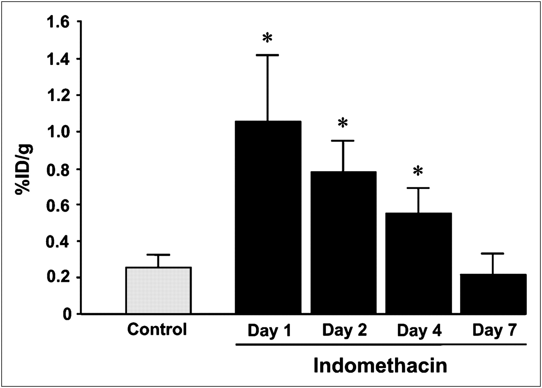
  • FIGURE 2. 
    • Download figure
    • Open in new tab
    • Download powerpoint
    FIGURE 2. 

    γ-counting study of 18F-FDG uptake in ulcerated intestine, showing 18F-FDG accumulation in small intestine at time points after indomethacin administration (days 1, 2, 4, and 7) and after vehicle injection (control). Data are shown as mean ± SD; n = 6–8 animals for each time point.

  • FIGURE 3. 
    • Download figure
    • Open in new tab
    • Download powerpoint
    FIGURE 3. 

    18F-FDG PET study of indomethacin-induced intestinal ulceration. (A) Abdominal PET images (coronal images) in same rat at different times after indomethacin administration. Arrowheads indicate characteristic accumulation of 18F-FDG. (B) Quantification of 18F-FDG uptake in PET study. Mean SUVs are shown as mean ± SD; n = 4 animals. *P < 0.05.
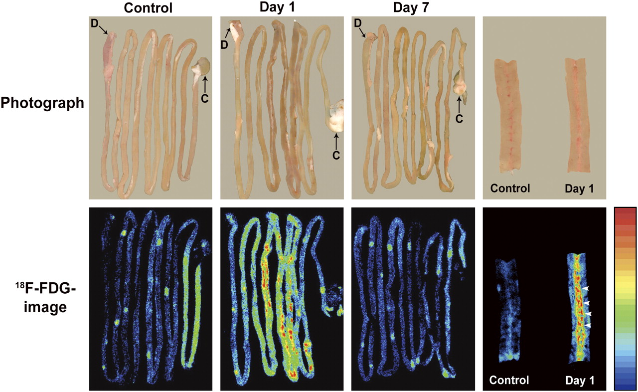
  • FIGURE 4. 
    • Download figure
    • Open in new tab
    • Download powerpoint
    FIGURE 4. 

    18F-FDG macroautoradiography of intestinal ulceration. Rats were intravenously injected with 18F-FDG and euthanized 45 min later. Discontinuous dotlike 18F-FDG accumulation was clearly identified mainly in ileum on day 1. 18F-FDG imaging profile was similar to that of control on day 7. Images at far right show plain photograph (upper) and 18F-FDG image (lower) of intestines opened along longitudinal axis for control and at day 1. D = duodenum; C = cecum.
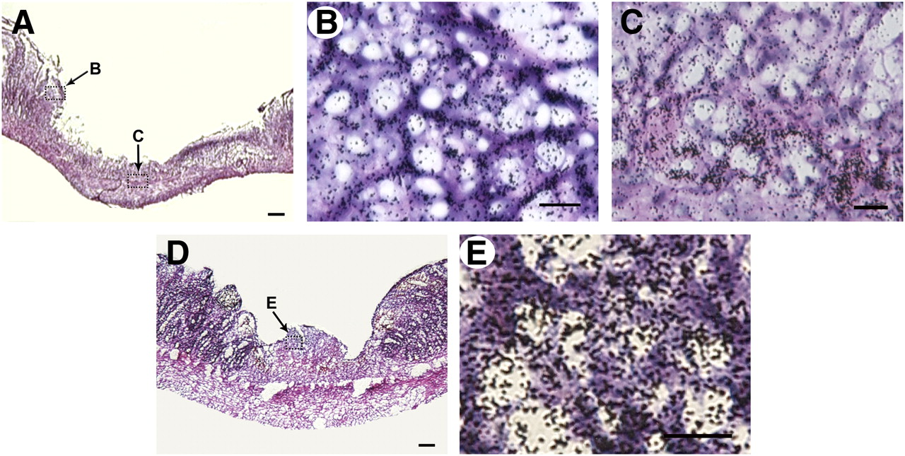
  • FIGURE 5. 
    • Download figure
    • Open in new tab
    • Download powerpoint
    FIGURE 5. 

    18F-FDG microautoradiography of indomethacin-induced intestinal lesions combined with hematoxylin and eosin staining. (A–C) At 1 d after administration of indomethacin, silver grains of high density were observed in ulcerated area, especially at ulcer margin (B) and submucosal and smooth muscle layer (C). B and C are magnified views of areas indicated by arrows B and C in A. (D and E) On day 4, grains were accumulated on cells forming granulation tissue. (E) Magnified view of the region indicated by arrow E in D. Scale bars: 100 μm (A and D) and 20 μm (B, C, and E).
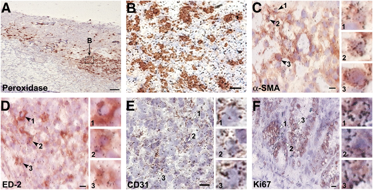
  • FIGURE 6. 
    • Download figure
    • Open in new tab
    • Download powerpoint
    FIGURE 6. 

    Microautoradiography combined with immunohistochemistry. In submucosal area at day 1 after indomethacin administration, 18F-FDG uptake was observed mainly in peroxidase-positive cells (A and B). On granulation tissue at day 4, myofibroblasts (C; α-SMA–positive), macrophages (D; ED2-positive), and endothelial cells (E; CD31-positive) were visualized. In normal tissue, silver grains of high density reflecting 18F-FDG radioactivity were observed in Ki67-positive crypt cells (F). Insets show magnified view of region indicated by arrowheads in each panel. Scale bars: 100 μm (A), 10 μm (B), and 5 μm (C–F).

Additional Files

  • Figures
  • Supplemental Data

    Files in this Data Supplement:

    • Supplemental Figures
PreviousNext
Back to top

In this issue

Journal of Nuclear Medicine: 50 (2)
Journal of Nuclear Medicine
Vol. 50, Issue 2
February 2009
  • Table of Contents
  • Table of Contents (PDF)
  • About the Cover
  • Index by author
Print
Download PDF
Article Alerts
Sign In to Email Alerts with your Email Address
Email Article

Thank you for your interest in spreading the word on Journal of Nuclear Medicine.

NOTE: We only request your email address so that the person you are recommending the page to knows that you wanted them to see it, and that it is not junk mail. We do not capture any email address.

Enter multiple addresses on separate lines or separate them with commas.
PET and Macro- and Microautoradiographic Studies Combined with Immunohistochemistry for Monitoring Rat Intestinal Ulceration and Healing Processes
(Your Name) has sent you a message from Journal of Nuclear Medicine
(Your Name) thought you would like to see the Journal of Nuclear Medicine web site.
Citation Tools
PET and Macro- and Microautoradiographic Studies Combined with Immunohistochemistry for Monitoring Rat Intestinal Ulceration and Healing Processes
Masanori Yamato, Yosky Kataoka, Hiroshi Mizuma, Yasuhiro Wada, Yasuyoshi Watanabe
Journal of Nuclear Medicine Feb 2009, 50 (2) 266-273; DOI: 10.2967/jnumed.108.057943

Citation Manager Formats

  • BibTeX
  • Bookends
  • EasyBib
  • EndNote (tagged)
  • EndNote 8 (xml)
  • Medlars
  • Mendeley
  • Papers
  • RefWorks Tagged
  • Ref Manager
  • RIS
  • Zotero
Share
PET and Macro- and Microautoradiographic Studies Combined with Immunohistochemistry for Monitoring Rat Intestinal Ulceration and Healing Processes
Masanori Yamato, Yosky Kataoka, Hiroshi Mizuma, Yasuhiro Wada, Yasuyoshi Watanabe
Journal of Nuclear Medicine Feb 2009, 50 (2) 266-273; DOI: 10.2967/jnumed.108.057943
Twitter logo Facebook logo LinkedIn logo Mendeley logo
  • Tweet Widget
  • Facebook Like
  • Google Plus One
Bookmark this article

Jump to section

  • Article
    • Abstract
    • MATERIALS AND METHODS
    • RESULTS
    • DISCUSSION
    • CONCLUSION
    • Acknowledgments
    • Footnotes
    • References
  • Figures & Data
  • Supplemental
  • Info & Metrics
  • PDF

Related Articles

  • This Month in JNM
  • PubMed
  • Google Scholar

Cited By...

  • PET Imaging Analysis with 64Cu in Disulfiram Treatment for Aberrant Copper Biodistribution in Menkes Disease Mouse Model
  • Google Scholar

More in this TOC Section

  • Design and Fabrication of Kidney Phantoms for Internal Radiation Dosimetry Using 3D Printing Technology
  • Synthesis and Biologic Evaluation of Novel 18F-Labeled Probes Targeting Prostate-Specific Membrane Antigen for PET of Prostate Cancer
  • Tumor-Specific Binding of Radiolabeled PEGylated GIRLRG Peptide: A Novel Agent for Targeting Cancers
Show more Basic Science Investigations

Similar Articles

SNMMI

© 2025 SNMMI

Powered by HighWire