


Article Figures & Data
Figures
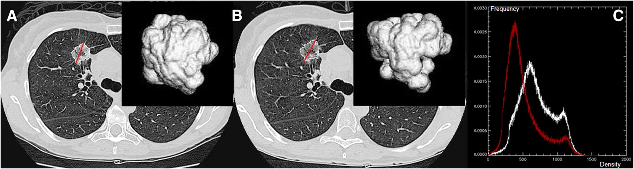
- FIGURE 1.
Images showing no changes in unidimensional or bidimensional measurements but enlargement of tumor along z-axis. (A) Baseline transverse CT image shows tumor contour (outlined in white), greatest diameter, and greatest perpendicular diameter (crossed lines in black) determined by semiautomated segmentation algorithm. (B) Three-dimensional view of segmented tumor on baseline. (C and D) On corresponding follow-up CT images obtained 24 d later, 3-dimensional tumor is seen from same angle along z-axis. Changes in unidimensional, bidimensional, and volumetric measurements are 0.4%, −4.4%, and 33.2%, respectively. (Reprinted with permission of (16).)
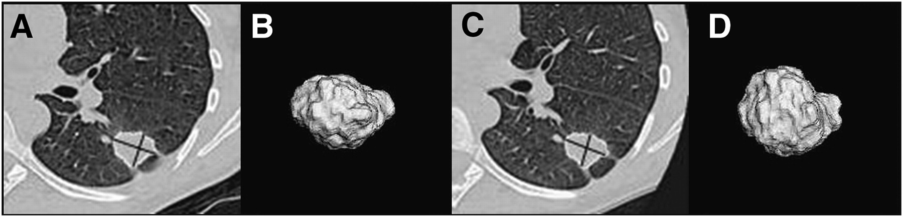
- FIGURE 2.
Images showing no change in tumor size but decrease in tumor density (ghost scenario). (A) Baseline transverse CT image with computer-generated tumor contour, greatest diameter, and 3-dimensional view. (B) On corresponding follow-up CT image obtained 21 d later, tumor is seen from same angle along z-axis. (C) Density histograms of tumor on baseline (white) and follow-up scans (red). Changes in unidimensional and volumetric measurements were 2.1% and −7.8%, respectively. Change in tumor average intensity was −189 Hounsfield unit.
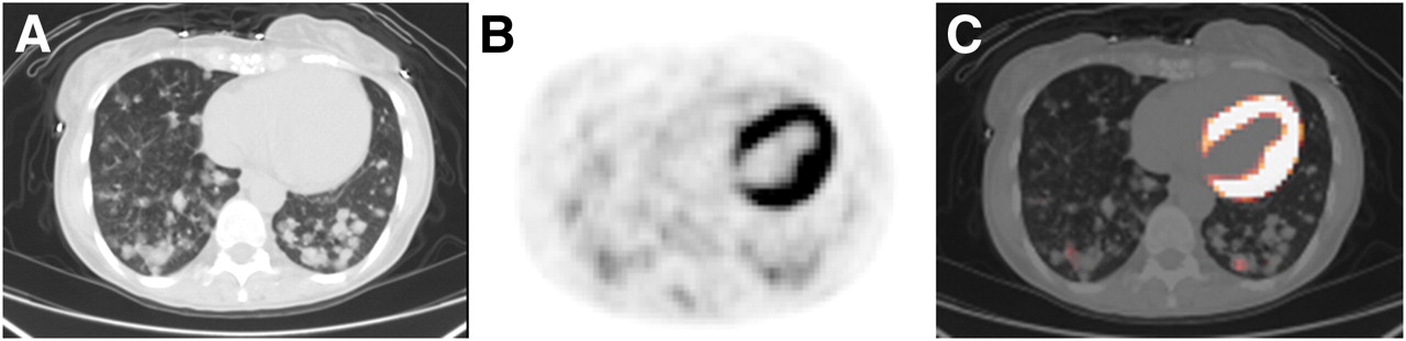
- FIGURE 3.
CT and 18F-FDG PET of bronchioalveolar cell carcinoma, with lesions on baseline CT (A), lesions on corresponding 18F-FDG PET (B), and CT fused with 18F-FDG PET (C). Only regions on PET containing pixels with SUVmax greater than or equal to 2 are visible on fused PET/CT image.
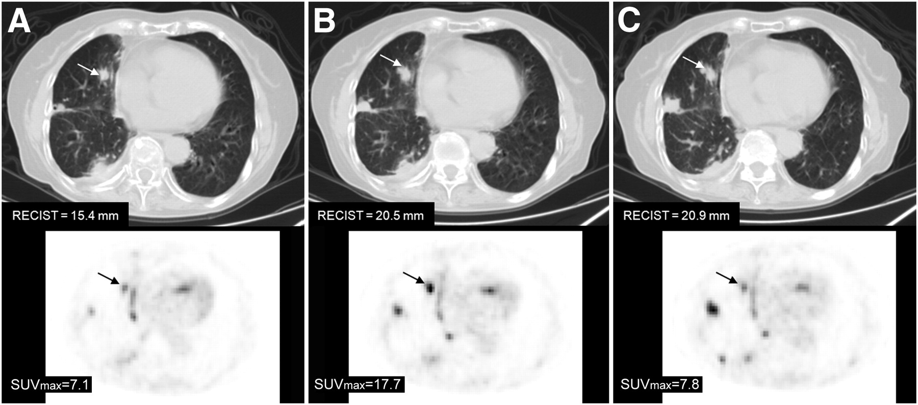
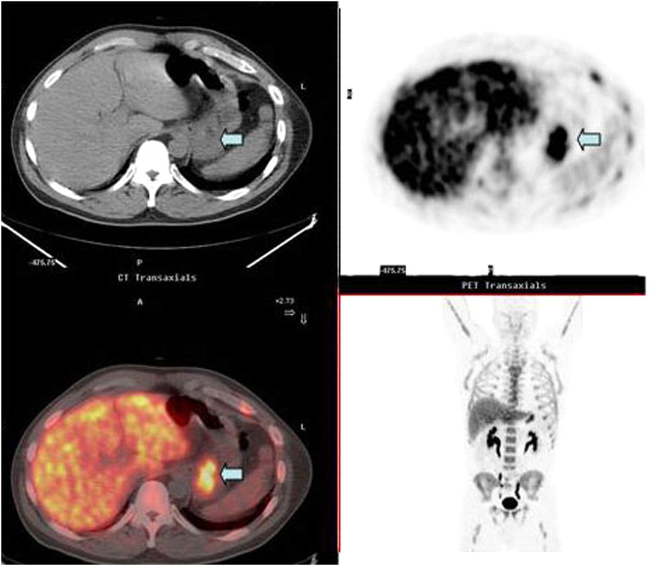
- FIGURE 4.
18F-FDG PET/CT of NSCLC tumor. (A) Baseline hybrid PET/CT performed on patient who had discontinued treatment with gefitinib. One target lesion (arrow) in right lung is indicated on CT (upper row) and on corresponding PET (lower row). (B) PET/CT after 3 wk of no gefitinib treatment. Both size (i.e., maximal tumor diameter) measured on CT and SUVmax measured on 18F-FDG PET increased. Patient then resumed treatment with gefitinib at dose patient was receiving before study entry. (C) PET/CT 3 wk after resumption of treatment. Size did not change, but SUVmax dropped.
- FIGURE 5.
18F-FLT uptake in gastric cancer (arrow) is indicator of rapid tumor cell proliferation: companion CT image through region of stomach mass (upper left), transaxial PET image showing 18F-FLT uptake in gastric mass (arrow; upper right), coronal PET image showing 18F-FLT uptake in gastric mass adjacent to left lobe of liver (bottom right), and fusion image of PET and CT showing 18F-FLT uptake in gastric mass (arrow; bottom left).
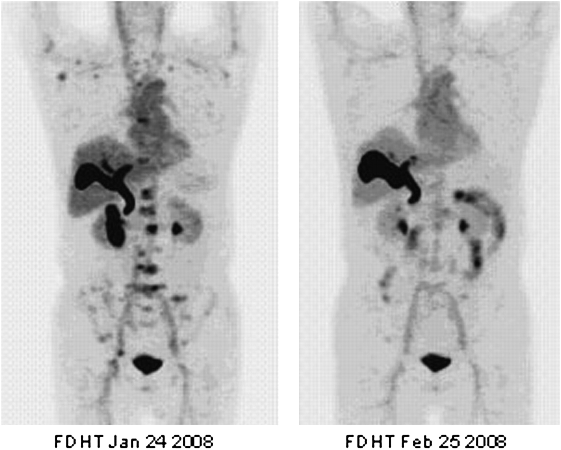
- FIGURE 6.
Actively metabolizing tumor with pre- and posttreatment uptake of 18F-FDHT, an androgen ligand radiotracer, in multiple lumbar vertebrae before (left) and after (right) treatment with Medivation, a high-affinity androgen receptor being studied under approved protocol at MSKCC.
- FIGURE 7.
Progression of molecular imaging radioprobe from laboratory to clinic for MSKCC radiotracer that targets HER 2 receptor: 68Ga-Fab′2 trastuzumab (75). Animal studies with human breast tumor xenografts are imaged with small-animal PET (left), and initial study in human patient with metastatic breast cancer (right) causing lytic skull lesion (companion CT image, leftmost image of right panel), with positive uptake on fusion image (rightmost image of right panel).


Tables
Radiotracer Function 18F-FDG (48) Glycolysis 18F-FLT Proliferation 11C-methionine (49–51), anti-1-amino-3-[18F]fluorocyclobutyl-carboxylic acid (52) Amino acid transport 18F-FES (53) Estrogen receptor 18F-FDHT (54–56) Androgen receptor Na 124I NIS 11C-acetate Krebs cycle, FA syn 18F-FMISO (57) Hypoxia 68Ga-Fab′2 trastuzumab HER 2 124I-cG250 Carbonic anhydrase IX 124I-A33 A33 antigen 124I-3F8 GD2 64Cu-trastuzumab HER 2 124I-Fluoroiodoarabinosylurideine* (58) Thymidine kinase (herpes virus) 18F-Fluoroethanylarabinosyluridine* (59) Thymidine kinase (herpes virus) ↵* Gene expression imaging.


Jump to section
- Article
- Abstract
- LIMITATION AND POTENTIAL IMPROVEMENT OF CONVENTIONAL RESPONSE-ASSESSMENT METHODS
- NEED FOR BETTER RESPONSE ASSESSMENT FOR NSCLC
- FUNCTIONAL RESPONSE ASSESSMENT IN NSCLC
- FUTURE APPLICATIONS OF MOLECULAR IMAGING WITH PET
- TRANSLATION FROM LABORATORY APPLICATIONS TO CLINICAL RESEARCH
- CONCLUSION
- Footnotes
- References
- Figures & Data
- Info & Metrics
Related Articles
Cited By...
- Serial Diffusion MRI to Monitor and Model Treatment Response of the Targeted Nanotherapy CRLX101
- 17{beta}-Estradiol Augments 18F-FDG Uptake and Glycolysis of T47D Breast Cancer Cells via Membrane-Initiated Rapid PI3K-Akt Activation
- Preparation, Biological Evaluation, and Pharmacokinetics of the Human Anti-HER1 Monoclonal Antibody Panitumumab Labeled with 86Y for Quantitative PET of Carcinoma
- Efficacy of PHA-848125, a Cyclin-Dependent Kinase Inhibitor, on the K-RasG12DLA2 Lung Adenocarcinoma Transgenic Mouse Model: Evaluation by Multimodality Imaging