
Main menu

  • Home
  • Content
    • Current
    • Ahead of print
    • Past Issues
    • JNM Supplement
    • SNMMI Annual Meeting Abstracts
    • Continuing Education
    • JNM Podcasts
  • Subscriptions
    • Subscribers
    • Institutional and Non-member
    • Rates
    • Journal Claims
    • Corporate & Special Sales
  • Authors
    • Submit to JNM
    • Information for Authors
    • Assignment of Copyright
    • AQARA requirements
  • Info
    • Reviewers
    • Permissions
    • Advertisers
  • About
    • About Us
    • Editorial Board
    • Contact Information
  • More
    • Alerts
    • Feedback
    • Help
    • SNMMI Journals
  • SNMMI
    • JNM
    • JNMT
    • SNMMI Journals
    • SNMMI

User menu

  • Subscribe
  • My alerts
  • Log in
  • My Cart

Search

  • Advanced search
Journal of Nuclear Medicine
  • SNMMI
    • JNM
    • JNMT
    • SNMMI Journals
    • SNMMI
  • Subscribe
  • My alerts
  • Log in
  • My Cart
Journal of Nuclear Medicine

Advanced Search

  • Home
  • Content
    • Current
    • Ahead of print
    • Past Issues
    • JNM Supplement
    • SNMMI Annual Meeting Abstracts
    • Continuing Education
    • JNM Podcasts
  • Subscriptions
    • Subscribers
    • Institutional and Non-member
    • Rates
    • Journal Claims
    • Corporate & Special Sales
  • Authors
    • Submit to JNM
    • Information for Authors
    • Assignment of Copyright
    • AQARA requirements
  • Info
    • Reviewers
    • Permissions
    • Advertisers
  • About
    • About Us
    • Editorial Board
    • Contact Information
  • More
    • Alerts
    • Feedback
    • Help
    • SNMMI Journals
  • View or Listen to JNM Podcast
  • Visit JNM on Facebook
  • Join JNM on LinkedIn
  • Follow JNM on Twitter
  • Subscribe to our RSS feeds
OtherBASIC SCIENCE INVESTIGATIONS

Targeting the αvβ3 Integrin for Small-Animal PET/CT of Osteolytic Bone Metastases

Thaddeus J. Wadas, Hongju Deng, Jennifer E. Sprague, Alexander Zheleznyak, Katherine N. Weilbaecher and Carolyn J. Anderson
Journal of Nuclear Medicine November 2009, 50 (11) 1873-1880; DOI: https://doi.org/10.2967/jnumed.109.067140
Thaddeus J. Wadas
  • Find this author on Google Scholar
  • Find this author on PubMed
  • Search for this author on this site
Hongju Deng
  • Find this author on Google Scholar
  • Find this author on PubMed
  • Search for this author on this site
Jennifer E. Sprague
  • Find this author on Google Scholar
  • Find this author on PubMed
  • Search for this author on this site
Alexander Zheleznyak
  • Find this author on Google Scholar
  • Find this author on PubMed
  • Search for this author on this site
Katherine N. Weilbaecher
  • Find this author on Google Scholar
  • Find this author on PubMed
  • Search for this author on this site
Carolyn J. Anderson
  • Find this author on Google Scholar
  • Find this author on PubMed
  • Search for this author on this site
  • Article
  • Figures & Data
  • Supplemental
  • Info & Metrics
  • PDF
Loading

Article Figures & Data

Figures

  • Additional Files
  • FIGURE 1. 
    • Download figure
    • Open in new tab
    • Download powerpoint
    FIGURE 1. 

    (A) Structure of CB-TE2A-c(RGDyK). (B) Isosurface image rendered from high-resolution CT data collected on Tax+ and WT mice. Isosurface images are rendered using same parameters and are equivalent. Arrows depict sites of osteolytic bone destruction in spinal vertebrae and femur of Tax+ mouse.

  • FIGURE 2. 
    • Download figure
    • Open in new tab
    • Download powerpoint
    FIGURE 2. 

    Biodistribution of 64Cu-RGD is different in Tax+ and WT mice. (A) Statistically significant differences are observed between accumulation of radioactivity in tails of 6- to 12-mo-old Tax+ (n = 12) and WT mice (n = 15). Additionally, uptake of 64Cu-RGD in 6- to 12-mo-old Tax+ mice is higher than in those older than 12 mo (P = 0.003). (B) However, statistically significant differences are not observed when radiopharmaceutical accumulation in tails of Tax+ mice that are older than 12 mo (n = 5) is compared with radiopharmaceutical accumulation in tails of WT mice in same age range (n = 6). (C) Positive correlation exists between accumulated tracer uptake in Tax+ mouse tails and measured serum TRAP levels in blood of these mice. High degree of variability reflects spontaneous nature of animal model. (D) Conversely, no correlation exists between accumulated tracer uptake in WT mouse tails and measured serum TRAP levels in blood of WT mice. Sera from both age groups were combined in parts C and D.
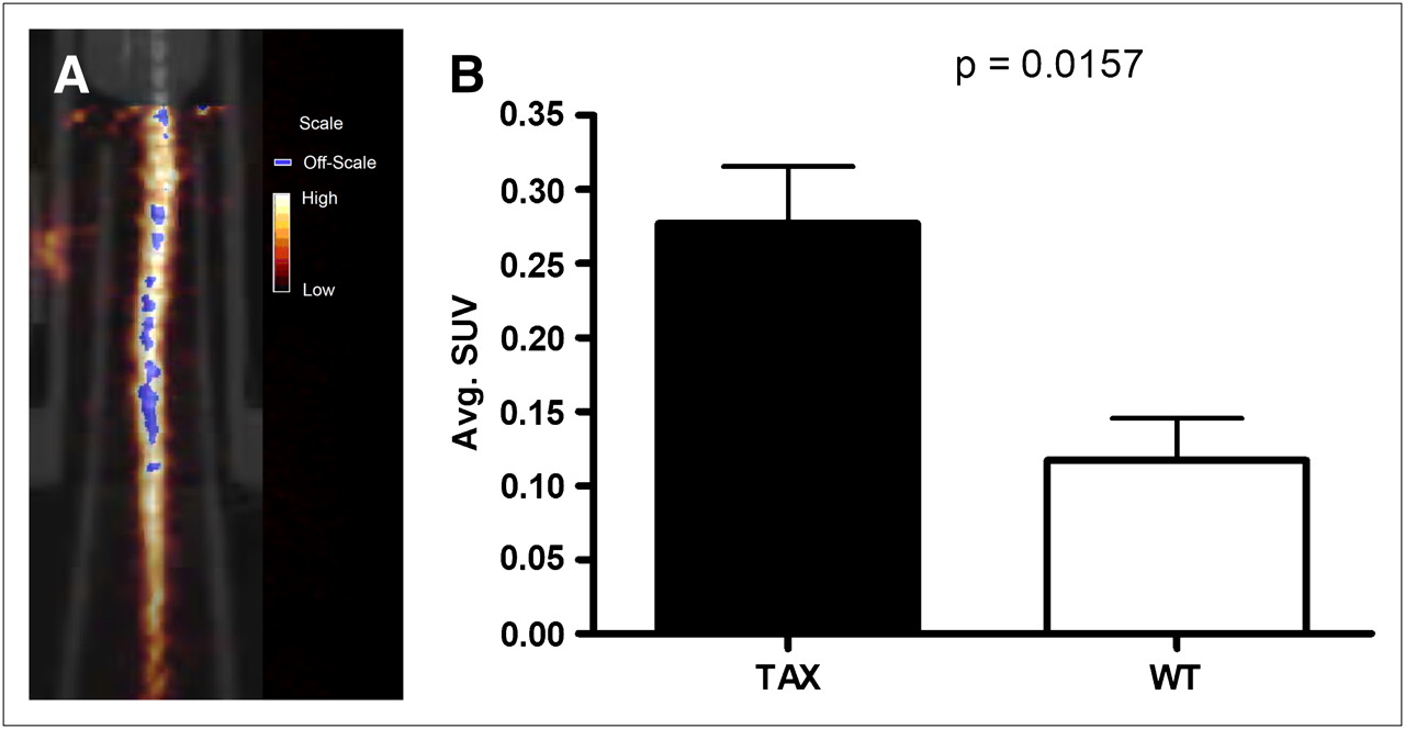
  • FIGURE 3. 
    • Download figure
    • Open in new tab
    • Download powerpoint
    FIGURE 3. 

    (A) Coregistered small-animal PET/CT image of Tax+ mouse tail that was imaged with 64Cu-RGD at 1 h after injection. Coronal slice presented here has numerous areas of focal uptake in tail vertebrae. (B) Graphic representation of observed differences in SUV between Tax+ (n = 5) and WT (n = 4) mice. On basis of SUV analysis, more activity is observed in Tax+ mouse tails.
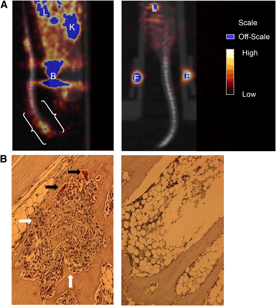
  • FIGURE 4. 
    • Download figure
    • Open in new tab
    • Download powerpoint
    FIGURE 4. 

    (A) Coregistered small-animal PET/CT images of Tax+ and WT mice (both 14 mo old) at 1 h after injection. OSEM reconstructions were used here, and only lower body of each mouse was placed in field of view. (A, left image) Sagittal slice of Tax+ mouse tail, which was held in place such that tail looped up onto ventral side of mouse. White brackets indicate where histologic sectioning was conducted. (A, right image) Coregistered small-animal PET/CT image (coronal slice) of WT mouse. Little activity is observed in tail vertebrae of this mouse. (B, left image) TRAP-stained section from tail of Tax+ mouse shown in A, which depicts osteolytic lesion. Several osteoclasts are indicated by black arrows, whereas tumor and proinflammatory cell infiltration is indicated by white arrows. (B, right image) TRAP-stained section from tail of WT mouse. Osteoclasts and proinflammatory cells are not observed in healthy marrow. B = bladder; F = fiducial marker; K = kidney; L = liver.

  • FIGURE 5. 
    • Download figure
    • Open in new tab
    • Download powerpoint
    FIGURE 5. 

    Small-animal PET/CT (with 64Cu-RGD) results before (left) and after (right) treatment with ZA. Three animals were used in this proof-of-principle study. Small-animal CT and small-animal PET data were rendered as whole-body reprojected images, which were summations of individual data slices. Notably, changes in bone due to administered therapy are observed in small-animal PET before being observed in the small-animal CT and can be quantified by determining SUVs before and after therapy. (A) Tax+ mouse tail demonstrates appreciable radiopharmaceutical uptake at time of imaging. SUV for whole tail of this animal before treatment was 0.47, but after therapy it was 0.13. (B) Different Tax+ mouse tail has developed large soft-tissue tumor. Here SUV analysis was conducted on 2 vertebrae separated by tumor. Before therapy, proximal vertebra (PV) and distal vertebra (DV) had SUVs of 0.38 and 0.43, respectively. After treatment with ZA, SUV of PV was 0.12 and SUV of DV was 0.12. F = fiducial marker; T = tumor.

Additional Files

  • Figures
  • Supplemental Data

    Files in this Data Supplement:

    • Supplemental Figures
PreviousNext
Back to top

In this issue

Journal of Nuclear Medicine: 50 (11)
Journal of Nuclear Medicine
Vol. 50, Issue 11
November 2009
  • Table of Contents
  • Table of Contents (PDF)
  • About the Cover
  • Index by author
Print
Download PDF
Article Alerts
Sign In to Email Alerts with your Email Address
Email Article

Thank you for your interest in spreading the word on Journal of Nuclear Medicine.

NOTE: We only request your email address so that the person you are recommending the page to knows that you wanted them to see it, and that it is not junk mail. We do not capture any email address.

Enter multiple addresses on separate lines or separate them with commas.
Targeting the αvβ3 Integrin for Small-Animal PET/CT of Osteolytic Bone Metastases
(Your Name) has sent you a message from Journal of Nuclear Medicine
(Your Name) thought you would like to see the Journal of Nuclear Medicine web site.
Citation Tools
Targeting the αvβ3 Integrin for Small-Animal PET/CT of Osteolytic Bone Metastases
Thaddeus J. Wadas, Hongju Deng, Jennifer E. Sprague, Alexander Zheleznyak, Katherine N. Weilbaecher, Carolyn J. Anderson
Journal of Nuclear Medicine Nov 2009, 50 (11) 1873-1880; DOI: 10.2967/jnumed.109.067140

Citation Manager Formats

  • BibTeX
  • Bookends
  • EasyBib
  • EndNote (tagged)
  • EndNote 8 (xml)
  • Medlars
  • Mendeley
  • Papers
  • RefWorks Tagged
  • Ref Manager
  • RIS
  • Zotero
Share
Targeting the αvβ3 Integrin for Small-Animal PET/CT of Osteolytic Bone Metastases
Thaddeus J. Wadas, Hongju Deng, Jennifer E. Sprague, Alexander Zheleznyak, Katherine N. Weilbaecher, Carolyn J. Anderson
Journal of Nuclear Medicine Nov 2009, 50 (11) 1873-1880; DOI: 10.2967/jnumed.109.067140
Twitter logo Facebook logo LinkedIn logo Mendeley logo
  • Tweet Widget
  • Facebook Like
  • Google Plus One
Bookmark this article

Jump to section

  • Article
    • Abstract
    • MATERIALS AND METHODS
    • RESULTS
    • DISCUSSION
    • CONCLUSION
    • Acknowledgments
    • Footnotes
    • References
  • Figures & Data
  • Supplemental
  • Info & Metrics
  • PDF

Related Articles

  • This Month in JNM
  • PubMed
  • Google Scholar

Cited By...

  • No citing articles found.
  • Google Scholar

More in this TOC Section

  • Tumor Uptake of Anti-CD20 Fabs Depends on Tumor Perfusion
  • How Sensitive Is the Upper Gastrointestinal Tract to 90Y Radioembolization? A Histologic and Dosimetric Analysis in a Porcine Model
  • 11C-Methionine PET of Myocardial Inflammation in a Rat Model of Experimental Autoimmune Myocarditis
Show more Basic Science Investigations

Similar Articles

SNMMI

© 2025 SNMMI

Powered by HighWire