


Article Figures & Data
Figures
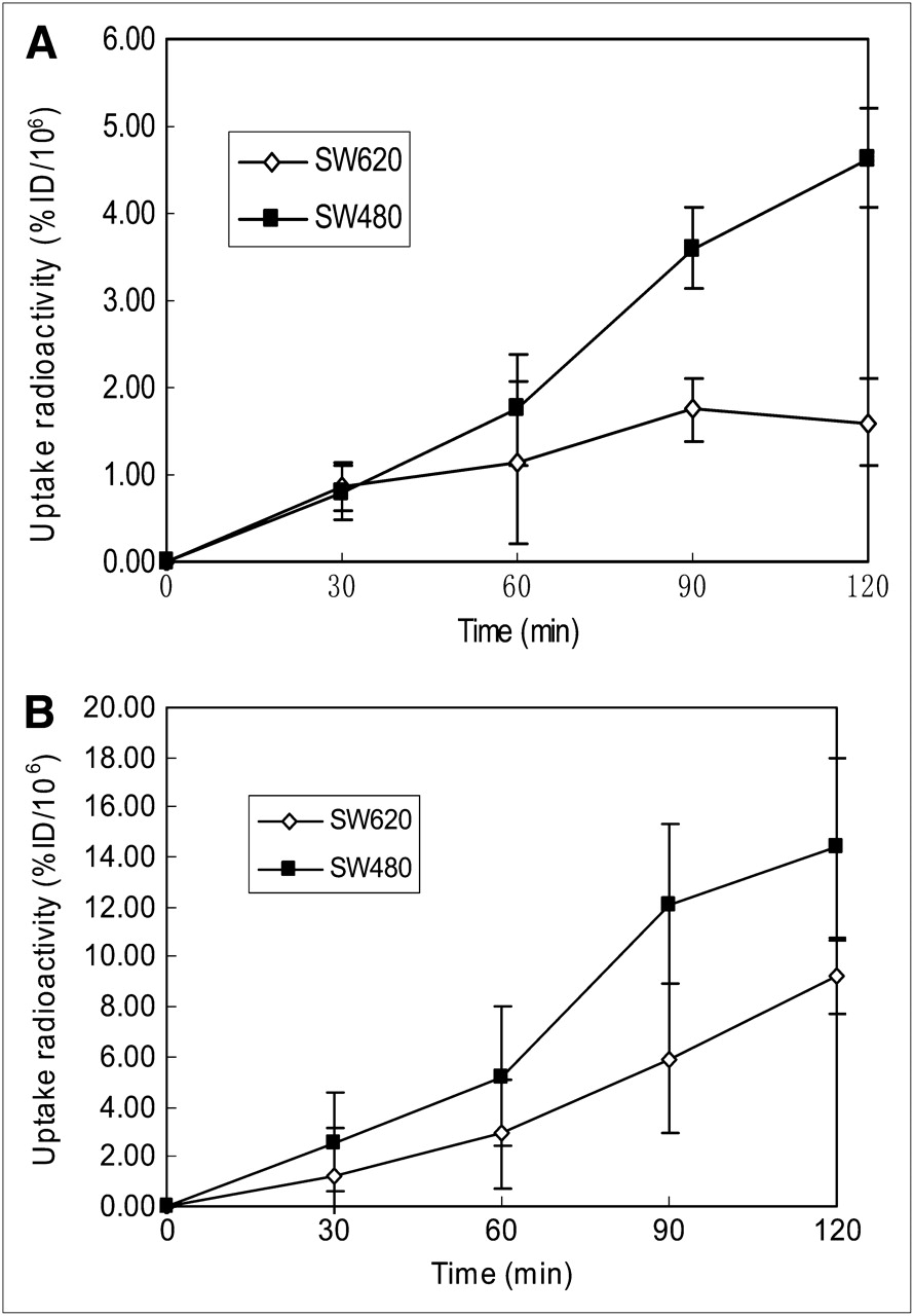
- FIGURE 1.
Cellular uptake of 18F-FDG and 18F-FLT in vitro. Data are expressed as percentage injected dose (%ID) of 18F-FDG (A) or 18F-FLT (B) per 106 cells. Error bar depicts SD (n = 10–15). After incubation for 60 min, uptake of 18F-FDG was different between SW480 and SW620 cells (t = −2.507, P = 0.021). At same time point, uptake of 18F-FLT was significantly different between SW480 and SW620 cells (t = 3.497, P = 0.002).
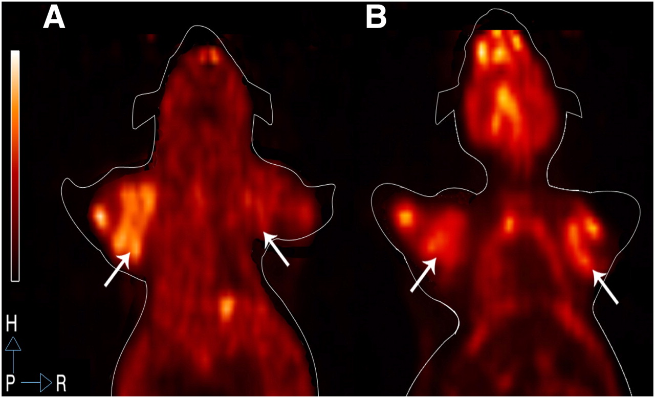
- FIGURE 2.
18F-FLT PET and 18F-FDG PET images of SW480 and SW620 tumors. (A) 18F-FLT PET image shows that uptake of 18F-FLT in SW480 tumors (left) was markedly stronger than that in SW620 tumors (right) (t = 6.491, P < 0.001). (B) 18F-FDG PET image shows that uptake of 18F-FDG in SW480 tumors (left) was weaker than that in SW620 tumors (right), with no significant difference (t = 0.657, P = 0.524) (n = 3 per group).
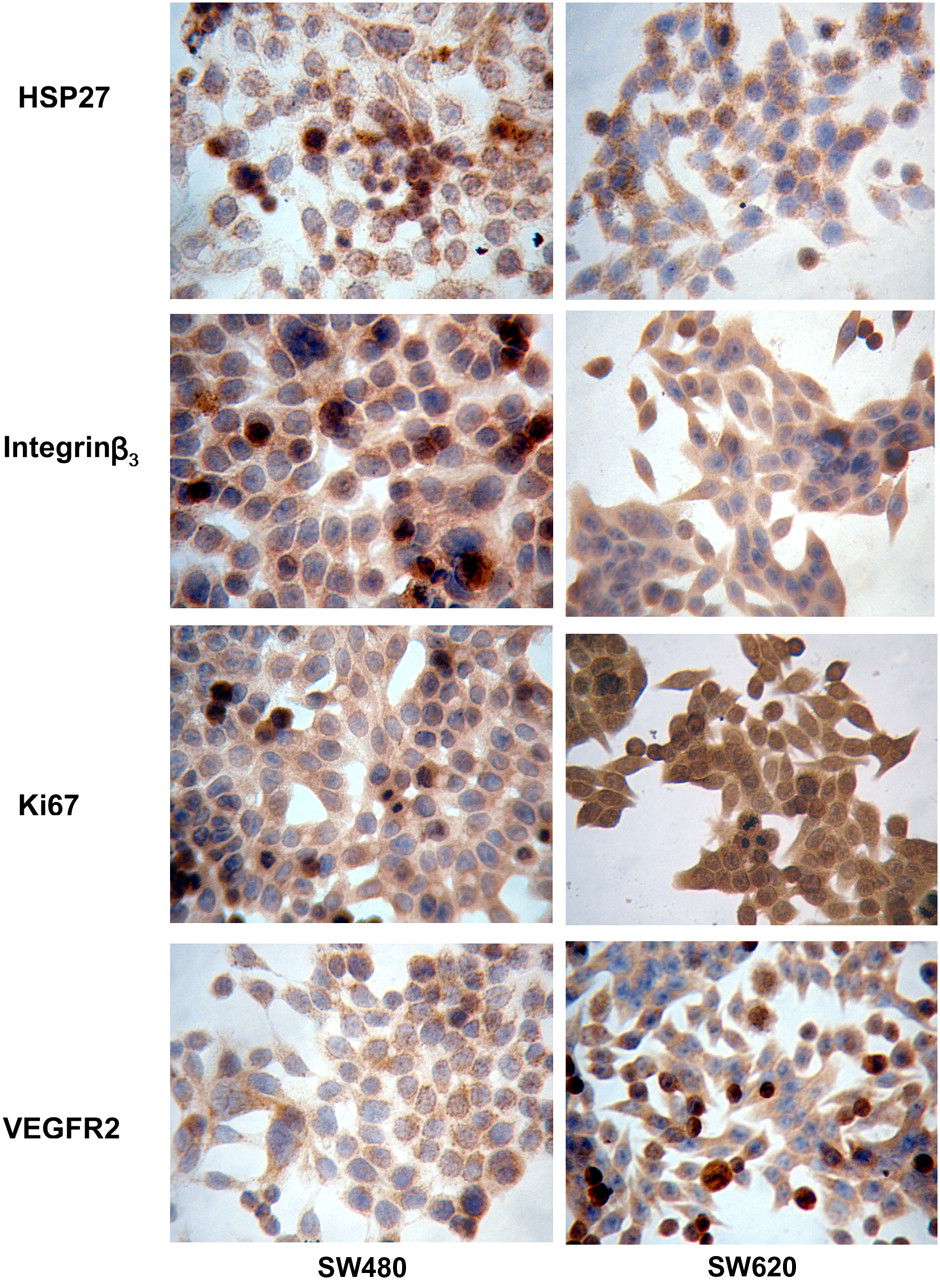
- FIGURE 3.
Expression of HSP27, integrin β3, Ki67, and VEGFR2 in SW480 (left column) and SW620 (right column) cell lines by immunocytochemistry detection (×400). HSP27 expression in cytoplasm and nucleus was significantly higher in SW480 than in SW620. Integrin β3 expression in cytoplasm and cytomembrane was higher in SW480 than in SW620. Ki67 expression in nucleus was higher in SW620 than in SW480. VEGFR2 expression in cytoplasm and cytomembrane was higher in SW620 than in SW480.
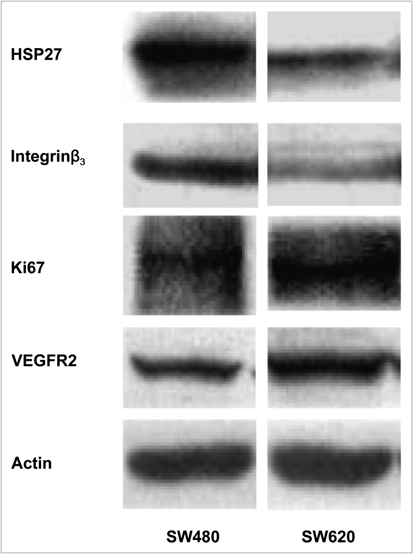
- FIGURE 4.
Western blot analysis of HSP27, integrin β3, Ki67, VEGFR2, and actin expression. Expression of HSP27 and integrin β3 was greater in SW480 tumors than in SW620 tumors. Expression of Ki67 and VEGFR2 was less in SW480 tumors than in SW620 tumors. Actin antibody is loading control.
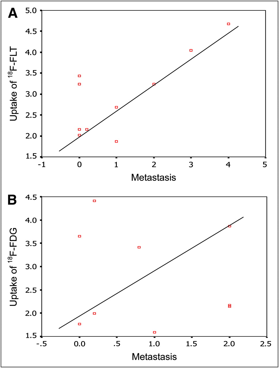
- FIGURE 5.
(A) Linear regression analysis shows significant correlation between metastatic activity and uptake of 18F-FLT PET in CRC (r = 0.763, P = 0.005). (B) No significant correlation is seen between metastatic activity and uptake of 18F-FDG PET in CRC (r = −0.111, P = 0.388).
- FIGURE 6.
Linear regression analysis shows significant correlation between HSP27 expression and T/NT for 18F-FLT PET in CRC (r = 0.924, P = 0.004) (A) and between integrin β3 expression and T/NT for 18F-FLT PET in CRC (r = 0.813, P = 0.025) (B).
- FIGURE 7.
(A) Linear regression analysis shows no significant correlation between survival of tumor-bearing mice and T/NT for 18F-FLT PET (r = 0.262, P = 0.182). (B) Significant correlation is seen between survival of tumor-bearing mice and T/NT for 18F-FDG PET (r = −0.500, P = 0.017).


