
Main menu

  • Home
  • Content
    • Current
    • Ahead of print
    • Past Issues
    • JNM Supplement
    • SNMMI Annual Meeting Abstracts
    • Continuing Education
    • JNM Podcasts
  • Subscriptions
    • Subscribers
    • Institutional and Non-member
    • Rates
    • Journal Claims
    • Corporate & Special Sales
  • Authors
    • Submit to JNM
    • Information for Authors
    • Assignment of Copyright
    • AQARA requirements
  • Info
    • Reviewers
    • Permissions
    • Advertisers
  • About
    • About Us
    • Editorial Board
    • Contact Information
  • More
    • Alerts
    • Feedback
    • Help
    • SNMMI Journals
  • SNMMI
    • JNM
    • JNMT
    • SNMMI Journals
    • SNMMI

User menu

  • Subscribe
  • My alerts
  • Log in
  • My Cart

Search

  • Advanced search
Journal of Nuclear Medicine
  • SNMMI
    • JNM
    • JNMT
    • SNMMI Journals
    • SNMMI
  • Subscribe
  • My alerts
  • Log in
  • My Cart
Journal of Nuclear Medicine

Advanced Search

  • Home
  • Content
    • Current
    • Ahead of print
    • Past Issues
    • JNM Supplement
    • SNMMI Annual Meeting Abstracts
    • Continuing Education
    • JNM Podcasts
  • Subscriptions
    • Subscribers
    • Institutional and Non-member
    • Rates
    • Journal Claims
    • Corporate & Special Sales
  • Authors
    • Submit to JNM
    • Information for Authors
    • Assignment of Copyright
    • AQARA requirements
  • Info
    • Reviewers
    • Permissions
    • Advertisers
  • About
    • About Us
    • Editorial Board
    • Contact Information
  • More
    • Alerts
    • Feedback
    • Help
    • SNMMI Journals
  • View or Listen to JNM Podcast
  • Visit JNM on Facebook
  • Join JNM on LinkedIn
  • Follow JNM on Twitter
  • Subscribe to our RSS feeds
Research ArticleCLINICAL INVESTIGATIONS

99mTc-MIBI Imaging in the Presurgical Characterization of Thyroid Follicular Neoplasms: Relationship to Multidrug Resistance Protein Expression

Enrico Saggiorato, Tiziana Angusti, Rosy Rosas, Morena Martinese, Monica Finessi, Federico Arecco, Edoardo Trevisiol, Nicoletta Bergero, Barbara Puligheddu, Marco Volante, Valerio Podio, Mauro Papotti and Fabio Orlandi
Journal of Nuclear Medicine November 2009, 50 (11) 1785-1793; DOI: https://doi.org/10.2967/jnumed.109.064980
Enrico Saggiorato
  • Find this author on Google Scholar
  • Find this author on PubMed
  • Search for this author on this site
Tiziana Angusti
  • Find this author on Google Scholar
  • Find this author on PubMed
  • Search for this author on this site
Rosy Rosas
  • Find this author on Google Scholar
  • Find this author on PubMed
  • Search for this author on this site
Morena Martinese
  • Find this author on Google Scholar
  • Find this author on PubMed
  • Search for this author on this site
Monica Finessi
  • Find this author on Google Scholar
  • Find this author on PubMed
  • Search for this author on this site
Federico Arecco
  • Find this author on Google Scholar
  • Find this author on PubMed
  • Search for this author on this site
Edoardo Trevisiol
  • Find this author on Google Scholar
  • Find this author on PubMed
  • Search for this author on this site
Nicoletta Bergero
  • Find this author on Google Scholar
  • Find this author on PubMed
  • Search for this author on this site
Barbara Puligheddu
  • Find this author on Google Scholar
  • Find this author on PubMed
  • Search for this author on this site
Marco Volante
  • Find this author on Google Scholar
  • Find this author on PubMed
  • Search for this author on this site
Valerio Podio
  • Find this author on Google Scholar
  • Find this author on PubMed
  • Search for this author on this site
Mauro Papotti
  • Find this author on Google Scholar
  • Find this author on PubMed
  • Search for this author on this site
Fabio Orlandi
  • Find this author on Google Scholar
  • Find this author on PubMed
  • Search for this author on this site
  • Article
  • Figures & Data
  • Info & Metrics
  • PDF
Loading

Article Figures & Data

Figures

  • Tables
  • FIGURE 1. 
    • Download figure
    • Open in new tab
    • Download powerpoint
    FIGURE 1. 

    Thyroid 99mTc-MIBI scan. (A and B) Representative positive case (RI, 71.76) of solitary cold thyroid nodule in right lobe histologically diagnosed as nononcocytic solid/trabecular poorly differentiated carcinoma. (A) Early image with tracer accumulation in nodule (ER, 2.62). (B) Delayed image with tracer retention in lesion (DR, 4.50). (C and D) Representative negative case (RI, −40.64) of solitary cold thyroid nodule in left lobe that proved to be microfollicular goiter. (C) Early image with tracer increase uptake in nodule (ER, 2.81). (D) Delayed image with nearly complete tracer washout from nodule (DR, 1.67).

  • FIGURE 2. 
    • Download figure
    • Open in new tab
    • Download powerpoint
    FIGURE 2. 

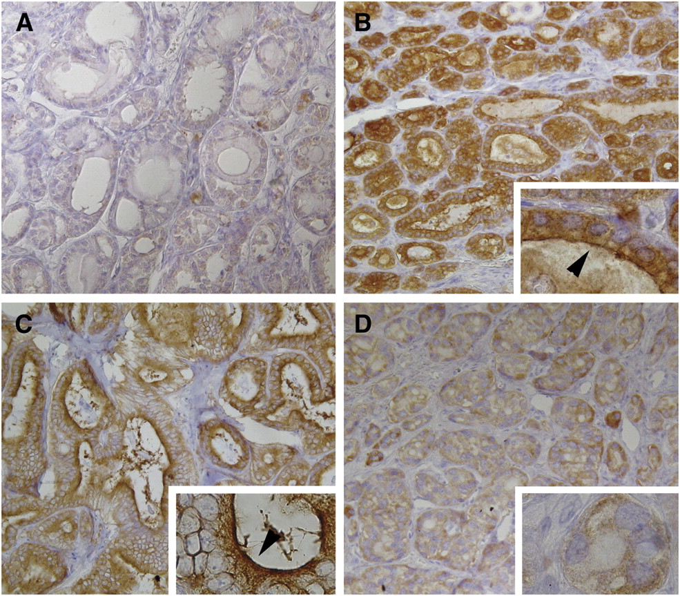
    MDR1 expression in thyroid nodules with cytologic result of follicular neoplasm. (A) No P-gp immunoreactivity of follicular cells in nononcocytic emblematic case (follicular adenoma). (B and C) MRP1 membranous apical immunoexpression in case of nononcocytic microfollicular goiter (B) and in case of nononcocytic follicular variant of papillary carcinoma (C), both with negative RI indices. Cytoplasmic immunoreactivity with granular pattern is seen. (D) No MRP1 membranous apical labeling in case of nononcocytic solid/trabecular papillary carcinoma with positive RI value. Cytoplasmic immunoreactivity with granular pattern is seen. Arrows show linear membranous apical immunoreactivity. Immunoperoxidase with Mayer solution counterstain was used (magnification, ×160; insets: magnification, ×630).

  • FIGURE 3. 
    • Download figure
    • Open in new tab
    • Download powerpoint
    FIGURE 3. 

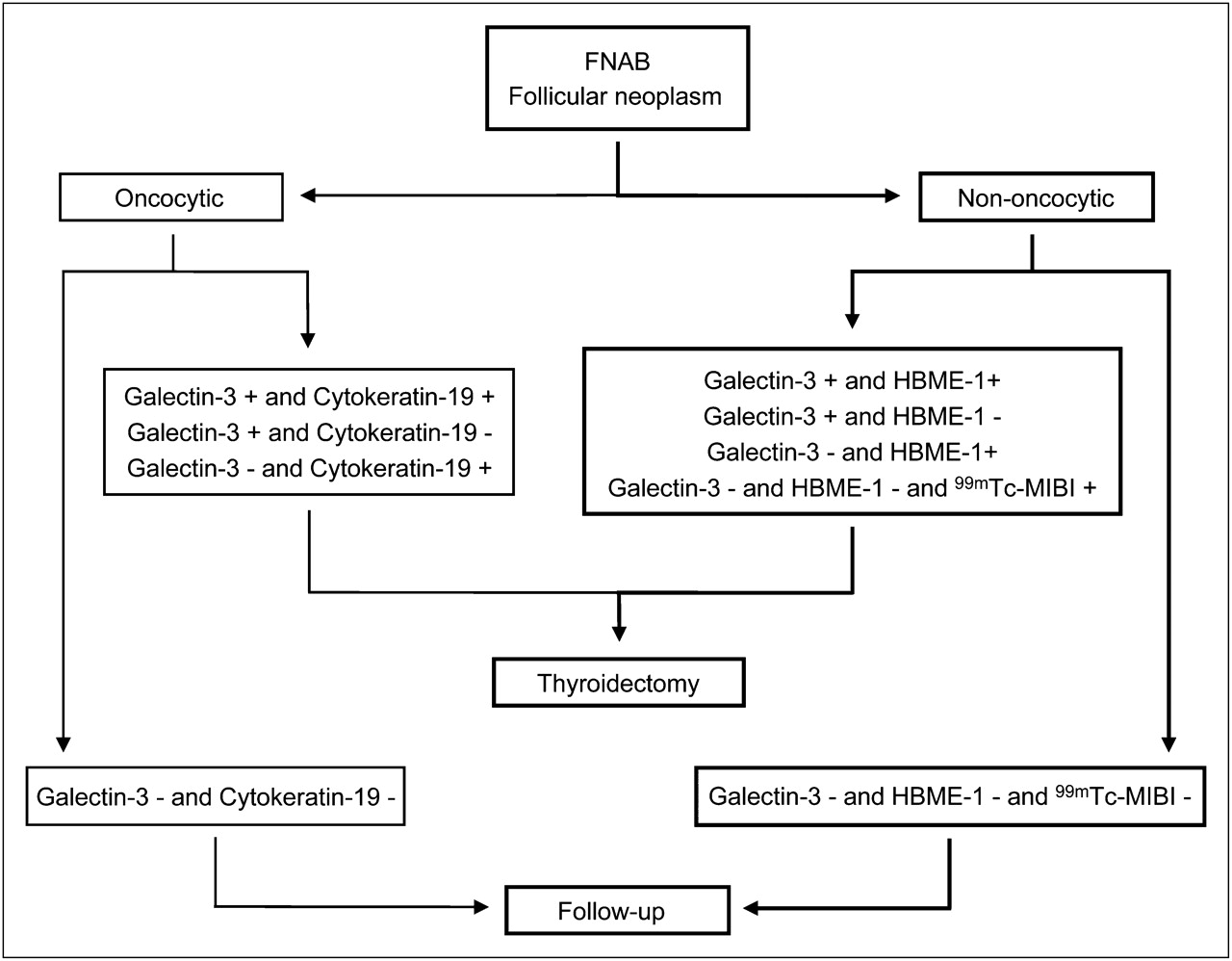
    Proposed decision tree for thyroid follicular neoplasms combining 99mTc-MIBI scanning with molecular marker panel that we previously described (8). For cytologic diagnosis of oncocytic follicular neoplasm, we propose evaluation of galectin-3 and cytokeratin-19. Conversely, for nononcocytic follicular neoplasms, semiquantitative 99mTc-MIBI scintigraphy could be performed when galectin-3 and HBME-1 are negative. Negative results for both markers (galectin-3 and HBME-1) and for 99mTc-MIBI scanning indicate that close follow-up is needed. If molecular testing or 99mTc-MIBI scintigraphy results are positive, patient should be referred for thyroidectomy. HBME-1 = cell surface mesothelial antigen HBME-1.

Tables

  • Figures
    • View popup
    TABLE 1

    Clinicopathologic Data of Patients with Thyroid Follicular Lesions, Visual 99mTc-MIBI Results, and MRP1 Expression

    Visual pattern*Apical expression†
    Sex (M/F)Median age and range (y)Median tumor size and range (cm)NegativePositiveNegativePositive
    Lesion typen0123−++++++
    Nononcocytic
     Microfollicular goiter93/659; 54–692.00; 1.20–4.00342—1152
     Follicular adenoma121/1148; 35–711.85; 1.00–3.50192—3261
     Minimally invasive follicular carcinoma20/239.5; 36–433.65; 3.50–3.80——2—1—1—
     Follicular variant of papillary carcinoma123/951; 28–621.35; 1.00–3.00—48—10—2—
     Poorly differentiated carcinoma11/0724.00——1—1———
    Oncocytic
     Microfollicular goiter51/460; 46–652.50; 1.00–3.00——414—1—
     Follicular adenoma61/546.5; 36–681.75; 1.00–2.90—1413—3—
     Follicular variant of papillary carcinoma30/369; 42–701.60; 1.10–1.60——3—3———
     Insular carcinoma11/0728.00——1—1———
    • ↵* Visual 99mTc-MIBI images were assessed using the following scoring system: pattern 0, no increased uptake of the nodule in either early or delayed images; pattern 1, increased uptake by nodule only in early images; pattern 2, increased uptake by nodule both in early and in delayed images; and pattern 3, increased uptake by nodule only in delayed images. For statistical analysis, visual scintigraphic patterns 0 or 1 and 2 or 3 were considered negative and positive, respectively.

    • ↵† MRP1 apical immunoreactivity was analyzed using the following scale: −, no reactivity; +, <25% positive follicular cells; ++, 25%−50% positive follicular cells; and +++, >50% positive follicular cells. For statistical analysis, MRP1 was considered positive when apical membrane immunoreactivity was observed in >25% of tumor cells (++ and +++).

    • View popup
    TABLE 2

    Discrimination Between Malignant and Benign Nononcocytic Thyroid Lesions by 99mTc-MIBI Analyses

    Type of analysisSensitivity (%)Specificity (%)Positive predictive value (%)Negative predictive valuePositive likelihood ratioAccuracy
    Visual73.33% (44.91–92.05)80.95% (58.08–94.44)73.33% (44.91–92.05)80.95% (58.08–94.44)3.85 (1.51–9.79)77.78%
    Semiquantitative*100% (78.03–100)90.48% (69.58–98.55)88.24% (63.52–98.20)100% (82.20–100)10.50 (2.81–39.24)94.44%
    • ↵* RI threshold level = −11.94.

    • Data in parentheses are confidence intervals.

    • View popup
    TABLE 3

    Semiquantitative 99mTc-MIBI Analysis Results

    BenignMalignant (carcinoma)
    CaseDiagnosisERDRRI*CaseDiagnosisERDRRI†
    Nononcocytic lesions
     TH-01Goiter1.191.3210.81TH-22Follicular1.832.7047.52
     TH-02Goiter2.722.25-17.28TH-23Follicular6.797.206.04
     TH-03Goiter2.811.67-40.64TH-24Papillary0.580.6817.97
     TH-04Goiter1.090.87-20.41TH-25Papillary0.400.4513.64
     TH-05Goiter2.501.40-44.00TH-26Papillary2.012.5325.82
     TH-06Goiter0.840.72-14.91TH-27Papillary2.823.8837.62
     TH-07Goiter1.501.27-15.15TH-28Papillary2.683.0012.15
     TH-08Goiter5.534.67-15.66TH-29Papillary1.942.5028.91
     TH-09Goiter9.753.75-61.54TH-30Papillary1.431.8831.92
     TH-10Adenoma2.324.67101.25TH-31Papillary1.361.8233.97
     TH-11Adenoma3.923.45-11.94TH-32Papillary1.491.6812.73
     TH-12Adenoma1.960.78-60.25TH-33Papillary2.183.0037.50
     TH-13Adenoma1.570.80-49.09TH-34Papillary1.241.23-0.81
     TH-14Adenoma1.800.83-53.70TH-35Papillary1.381.34-2.89
     TH-15Adenoma0.930.81-12.72TH-36Poorly differentiated2.624.5071.76
     TH-16Adenoma0.680.58-14.68
     TH-17Adenoma1.351.18-12.78
     TH-18Adenoma0.440.25-43.75
     TH-19Adenoma1.541.15-25.10
     TH-20Adenoma1.551.25-19.12
     TH-21Adenoma1.140.89-22.22
    Oncocytic lesions
     TH-37Goiter3.003.258.33TH-48Papillary1.241.4113.63
     TH-38Goiter1.692.0018.68TH-49Papillary2.252.365.05
     TH-39Goiter2.123.0041.67TH-50Papillary0.701.0043.33
     TH-40Goiter0.390.5539.39TH-51Insular3.103.8423.84
     TH-41Goiter1.201.4217.88
     TH-42Adenoma1.171.6541.02
     TH-43Adenoma2.905.6795.29
     TH-44Adenoma2.682.07-22.67
     TH-45Adenoma4.003.00-25.00
     TH-46Adenoma2.234.1786.78
     TH-47Adenoma1.761.9611.65
    • ↵* For benign nononcocytic lesions, all but 2 cases showed negative RI values. The 2 RI-positive cases had negative MRP1 apical expression (−). For benign oncocytic lesions, all but 2 cases showed positive RI values. The 2 RI-negative cases had positive MRP1 apical expression (++).

    • ↵† For malignant nononcocytic lesions, all but 2 cases showed positive RI values. The 2 RI-negative cases had positive MRP1 apical expression (++). For malignant oncocytic lesions, all cases showed positive RI values and had negative MRP1 apical expression (−).

PreviousNext
Back to top

In this issue

Journal of Nuclear Medicine: 50 (11)
Journal of Nuclear Medicine
Vol. 50, Issue 11
November 2009
  • Table of Contents
  • Table of Contents (PDF)
  • About the Cover
  • Index by author
Print
Download PDF
Article Alerts
Sign In to Email Alerts with your Email Address
Email Article

Thank you for your interest in spreading the word on Journal of Nuclear Medicine.

NOTE: We only request your email address so that the person you are recommending the page to knows that you wanted them to see it, and that it is not junk mail. We do not capture any email address.

Enter multiple addresses on separate lines or separate them with commas.
99mTc-MIBI Imaging in the Presurgical Characterization of Thyroid Follicular Neoplasms: Relationship to Multidrug Resistance Protein Expression
(Your Name) has sent you a message from Journal of Nuclear Medicine
(Your Name) thought you would like to see the Journal of Nuclear Medicine web site.
Citation Tools
99mTc-MIBI Imaging in the Presurgical Characterization of Thyroid Follicular Neoplasms: Relationship to Multidrug Resistance Protein Expression
Enrico Saggiorato, Tiziana Angusti, Rosy Rosas, Morena Martinese, Monica Finessi, Federico Arecco, Edoardo Trevisiol, Nicoletta Bergero, Barbara Puligheddu, Marco Volante, Valerio Podio, Mauro Papotti, Fabio Orlandi
Journal of Nuclear Medicine Nov 2009, 50 (11) 1785-1793; DOI: 10.2967/jnumed.109.064980

Citation Manager Formats

  • BibTeX
  • Bookends
  • EasyBib
  • EndNote (tagged)
  • EndNote 8 (xml)
  • Medlars
  • Mendeley
  • Papers
  • RefWorks Tagged
  • Ref Manager
  • RIS
  • Zotero
Share
99mTc-MIBI Imaging in the Presurgical Characterization of Thyroid Follicular Neoplasms: Relationship to Multidrug Resistance Protein Expression
Enrico Saggiorato, Tiziana Angusti, Rosy Rosas, Morena Martinese, Monica Finessi, Federico Arecco, Edoardo Trevisiol, Nicoletta Bergero, Barbara Puligheddu, Marco Volante, Valerio Podio, Mauro Papotti, Fabio Orlandi
Journal of Nuclear Medicine Nov 2009, 50 (11) 1785-1793; DOI: 10.2967/jnumed.109.064980
Twitter logo Facebook logo LinkedIn logo Mendeley logo
  • Tweet Widget
  • Facebook Like
  • Google Plus One
Bookmark this article

Jump to section

  • Article
    • Abstract
    • MATERIALS AND METHODS
    • RESULTS
    • DISCUSSION
    • CONCLUSION
    • Acknowledgments
    • Footnotes
    • References
  • Figures & Data
  • Info & Metrics
  • PDF

Related Articles

  • This Month in JNM
  • PubMed
  • Google Scholar

Cited By...

  • Thyroid nodules with indeterminate cytology: prospective comparison between 18F-FDG-PET/CT, multiparametric neck ultrasonography, 99mTc-MIBI scintigraphy and histology
  • DIAGNOSIS OF ENDOCRINE DISEASE: High-yield thyroid fine-needle aspiration cytology: an update focused on ancillary techniques improving its accuracy
  • Google Scholar

More in this TOC Section

  • Feasibility of Ultra-Low-Activity 18F-FDG PET/CT Imaging Using a Long–Axial-Field-of-View PET/CT System
  • Cardiac Presynaptic Sympathetic Nervous Function Evaluated by Cardiac PET in Patients with Chronotropic Incompetence Without Heart Failure
  • Validation and Evaluation of a Vendor-Provided Head Motion Correction Algorithm on the uMI Panorama PET/CT System
Show more Clinical Investigations

Similar Articles

SNMMI

© 2025 SNMMI

Powered by HighWire