


Article Figures & Data
Figures
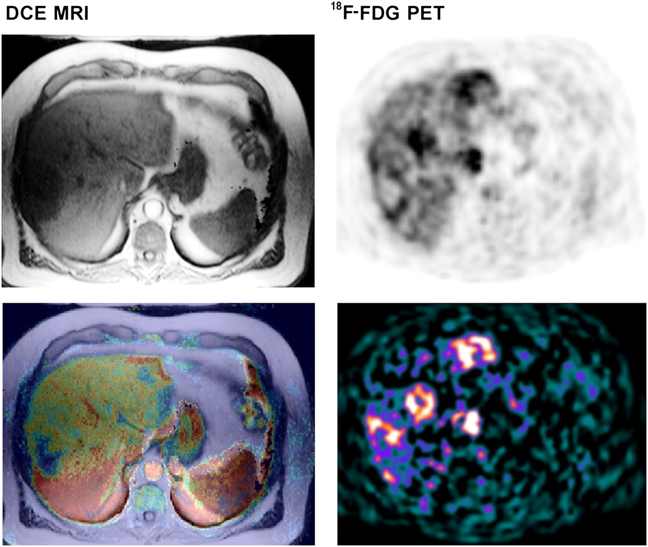
- FIGURE 1.
Example of 78-y-old man with intermediately differentiated adenocarcinoma of sigmoid (T3N1M1) with metachronous liver metastases. (Left, top to bottom) Conventional T1-weighted MR image (before Gd-DTPA) and fused parametric image of kep values with T1-weighted MR image. (Right, top to bottom) 18F-FDG PET uptake image (20–50 min after injection) and parametric image of MRglc.
Tables
Characteristic Value No. of patients 23 Demographic Mean age (y) 61.5 Range (y) 44.8–78.9 No. of men 17 (74%) Matched lesions per patient Mean 1.3 Range 1–5 Patients > 1 matched lesion 5 (22%) Median lesion MAD (mm) 56 IQR (mm) 38–75 Histology Adenocarcinoma 22 (96%) Mucinous adenocarcinoma 1 (4%) Location of primary tumor Sigmoid 9 (39%) Rectum 6 (26%) Colon 5 (22%) Colon and rectum 3 (13%) Presenting stage Stage II 3 (13%) Stage III 4 (17%) Stage IV 16 (70%) - TABLE 2
Predictive Value of Pretreatment Parameters to Survival Assessed by Cox Regression Analysis
OS PFS Parameter HR CI P HR CI P Univariate MRglc* 3.61 1.58–8.26 0.002† 3.11 1.41–6.86 0.005† MAD‡ 1.03 1.00–1.06 0.032† 1.03 1.00–1.07 0.039† Chemotherapy line§ 2.19 0.87–5.51 0.097 2.40 0.96–6.03 0.062 Multivariate MRglc* 4.29 1.72–10.67 0.002† 3.19 1.39–7.35 0.006† MAD‡ 1.04 1.01–1.07 0.020† 1.03 1.00–1.07 0.059 Chemotherapy line§ — — — — — — - TABLE 3
Baseline and Follow-up Values of Vascular and Metabolic Parameters (n = 23 Patients)
Baseline Follow-up Parameter Median IQR Median IQR P DCE-MRI kep (s−1) 0.014 0.005–0.034 0.022 0.012–0.049 0.056 Ktrans (s−1) 0.009 0.003–0.020 0.016 0.008–0.033 0.035* ve 0.638 0.516–0.698 0.614 0.566–0.744 0.893 18F-FDG PET MRglc (μmol·mL−1·min−1) 0.128 0.108–0.160 0.054 0.041–0.122 <0.001* T1-weighted MRI MAD (mm) 56 50–92 54 47–109 0.268 ↵* P < 0.05 (Wilcoxon signed rank test for paired samples).
- TABLE 4
Predictive Value of Therapy-Induced Parameter Changes for Early Response Evaluation, Assessed by Cox Regression Analysis
OS PFS Parameter HR CI P* HR CI P* Univariate ΔMRglc (all in FOV)† 1.15 1.01–1.32 0.041 — — — ΔMAD† 1.40 1.06–1.85 0.023 1.34 1.04–1.74 0.026 Chemotherapy line‡ — — — — — — Multivariate ΔMRglc (all in FOV)† 1.22 1.05–1.41 0.008 — — — Δkep† 0.99 0.98–1.00 0.100 — — — ΔMAD† — — — 1.48 1.10–1.99 0.010 Chemotherapy line‡ — — — 3.15 1.17–8.49 0.023


Jump to section
Related Articles
Cited By...
- No citing articles found.