


Article Figures & Data
Figures
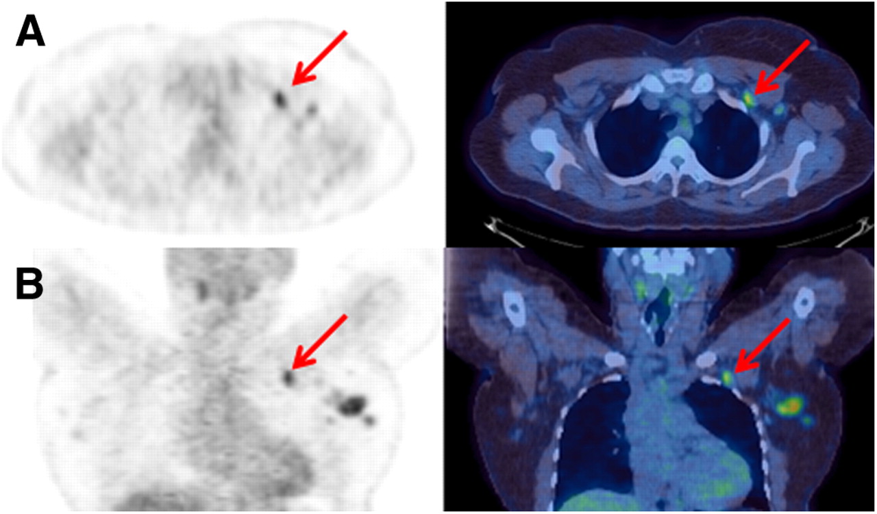
- FIGURE 1.
Axial (A) and coronal (B) PET and PET/CT images demonstrating focal increased metabolic activity in subcentimeter node (arrow) that was variably reported as supraclavicular, infraclavicular, or axillary. This did not result in a change in overall stage due to the presence of widespread disease, and this variability would therefore not change management.
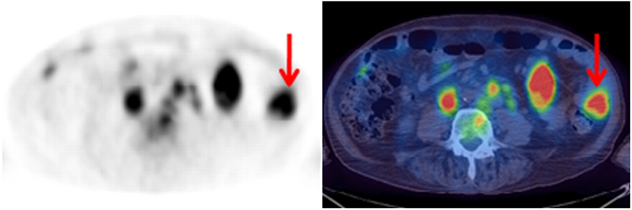
- FIGURE 2.
Axial PET and PET/CT images demonstrating large-bowel lymphomatous involvement (arrow) in addition to mesenteric nodal involvement. Some reviewers reported only mesenteric nodal involvement, but involvement of bowel wall was clearly evident on review.
Tables
Subtype n Diffuse large B cell 40 Hodgkin 32 Follicular 9 T cell 9 High-grade non-Hodgkin, unspecified 3 Burkitt 1 Anaplastic large cell 2 Mantle cell 2 Uncertain 1 Posttransplant lymphoproliferative disorder 1 - TABLE 2
Percentage of Patients with Lymphomatous Involvement at Specific Nodal and Extranodal Sites
Site Percentage Nodal Cervical 55.8 (L); 47.4 (R) Axillary 28.8 (L); 28.0 (R) Infraclavicular 6.4 (L); 4.2 (R) Hilar 23.0 Mediastinal 38.8 Periaortic 37.8 Pelvic 31.4 (L); 31.6 (R) Inguinal 33.6 (L); 28.6 (R) Extranodal Spleen 17.8 Bone marrow or bone 20.0 Lung 5.4 Liver 10.0 Bowel or gastric 5.4 Other nodal sites* 8.0 ↵* Muscle, subcutaneous tissue, breast, and uterus.
Intraobserver Interobserver Parameter 1 2 1 vs. 2 2 vs. 3 1 vs. 3 Ann Arbor 0.91 (0.82–0.97) 0.88 (0.80–0.95) 0.81 (0.69–0.90) 0.79 (0.68–0.86) 0.87 (0.77–0.94) Extranodal 0.82 (0.70–0.93) 0.86 (0.76–0.96) 0.74 (0.61–0.87) 0.82 (0.71–0.93) 0.76 (0.63–0.89) No. of nodal groups 0.93 (0.90–0.96) 0.91 (0.87–0.94) 0.83 (0.76–0.89) 0.92 (0.89–0.95) 0.88 (0.83–0.92) Nodal sites Cervical* 0.84 (0.72–0.92) 0.86 (0.78–0.93) 0.81 (0.71–0.90) 0.77 (0.66–0.86) 0.79 (0.68–0.88) Axillary 0.80 (0.68–0.89) 0.74 (0.61–0.85) 0.69 (0.56–0.81) 0.69 (0.53–0.83) 0.73 (0.60–0.84) Infraclavicular 0.55 (−0.01–0.89) 0.39 (0.10–0.73) 0.23 (−0.02–0.55) 0.37 (−0.01–0.68) 0.14 (−0.04–0.50) Hilar 0.82 (0.70–0.95) 0.65 (0.48–0.83) 0.56 (0.37–0.75) 0.58 (0.36–0.79) 0.63 (0.45–0.81) Pelvic 0.82 (0.70–0.91) 0.68 (0.53–0.81) 0.65 (0.53–0.76) 0.71 (0.60–0.82) 0.68 (0.55–0.79) Inguinal or femoral 0.82 (0.72–0.91) 0.69 (0.55–0.82) 0.71 (0.59–0.82) 0.76 (0.64–0.87) 0.69 (0.55–0.82) Mediastinal 0.75 (0.61–0.88) 0.75 (0.61–0.88) 0.75 (0.61–0.88) 0.77 (0.64–0.90) 0.73 (0.59–0.87) Periaortic 0.77 (0.65–0.90) 0.76 (0.63–0.89) 0.75 (0.62–0.88) 0.81 (0.69–0.93) 0.78 (0.65–0.90) Mesentery 0.65 (0.47–0.83) 0.61 (0.41–0.81) 0.63 (0.44–0.81) 0.61 (0.41–0.81) 0.67 (0.49–0.85) Extranodal sites Spleen 0.84 (0.69–0.99) 0.81 (0.67–0.96) 0.75 (0.57–0.92) 0.81 (0.67–0.96) 0.69 (0.51–0.88) Bone marrow 0.94 (0.85–0.99) 0.81 (0.67–0.96) 0.76 (0.60–0.92) 0.76 (0.60–0.92) 0.93 (0.84–0.99) Lung 0.82 (0.58–0.99) 0.90 (0.72–0.99) 0.58 (0.21–0.95) 0.58 (0.21–0.95) 0.65 (0.32–0.97) Liver 0.95 (0.84–0.99) 0.88 (0.71–0.99) 0.84 (0.66–0.99) 0.78 (0.57–0.99) 0.59 (0.32–0.87) Bowel 0.71 (0.40–0.99) 0.56 (0.11–0.99) 0.64 (0.35–0.93) 0.59 (0.28–0.91) 0.37 (−0.03–0.76) ↵* Cervical includes supraclavicular and all head and neck nodal stations.
Data are intraclass correlation coefficient for number of nodal groups and κw for all other variables, with 95% confidence intervals in parentheses.


Jump to section
Related Articles
Cited By...
- Interobserver Agreement of Interim and End-of-Treatment 18F-FDG PET/CT in Diffuse Large B-Cell Lymphoma: Impact on Clinical Practice and Trials
- In Newly Diagnosed Diffuse Large B-Cell Lymphoma, Determination of Bone Marrow Involvement with 18F-FDG PET/CT Provides Better Diagnostic Performance and Prognostic Stratification Than Does Biopsy
- PET-CT staging of DLBCL accurately identifies and provides new insight into the clinical significance of bone marrow involvement